


 Close
Close
 29th May 2023
Common Haematological Malignancies
Acute leukaemia
Acute myeloblastic leukaemia (AML)
Overview
AML is a life-threatening malignancy in which a clonal population of early myeloid progenitors called blasts proliferate in the bone marrow, blood and sometimes extramedullary tissues. It is characterised particularly by cytogenetic abnormalities, but also genetic and epigenetic mutations. It is the most common type of acute leukaemia. The incidence in the UK is 4.05 per 100,000 people per year. It can present at any age, but incidence increases with age and the median age at diagnosis is 67.
As the blast population increases, there is progressively less room for healthy blood cell maturation, leading to a drop (usually) in red cells, normal white cells and platelets. Most symptoms of AML are attributable to these cytopenias. AML is a rapidly progressive condition, and the symptoms often develop quickly.
- Symptomatic anaemia
- Bleeding – from low platelets but also DIC
- Infection
- Hepatosplenomegaly (sometimes)
- Bone pain
- Gum infiltration
The white cell count (WCC) may be high or low at presentation. If the WCC is markedly raised (>100), this can cause hyperviscosity and leukostasis (sluggish blood flow). This tends to present with pulmonary infiltrates and hypoxia. This is a medical emergency and needs immediate action with leukapheresis and cytoreductive therapy.
Investigations
- Full panel of bloods, especially
- FBC – often anaemia and thrombocytopenia. WCC may be raised (due to blasts), normal or low. Neutropenia is common
- Coagulation screen – to look for DIC
- Blood film – often shows circulating blasts. Can be hard to distinguish from ALL but presence of Auer rods in blasts confirms AML
- It may be possible to perform flow cytometry (immunophenotyping) on peripheral blood to confirm the diagnosis, plus cytogenetics and molecular genetic analysis
- Bone marrow aspirate and trephine are essential to confirm the presence of blasts – diagnosis usually requires 20% blast percentage in the marrow. Flow, cytogenetics and molecular studies are also performed on this sample
- Further tests can include echo/multigated acquisition (MUGA) scan pre-chemo
Treatment
AML is potentially curable. However, the treatment is highly intense, with significant toxicity and so is largely delivered as an inpatient. Patients who are older in age (>65 as a general guide) or with significant comorbidities may not be fit enough for this treatment.
For patients treated with curative intent, this involves highly myelotoxic chemotherapy delivered in cycles – first in an induction phase and then a consolidation phase. Treatment and diagnostics have improved considerably in AML and patients are stratified into favourable, standard and unfavourable risk groups based on cytogenetic, genetic and epigenetic profile. This not only impacts prognosis but also treatment strategy. Most patients achieve remission with treatment, but patients with unfavourable risk profiles are much more likely to relapse. These patients may be offered allogeneic stem cell transplant after induction chemotherapy. Patients with a favourable risk profile would normally only be offered a transplant if they relapse, as the risks of a transplant outweigh the potential benefit given their lower chance of relapse.
Prognostic factors
The following are associated with a worse prognosis:
- Increasing age
- High risk features from cytogenetics or genetic analysis
- Progression to AML from pre-existing haematological disorder e.g. myelodysplastic syndrome (MDS), myeloproliferative neoplasm (MPN)
- Development of AML in context of previous chemotherapy (treatment associated)
Acute promyelocytic leukaemia
This is a variant of AML that is important to recognise as it commonly presents as an emergency with DIC and can lead to catastrophic bleeds. It is associated with a t(15;17) PML-RARA translocation gene, which fuses the promyelocytic leukaemia gene with the retinoic acid receptor alpha gene, leading to a block in cellular maturation and marked proliferation. Blasts in APML are promyelocytes and they have distinctive morphological features (pronounced cytoplasmic granulation and Auer rods with prominent nucleoli).
The treatment for APML is very different. It is based around all-trans retinoic acid (ATRA) and sometimes arsenic. It is imperative that treatment is started as soon as possible, and very close supportive care is necessary. If the diagnosis is suspected from morphology, treatment should be initiated without awaiting confirmation of genetics.
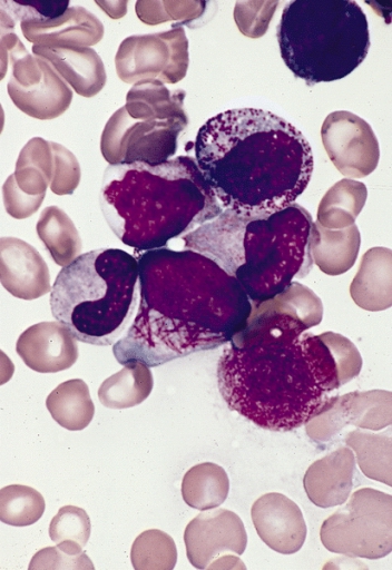
Blood film showing promyelocyte with Auer rods.
Image Source: Wikimedia Commons (2008). Faggot cell in AML-M3, Licensed under a creative commons licence. Available for free at: https://commons.wikimedia.org/wiki/File:Faggot_cell_in_AML-M3.jpg (Accessed on: 10 January 2023).

Acute lymphoblastic leukaemia (ALL)
Overview
Acute lymphoblastic leukaemia is similar to AML, but the progenitor cells are lymphoid blast cells rather than myeloid. Mostly, these cells are B-cells in origin, but they can also be T-cells. In contrast to AML, most cases present in childhood, and it is the most common cancer in children. The peak age is between 2-4 years old.
Like AML, as the blast clone increases in size it causes progressive cytopenia. However, in ALL it is more common to see lymphadenopathy and extramedullary tissue infiltration, especially testicular. Central nervous system (CNS) disease is also significantly more common in ALL.
Investigations
- Full panel of bloods, especially
- FBC – often anaemia and thrombocytopenia. WCC may be raised (due to blasts), normal or low. Neutropenia is common
- Coagulation screen – to look for DIC
- Blood film – often shows circulating blasts. Can be hard to distinguish from AML but cytoplasm is more basophilic and less granular, and Auer rods will not be present
- It may be possible to perform flow cytometry (immunophenotyping) on peripheral blood to confirm the diagnosis, plus cytogenetics and molecular genetic analysis
- Bone marrow aspirate and trephine are essential to confirm the presence of blasts – diagnosis usually requires 20% blast percentage in the marrow. Flow, cytogenetics, and molecular studies are also performed on this sample
- Lumbar puncture – CNS disease is more common at presentation than with AML
Further tests can include echo/MUGA scan pre-chemo.
Treatment
Treatment consists of intense chemotherapy, with much of it being delivered as an inpatient. It consists of an induction phase, a consolidation phase and then maintenance treatment. This also incorporates treatment that can penetrate the blood brain barrier (for CNS disease prophylaxis). The regimens differ depending on the age of the patient and the results of cytogenetic analysis. In children over the age of 1, cure rates are up to 90%, but this is considerably lower in adults and infants. Higher WCC at diagnosis is associated with poor outcomes. Also, certain cytogenetic abnormalities confer a worse prognosis, such as t(9;22) Philadelphia chromosome.
Chronic leukaemia
Chronic myeloid leukaemia (CML)
Overview
CML is a type of (usually) slow-growing myeloproliferative neoplasm. The affected cells are haematological pluripotent stem cells, and these cells continue to undergo maturation into all three subsequent myeloid lineages – erythroid, platelet and granulocytes.
More than 90% of CML is associated with the Philadelphia chromosome BCR-ABL, which is caused by the gene translocation t(9;22). This fusion gene codes for the synthesis of a constitutively active tyrosine kinase signalling protein which leads to uncontrolled cell division and loss of cell cycle regulation in affected cells.
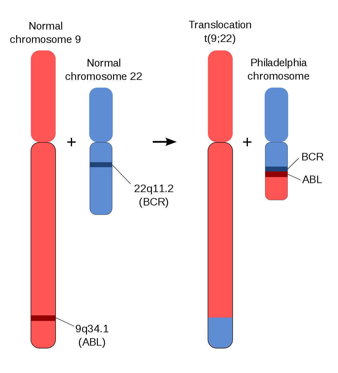
Schematic of Philadelphia chromosome translocation.
Image Source: Wikimedia Commons (2012). Schematic of the Philadelphia Chromosome, Licensed under a creative commons licence. Available for free at: https://commons.wikimedia.org/wiki/File:Schematic_of_the_Philadelphia_Chromosome.svg (Accessed on: 10 January 2023).

The incidence is 4.2 per 100,000 people per year. It can present at any age but the median age at diagnosis is 72 years.
Most cases are detected asymptomatically on routine bloods, but features can include:
- Constitutional symptoms
- Splenomegaly
- Symptomatic cytopenia – anaemia, bleeding, infections
- Gout (due to high cell turnover)
- Hyperviscosity (due to high WCC)
The following tests are usually performed during workup for suspected CML:
- Bloods – full panel, especially:
- FBC – leukocytosis is common with neutrophils, basophils and eosinophils. Thrombocytopenia and anaemia (normocytic) develop over time usually
- Blood film shows granulocytes of all stages of maturation
- BCR:ABL analysis can often be run on peripheral blood and is quantifiable – the percentage of BCR:ABL positive cells can be monitored as response to treatment
- Bone marrow is essential for evaluating blast percentages and staging disease and cytogenetics can also be sent
CML can be divided into three stages:
- Chronic phase – Most people are diagnosed in the chronic stage – counts are relatively well maintained and symptoms are minimal. This often lasts years.
- Accelerated phase – With rising blast counts (up to 29%), falling RBC and platelets and increasing symptoms.
- Blast phase – Finally in the blast phase the blast percentage rises to over 30% and the disease behaves like AML but with a very poor response to chemotherapy. The blast phase of CML is often rapidly fatal.
Treatment for CML has been revolutionised by the development of targeted therapy. The target is the tyrosine kinase protein produced by the BCR:ABL gene. These drugs are known as tyrosine kinase inhibitors; the first example of this class of drug was imatinib but there are now also second and third generation forms. They are generally well tolerated and have drastically changed prognosis for patients diagnosed with CML. The ten-year life expectancy has risen from around 11% to 84%, and patients diagnosed in the chronic phase have an almost normal life expectancy.
Chronic lymphocytic leukaemia (CLL)
Overview
Chronic lymphocytic leukaemia (CLL) is a malignant clonal expansion of immature B-lymphocytes. It is typically a slowly progressive condition and is increasingly common with age. The median age of diagnosis is 72 years. Only 10% are diagnosed under 55 years.
It is most commonly diagnosed on routine blood tests and is asymptomatic at presentation. Symptoms can include:
- Constitutional symptoms
- Enlarged lymph nodes or spleen
- Anaemia
- Bleeding symptoms from thrombocytopenia
- Increased susceptibility to infection
Diagnostic criteria:
- Persistent clonal lymphocytes >5 x 10⁹/L for more than 3 months
- Immunophenotype consistent with CLL
- Morphologically consistent with lymphocytes
Lymphadenopathy may or may not be present. There is an association of CLL with autoimmune phenomena such as Immune thrombocytopenia (ITP) and autoimmune haemolytic anaemia (AIHA).
Investigations for suspected CLL:
- Bloods – full panel, especially:
- FBC to look for cytopenia
- Blood film to evaluate morphology of lymphocytes
- Immunoglobulins, viral screen, DAT screen (AIHA)
- Flow cytometry to confirm CLL plus analysis for TP53 mutations and IGHV mutations (relevant for prognosis +/- treatment)
- CT scan – only if concerns about lymphadenopathy or splenomegaly
- Lymph node biopsy – only if large or rapidly growing lymph nodes to look for Richter’s transformation*
- Bone marrow biopsy – not always performed
*In a small proportion of cases, CLL can undergo a phenomenon known as Richter’s transformation. A proportion of clonal lymphocytes will mutate and take on the phenotype of a diffuse large B cell lymphoma. This carries a very poor prognosis.

Blood film showing typical appearance of CLL lymphocytes.
Image Source: Wikimedia Commons (2006). Chronic lymphocytic leukemia, Licensed under a creative commons licence. Available for free at: https://commons.wikimedia.org/wiki/File:Chronic_lymphocytic_leukemia.jpg (Accessed on: 10 January 2023).

Staging
Traditionally the Binet system was used in Europe, whereas the Rai system was used in the US. It is now more informative to use CLL-IPI, although the Rai-Binet system is still in use.
Binet A –Hb >100 g/L, Platelets >100 x 10⁹/L, <3 lymph node areas involved
Binet B – Hb and platelets as Binet A and >3 lymph node areas
Binet C – Hb <100 g/L or platelets <100 x 10⁹/L
Treatment
Not everyone needs treatment. CLL is a slow growing cancer and treatment carries significant morbidity risk. Most patients are managed with a ‘watch and wait’ approach to begin with. Treatment is not considered curative, and the disease course is of a relapsing and remitting nature.
Indications for treatment include:
- Progressive or troublesome lymphadenopathy
- Progressive splenomegaly
- Constitutional symptoms
- Cytopenias (Hb <100 g/L, Platelets <100 x 10⁹/L)
- Recurrent infections
- Associated immune phenomena e.g. AIHA not responsive to other treatment
- Lymphocyte doubling time < 6 months*
*This is a relative indication only
Treatment options themselves are beyond the scope of this course but include a combination of immunotherapy and conventional chemotherapy such as Fludarabine, Cyclophosphamide and Rituximab. Newer options such as Bruton Tyrosine Kinase inhibitors e.g. ibrutinib can also be used. The decision for treatment is determined by factors such as fitness for treatment, previous therapy, and mutation status of TP53. Some treatment courses can be quite intense (such as FCR) whereas ibrutinib is usually well tolerated.
Stem cell transplantation
Types of stem cell transplant
Autologous stem cell transplant
The patient’s own stem cells are harvested (usually during a period of remission) and then returned to the patient after conditioning chemotherapy. The principle for this is to allow higher doses of chemotherapy to be given that would normally risk permanent bone marrow suppression. As the stem cells returned are the patient’s own, there is no risk of Graft Versus Host Disease. This procedure carries a mortality of 1-2%.
Allogeneic stem cell transplant
Stem cells are harvested from either a matched sibling donor or an unrelated but matched donor. They are then infused into the patient after conditioning chemotherapy. This both allows for high dose chemotherapy to be given but also can lead to a Graft Versus Leukaemia effect which is believed to reduce the risk of recurrence of the disease. There is also the possibility for Graft Versus Host Disease. The mortality is higher for this procedure, in the region of 10-20%.
Indications
Autologous stem cell transplant:
- 1st line treatment for fit patients with myeloma
- 1st line treatment for certain lymphomas
- After first relapse from high grade lymphoma
- Some solid tumours such as germ cell tumours
Allogeneic stem cell transplant:
- High risk acute leukaemia after induction chemotherapy
- Relapsed acute leukaemia
- Relapsed lymphoma
- Chronic leukaemia
- Non-haematological conditions e.g. immunodeficiency syndromes
Complications of stem cell transplant
- Immunosuppression leading to infection
- Cytopenias
- Failure of stem cells to engraft
- Chemo side effects
- Graft Versus Host Disease
- Death
Lymphomas
Hodgkin’s lymphoma
Overview
Hodgkin’s disease is a B cell lymphoproliferative malignancy characterised by the presence of Reed Sternberg and Hodgkin cells surrounded by an inflammatory infiltrate. It has an incidence of 2.7 cases per 100,000 people per year in the UK. There is a bimodal distribution in Hodgkin’s with peaks in the 15-34 age group and over 70s.
The aetiology is not completely understood but is thought to be linked to infection, and 50% of cases show infection with EBV. HIV and immunosuppression are also risk factors.
Most people present with painless lymphadenopathy, typically in the lower neck or supraclavicular region. Mediastinal involvement is common and splenomegaly or distant lymphadenopathy is possible. B symptoms are present in only 25% (fevers, 10% weight loss in 6 months and drenching night sweats). Pruritus and alcohol induced lymph node pain is also possible.
Histological types of Hodgkin’s lymphoma
Hodgkin’s can be divided into classical and non-classical subtypes. Classical Hodgkin’s subtypes all show evidence of Reed Sternberg and Hodgkin cells and behave in a similar fashion. They are treated the same way and carry a similar prognosis.

Close up of tissue specimen showing a Reed-Sternberg cell.
Image Source: Wikimedia Commons (2008). Reed-sternberg cell, Licensed under a creative commons licence. Available for free at: https://commons.wikimedia.org/wiki/File:Reed-sternberg_cell.jpg (Accessed on: 10 January 2023).

Classical subtypes include
- Nodular sclerosis (70%)
- Mixed cellularity (25%)
- Lymphocyte-rich (5%)
- Lymphocyte-depleted (<1%)
Nodular lymphocyte predominant HD (Non-classical) makes up 5% of all Hodgkin’s disease. It behaves quite differently to Classical Hodgkin’s and does not show evidence of Reed Sternberg cells. It tends to progress more slowly.
Diagnosis
- Bloods
- Full panel to look for cytopenias, renal and liver dysfunction pre-chemo
- HBV, HCV, HIV prior to chemo (risk of reactivation)
- Imaging – PET CT is first line now
- Biopsy – ideally excision biopsy, but at least core biopsy. No role for FNA.
Staging (Lugano system)
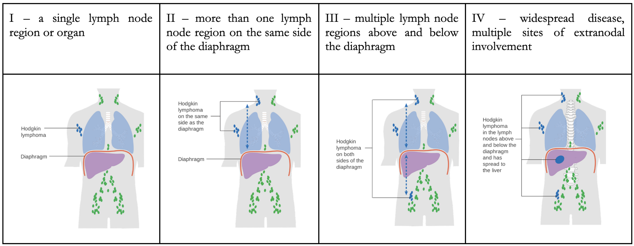
Adapted image of Lugano staging system.
Image Sources:
- Wikimedia Commons (2014). Diagram showing stage 1 Hogkin’s lymphoma CRUK 191, Licensed under a creative commons licence. Available for free at: https://commons.wikimedia.org/wiki/File:Diagram_showing_stage_1_Hogkin’s_lymphoma_CRUK_191.svg (Accessed on: 10 January 2023).
- Wikimedia Commons (2014). Diagram showing stage 2 Hodgkin’s lymphoma CRUK 208, Licensed under a creative commons licence. Available for free at: https://commons.wikimedia.org/wiki/File:Diagram_showing_stage_2_Hodgkin’s_lymphoma_CRUK_208.svg (Accessed on: 10 January 2023).
- Wikimedia Commons (2014). Diagram showing stage 3 Hodgkin’s lymphoma CRUK 221, Licensed under a creative commons licence. Available for free at: https://en.wikipedia.org/wiki/File:Diagram_showing_stage_3_Hodgkin’s_lymphoma_CRUK_221.svg (Accessed on: 10 January 2023).
- Wikimedia Commons (2014). Diagram showing stage 4 Hodgkin’s lymphoma CRUK 230, Licensed under a creative commons licence. Available for free at: https://commons.wikimedia.org/wiki/File:Diagram_showing_stage_4_Hodgkin’s_lymphoma_CRUK_230.svg (Accessed on: 10 January 2023).


Treatment
Treatment for Hodgkin’s disease aims to achieve a balance between the risk of remission and the risk of long-term complications from toxicity from chemotherapy.
Treatment is determined by whether the disease is early or advanced.
Early disease (stage I or II) can be divided into favourable or unfavourable, based upon factors such as ESR, presence of B symptoms, age, and bulk of disease. Early-stage disease is usually treated with a combination of chemotherapy and radiotherapy and treatment can be escalated or de-escalated in intensity depending on the response to treatment, which is measured through interval PET-CT scan.
Advanced disease is treated with chemotherapy, which is more intense than with early disease.
A cure is possible for both early and advanced disease. Long term remission is achieved in around 90% of early-stage disease and 60-80% of advanced-stage disease.
One of the major complications of treatment for Hodgkin’s disease is the long-term sequelae of the treatment itself. This includes:
- Second malignancy – skin cancer, MDS, AML, breast cancer, thyroid cancer
- Cardiovascular disease – MI, angina, CHF, valvular disorders
- Pulmonary toxicity
- Impaired fertility
Patients with Hodgkin’s disease need irradiated blood products for life.
Non-Hodgkin’s lymphoma
Overview
Non-Hodgkin’s lymphoma (NHL) is an umbrella term for all other forms of lymphoma and comprises a phenotypically disparate group of diseases, which all arise from a malignant lymphoid proliferation. There are more than 30 different types of NHL.
NHL can be divided in a number of ways, most commonly into high or low grade. They can also be divided into B- or T-cell lymphomas. In the UK, 9 out of 10 lymphomas are B-cell in origin.
High-grade lymphomas grow quickly and cause significant morbidity. They do however tend to be more sensitive to chemotherapy and this offers the possibility of a cure. Low-grade lymphomas tend to behave quite indolently, and some may never need treatment. If treatment is given, it is with the aim to produce a period of remission, with relapse at some point in the future likely. Low grade lymphoma may occasionally transform into a high-grade phenotype.
There is a huge variety in how NHL can present with almost any symptoms possible.
- Progressive painless lymphadenopathy is the most common feature
- Compression of surrounding structures is frequent, particularly with high grade lymphomas:
- Renal/ureteric obstruction
- Bowel obstruction
- SVC obstruction
- ‘B’ symptoms are common (drenching night sweats, fevers, >10% weight loss in a 6-month period)
- Hepatomegaly and/or splenomegaly
- Cytopenias if bone marrow involvement
- Extranodal involvement can affect almost anywhere including CNS disease
- Skin involvement is most commonly seen in T-cell lymphoma
Subtypes of NHL
Examples of high-grade lymphoma:
- Diffuse large B cell lymphoma (DLBCL) – most common (40%) type of NHL
- Burkitt’s lymphoma
- Mantle cell lymphoma
- Post-transplant lymphoproliferative disorder (PTLD)
- Primary CNS lymphoma
- Enteropathy associated T cell lymphoma (EATL)
- Peripheral T cell lymphoma NOS
- Anaplastic large cell lymphoma
Examples of low-grade lymphoma:
- Follicular lymphoma
- CLL/Small lymphocytic lymphoma
- Lymphoplasmacytic lymphoma (LPL)
- Marginal zone lymphoma including MALT lymphoma
- Hairy cell leukaemia
- Mycosis fungoides
Many types of NHL are closely linked with infectious/inflammatory conditions and immunodeficiency syndromes.
| Infections | EBV | Burkitt’s, PTLD |
| HIV | Multiple types, especially primary CNS lymphoma | |
| H. pylori | Gastric MALT lymphoma | |
| C. psittaci | Ocular MALT lymphoma | |
| HTLV-1 | T cell lymphoma | |
| Hepatitis C | Multiple types NHL | |
| Autoimmune | Hashimoto’s | Thyroid lymphoma |
| Sjogren’s | Salivary gland lymphoma | |
| Coeliac | EATL | |
PTLD is an umbrella term for a spectrum of lymphoproliferative disorders occurring due to immunosuppression following both solid organ and stem cell transplant. It is uncommon but well recognised. It is often, but not always, related to uncontrolled EBV-infected B-lymphocyte proliferation, which is normally controlled by cytotoxic T-lymphocytes (depleted with immunosuppressive therapy). It progresses through several stages, with initial stages showing more of a reactive hyperplasia before evolving into full blown lymphoma, with appearances and behaviour similar to DLBCL or Burkitt’s. In the early stages, a simple reduction of immunosuppression may result in complete remission, but later stages often need treatment with chemotherapy.
In a similar fashion, some forms of lymphoma which are strongly linked with infection e.g. gastric MALT lymphoma and H. pylori will sometimes regress spontaneously with the resolution of infection.
Treatment
There are large numbers of treatment regimes for NHL which are beyond the scope of MRCP. However, it is worth knowing about R-CHOP for DLBCL (and several other lymphoma types). This regimen consists of:
R – rituximab (anti-CD20 MAb expressed on B-cells)
C – cyclophosphamide
H – doxorubicin
O – vincristine
P – prednisolone
Many lymphoma regimens are based around a combination of steroids, conventional chemotherapy, and immunotherapy. Radiotherapy often plays a role, either as an adjunct in bulky disease or as a curative option in localised disease.
NHL is staged according to the Lugano system like with Hodgkin’s lymphoma.
Plasma cell neoplasms
Multiple myeloma
Overview
Multiple myeloma is the second most common type of haematological malignancy (after non-Hodgkin’s lymphoma). Nearly 6000 people per year are diagnosed with myeloma in the UK. The median age at presentation is 70 years of age. Overall, the risk for myeloma increases with age. It is slightly more common in men (57%) and in the Afro-Caribbean population.
Myeloma is a condition in which there is a malignant proliferation of plasma cells, which are terminally differentiated from B cells and produce antibodies. These malignant plasma cells invade and destroy the bone marrow and secrete a monoclonal protein/antibody called a paraprotein. Myeloma is subclassified by the subtype of antibody which they produce e.g. IgG or IgA. Together, the destruction of the bone marrow and release of paraprotein are responsible for the complications and symptoms seen in myeloma.
Presentation
Multiple myeloma classically presents with ‘CRAB’ symptoms; indeed these symptoms are considered a hallmark of the disease.
C – hypercalcaemia
R – renal impairment
A – anaemia
B – bone lesions
In order to establish a diagnosis of myeloma, there needs to be a myeloma-defining event plus evidence of a clonal population of plasma cells >10% in bone marrow.
Myeloma defining event:
- >60% plasma cells in bone marrow
- Light chain ratio >100
- Focal bone lesions on MRI/CT
- Hypercalcaemia >2.75
- Hb <100 without other causes
- Renal insufficiency CrCl <40 without other causes
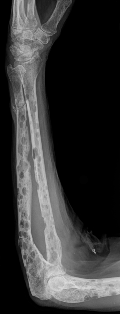
XR of forearm showing extensive lytic lesions in a patient with myeloma.
Image Source: Wikimedia Commons (2019). Multiple myeloma, Licensed under a creative commons licence. Available for free at: https://commons.wikimedia.org/wiki/Category:Multiple_myeloma (Accessed on: 10 January 2023).
Myeloma can affect multiple organ systems, most commonly the bones, blood, kidneys, and immune system. It can also be associated with cardiac impairment if there is concurrent AL-type amyloidosis, which is closely linked in a minority of myeloma presentations.
Standard investigations for myeloma include:
- Bloods
- FBC, renal function, calcium, serum Ig electrophoresis* and serum free light chains plus 𝛽-2-microglobulin
- Imaging – whole body MRI is the gold standard investigation to evaluate for bony lesions
- Bone marrow aspirate and trephine
*Interpretation of serum electrophoresis and serum free light chains – myeloma is a clonal disorder, and all clonal plasma cells will secrete an identical immunoglobulin which will be associated with either kappa or lambda light chains. The electrophoresis should show a monoclonal band if a paraprotein is present, which can be quantified. Polyclonal bands can be present during infection or inflammation and are normal. Likewise, a rise in both kappa and lambda light chains can occur in renal dysfunction or inflammatory conditions.
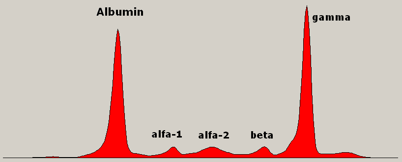
Serum immunoglobulin electrophoresis showing a monoclonal spike in gamma region.
Image Source: Wikimedia Commons (2009). Monoclonal gammopathy Multiple Myeloma, Licensed under a creative commons licence. Available for free at: https://commons.wikimedia.org/wiki/File:Monoclonal_gammopathy_Multiple_Myeloma.png (Accessed on: 10 January 2023).

Treatment
Myeloma is not considered a curable condition, but treatment has improved significantly in the last few decades. The details of treatment are beyond the scope of knowledge required for MRCP Part 1. However, treatment strategies are generally divided based on whether a patient is fit enough to undergo an autologous stem cell transplant or not. Eligible patients will undergo induction therapy followed by autologous stem cell transplant with the aim being as long a period of disease-free interval as possible. Most treatment is based on triple therapy with a combination of targeted treatment, traditional chemotherapy drugs and steroids.
When patients relapse, there are multiple further lines of treatment available, if well enough to continue. The chance of achieving remission and the duration of this decreases with each line of therapy. Some patients will receive 4th or even 5th line therapy.
10% of people will die within one year of presentation but some will live > 20 years. Overall 5-year survival is just over 50%.
Prognostic factors
Previously based on ISS scoring system. This is not as useful with improved treatments as prognosis has changed. A revised scoring system incorporates cytogenetics.
| Stage | I | II | III |
| Criteria | 𝛽-2-microglobulin<3.5 and albumin >35 | Not fitting I or II | 𝛽-2-microglobulin >5.5 |
Increasing age and or frailty are also poor prognostic factors, although not formally incorporated into scoring systems.
Pre-malignant disorders of plasma cells
Monoclonal gammopathy of undetermined significance (MGUS)
All multiple myeloma is now believed to progress through an asymptomatic premalignant stage called MGUS. Patients with MGUS have a clonal population of plasma cells producing a paraprotein (<30 g/L). There is a 1% risk of progression to myeloma per year. By definition, there must not be evidence of end organ damage.
Smouldering myeloma
An intermediate stage with a paraprotein >30 g/L or plasma cells >10% and <60% in bone marrow but without evidence of end organ damage. The risk of progression to overt myeloma is around 10% per year in the first 5 years but actually decreases after this point.
Waldenstrom’s macroglobulinaemia
Waldenstrom’s is a type of indolent B-cell lymphoma called lymphoplasmacytic lymphoma (LPL), but one which secretes IgM (monoclonal) paraprotein. The malignant cells share features of both lymphoid cells and plasma cells. It is relatively rare and mostly presents in >70 years. There is an association with Sjogren’s syndrome and autoimmune haemolytic anaemia.
There is usually bone marrow infiltration and there may be lymphadenopathy or splenomegaly. Symptoms can be diverse and may be related to anaemia from marrow infiltration, constitutional symptoms or lymphadenopathy or directly related to the IgM paraprotein (due to it being a large and ‘sticky’ protein):
- Hyperviscosity syndrome – visual disturbances, neurological deficits, and skin/mucosal bleeding. Treatment is with fluids and urgent plasmapheresis.
- Cryoglobulinaemia
- Coagulation abnormalities
- Cold haemagglutinin disease
- Peripheral neuropathy
It is preceded by a premalignant IgM MGUS condition, with parallels to MGUS and myeloma.
Investigations
- Bloods
- To include full panel plus serum Ig electrophoresis, may need hyperviscosity screen and DAT screen
- Bone marrow – essential for diagnosis
- Imaging – if symptomatic, CT CAP for staging should be performed
Treatment
Treatment is not always needed immediately. A ‘watch and wait’ approach is often taken initially. The following are indications to start treatment:
- Constitutional symptoms
- Symptomatic lymphadenopathy/splenomegaly
- Hyperviscosity syndrome
- Cytopenias due to marrow infiltration
- IgM related syndromes – CHAD, peripheral neuropathy
Treatment is not generally considered curative. Overall median survival is 5 years from diagnosis.
Myeloproliferative disorders
Myeloproliferative disorders are characterised by excessive proliferation of one or more cells originating from the myeloid lineage – RBC, platelets, or monocytes/neutrophils. They are clonal proliferations, often with characteristic genetic mutations and as such are classified as malignancies. They tend to be very slow growing conditions, but all carry a risk of progression to AML. There are three major forms of myeloproliferative disorders:
- Polycythaemia vera (PV)
- Essential thrombocytosis (ET)
- Myelofibrosis (MF)
CML is also sometimes considered a type of myeloproliferative disorder.
It can be helpful to consider these disorders by looking at changes in blood counts, however, it is worth remembering that in reality the situation is more complex than this and there is considerable overlap between these conditions.
Polycythaemia
Polycythaemia is defined as Hb >185 in males and >165 in women. In practice, haematocrit (HCT) is a more reliable indicator of polycythaemia (HCT >0.52 in males, >0.48 in females)
Primary polycythaemia
This includes rare congenital forms but is mostly synonymous with an acquired malignant condition known as polycythaemia vera. This is due to a somatic mutation in a hematopoietic stem cell. In over 95% of cases, this is a mutation in the JAK2 gene.
Because it is the stem cell that is mutated, there can be an increase in all three myeloid cell lines (RBC, platelets, and granulocytes). The erythropoietin (EPO) level will be low due to negative feedback.
The incidence is 2 cases per 100,000 people per year. The median age at presentation is 65-75 years.
Features:
- Splenomegaly
- Arterial and venous thrombosis including Budd-Chiari syndrome
- Pruritus, particularly after a bath or shower
- Erythromelalgia – itching or burning in extremities (see image)
- Haemorrhage
- Gout
- Constitutional symptoms

Photo showing characteristic changes during an episode of erythromelalgia.
Image Source: Wikimedia Commons (2017). Erythromelalgia in left hand, Licensed under a creative commons licence. Available for free at: https://commons.wikimedia.org/wiki/File:Erythromelalgia_in_left_hand.jpg (Accessed on: 10 January 2023).

Confirmation of the diagnosis requires JAK2 positive mutation with a persistently elevated haematocrit >0.52 in males and >0.48 in females.
Without treatment, life expectancy is around 18 months – death is most commonly due to stroke or MI. Median survival is 14 years with treatment. There is a 5-15% risk of transformation to myelofibrosis over 10 years and the risk of progression to AML is ~2% at 10 years and 8% at 20 years.
Management:
- Reduce cardiovascular risk factors
- Low dose aspirin for all patients
- Venesection to reduce the HCT to below 0.45 target
- Cytoreductive therapy (usually hydroxycarbamide or interferon first line) for high-risk patients (>65 years or history of thrombosis)
Secondary polycythaemia
Secondary polycythaemia is a much more common cause of a raised haematocrit than polycythaemia vera. These conditions will show a raised EPO level. They are summarised in the table below:
| Central hypoxia | Smoking, chronic lung disease, OSA, right to left shunts, carbon monoxide poisoning |
| Renal hypoxia | Renal artery stenosis, PKD, ESRF |
| Alcohol excess | |
| Drugs | Diuretics, EPO, androgens, SGLT2 inhibitors |
| Pathological | Hepatocellular carcinoma, renal cell carcinoma, cerebellar haemangioma, parathyroid cancer, phaeochromocytoma |
Management is generally aimed at identifying and treating the underlying cause.
Relative polycythaemia
This is caused by decreased plasma volume causing a proportional increase in HCT but without an increase in absolute red cell mass. It corrects on normalisation of intravascular plasma volume.
Thrombocytosis
As with polycythaemia, thrombocytosis can be split into primary and secondary causes. Primary thrombocytosis can be associated with a number of haematological disorders, mostly myeloproliferative diseases. This includes PV, MF, CML, CMML and most commonly ET.
Essential thrombocytosis (ET)
This is the most common form of myeloproliferative neoplasm (MPN). Unlike PV, only platelet count is increased. Most patients present between 50-60 years.
It is associated with the following common mutations:
- JAK2 ~50%
- CALR ~30%
- MPL ~10%
The remainder of cases are classed as triple negative. If one of the above mutations is present, along with a platelet count over 450 x 10⁹/L, this is sufficient to establish a diagnosis, providing there is no evidence of another MPN. Without one of the above three mutations, bone marrow investigation is usually necessary to confirm the diagnosis.
Features:
- Increased risk of venous and arterial thrombosis
- Increased risk of haemorrhage
- Constitutional symptoms
- Hepatosplenomegaly may be present
- Headache and dizziness
- Erythromelalgia
- Pregnancy complications
Prognosis
This is generally better than PV. Life expectancy is not affected for the first 10 years but is difficult to quantify afterwards. There is a risk of progression to MF and AML, but this is lower than in PV.
Treatment is similar to PV:
- Manage CV risk factors
- Aspirin for all patients
- Cytoreductive patients only for high-risk patients (age >60, plt >1500, thrombotic or haemorrhagic complication)
Reactive thrombocytosis
There are a multitude of causes for this.
- Infection or inflammation/tissue damage
- Iron deficiency
- Hyposplenism
- Haemorrhage
- Drugs e.g. steroids
- Other malignancy e.g. sarcoma
Myelofibrosis
This is a condition characterised by progressive fibrosis of the marrow secondary to a clonal population of megakaryocytes; these cells interact with the surrounding bone marrow stroma and release pro-inflammatory and fibrotic cytokines. This causes progressive bone marrow failure, plus splenomegaly and constitutional symptoms. Pulmonary hypertension may also develop over time. The blood film may show a leukoerythroblastic picture. It can arise de novo or as a result of progression from PV or ET. Mutations in JAK2, CALR and MPL can be seen (as with ET), plus other somatic mutations are also seen in AML.
In order to make the diagnosis, a bone marrow is performed. This must show significant fibrosis and megakaryocyte proliferation without another causative explanation. A combination of this plus at least one other feature such as anaemia or splenomegaly is usually adequate. The presence of typical mutations (JAK2, CALR or MPL) is supportive.
The prognosis is variable but generally poor. Compared with PV and ET, there is a significantly higher risk of transformation to AML.
A very small minority of patients may be cured with allogeneic stem cell transplant. Many patients are either not fit enough for this or else their disease is mild, and the risk of such intensive treatment outweighs the benefit. Other treatment options include supportive care, cytoreductive therapy or newer drugs such as ruxolitinib (JAK 2 inhibitor).
Myelodysplastic syndromes (MDS)
Myelodysplastic syndromes comprise a group of heterogeneous conditions which share similar underlying features:
- Malignant clonal proliferation of abnormal cells within the marrow
- Dysplasia in at least one myeloid cell lineage
- Ineffective haematopoiesis
- Propensity to progression to AML
The bone marrow itself is often hypercellular but with ineffective haematopoiesis there is a reduction in healthy cell maturation and so cytopenias result.
It is generally considered a disease of the elderly and prevalence is increasing. 10% of MDS is secondary to exposure to mutagens, including chemo and radiotherapy but also environmental such as benzenes.
Symptoms are usually due to cytopenias:
- Erythroid – anaemia
- Platelets – bleeding
- Granulocytes – infection
Splenomegaly is possible.
Investigations
- Bloods – rule out other causes for cytopenias. Anaemia can be normocytic or macrocytic.
- Blood film shows dysplastic features in at least one lineage (see image of a dysplastic neutrophil)
- Bone marrow – usually hypercellular with evidence of dysplasia. Blasts may be present (<20%)
- Cytogenetic analysis is important for risk stratification.

Blood film showing a dysplastic neutrophil – the cytoplasm is hypogranular and there is a pseudo-Pelger-Huet nucleus. The accompanying red cell changes in this case are actually a result of splenectomy.
Image Source: Wikimedia Commons (2021). Myelodysplasia, Licensed under a creative commons licence. Available for free at: https://commons.wikimedia.org/wiki/File:Myelodysplasia.jpg (Accessed on: 10 January 2023).

Subtypes of MDS:
- MDS with single lineage dysplasia
- MDS with multi lineage dysplasia
- MDS with ring sideroblasts
- MDS with excess blasts 5-9%
- MDS with excess blasts 10-19%
- MDS with isolated del 5q
Some variants of MDS overlap with proliferative phenotypes (MDS/MPN overlap). They show features of both dysplasia and proliferation. Chronic myelomonocytic leukaemia (CMML) is an example.
Prognosis
The IPSS-R is a scoring system which guides prognosis. It is based on the severity of cytopenias, cytogenetic abnormalities and blast percentage. The risk of transformation to AML increases with each risk category.
| Risk category | Median survival |
|---|---|
| Very low | 8.8 years |
| Low | 5.3 years |
| Intermediate | 3 years |
| High | 1.6 years |
| Very high | 0.8 years |
Management
This is highly individualised and takes into account the severity of MDS, patient age and fitness for treatment. Elderly patients with low risk MDS are more likely to die with MDS than from it.
- The only truly curative option for MDS is high dose chemotherapy, usually supported by allogeneic stem cell transplant. This option is reserved for high risk MDS and only in those fit enough.
- Lower intensity chemotherapy such as azacytidine or lenalidomide
- Supportive therapy
- Blood and platelet transfusions
- Growth factor support
- Antibiotics as necessary
Patients receiving regular transfusions of RBC are at risk for transfusion-associated iron overload and may require iron chelation therapy.
Complete Guide to Clinical Genetics ST3 Application and Recruitment (2027)
Written by Medibuddy. Last updated: October 2026. Clinical Genetics is

Complete Guide to Clinical Radiology ST3 Application and Recruitment (2027)
Recruitment paused: NHS England is not running Clinical Radiology ST3+ recru

Complete Guide to Paediatric Surgery ST3 Application and Recruitment in United Kingdom (2026)
Written by Medibuddy. Last updated: October 2026. If you’re planning


Take your subscriptions with you
Our mobile app allows you to access your interview and exam question banks wherever you are.

