


 Close
Close
 7th June 2023
Haemorrhagic Stroke
Intracerebral haemorrhage
Intracerebral haemorrhage accounts for approximately 15% of strokes and causes include hypertension, therapeutic anticoagulants, increasing age, recreational drug use (e.g. cocaine), vascular anomalies, venous sinus thrombosis and brain tumours.
Symptoms can be gradual or sudden in onset and are related either to the part of the brain involved or to raised intracranial pressure, such as nausea, vomiting, headache, drowsiness, and seizures. Investigation is with non-contrast CT or MRI brain.
Conservative management consists of stopping the bleeding by reversing anticoagulants and blood pressure control. Haemorrhages in the basal ganglia and thalamus are generally managed conservatively as there is no evidence that surgery improves outcomes. Surgical treatment is considered for patients with haemorrhage in particular locations, such as cerebellar and lobar, as well as for complications, such as hydrocephalus.
Subarachnoid Haemorrhage
Subarachnoid haemorrhage is bleeding into the subarachnoid space between the arachnoid membrane and the pia mater. It causes sudden onset of severe headache (known as ‘thunderclap headache’), decreased consciousness, focal neurology and meningism (nuchal rigidity, photophobia). It is most often due to rupture of an intracranial aneurysm (Berry aneurysm) which can be sporadic or occur secondary to genetic conditions such as autosomal dominant polycystic kidney disease, Ehlers-Danlos syndrome, Marfan syndrome and neurofibromatosis.
Diagnosis is from a CT brain scan. If this is negative, cerebrospinal fluid (CSF) should be examined for xanthochromia (discolouration of the CSF due to lysis of blood).
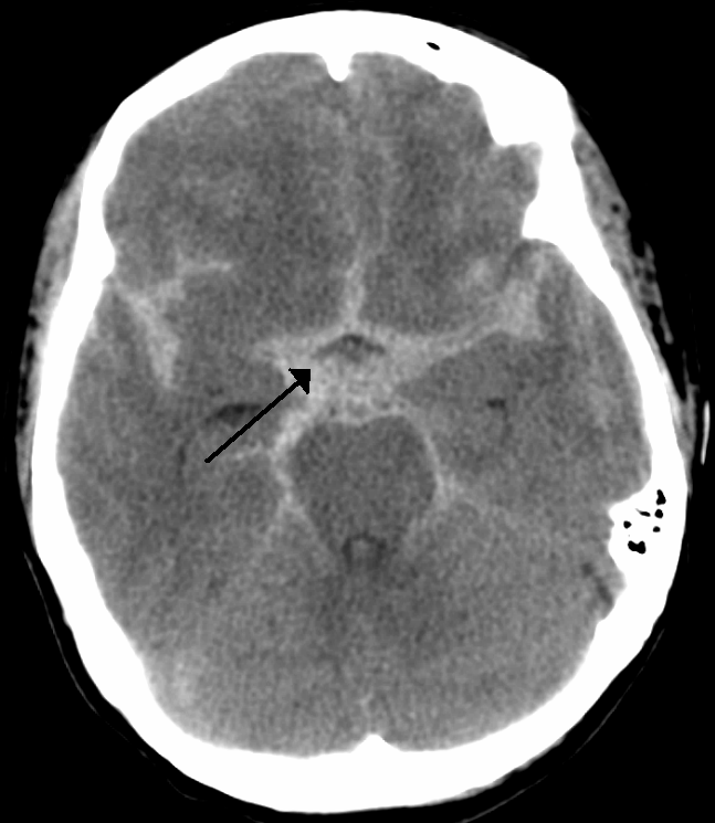
Subarachnoid haemorrhage on CT head
Image Source: Wikimedia Commons (2010). Circle of Willis en, Licensed under a creative commons licence. Available for free at: https://commons.wikimedia.org/wiki/File:SubarachnoidP.png (Accessed on: 11 January 2023).

Cerebral Venous Sinus Thrombosis
This is a rare cause of stroke, produced by a clot in the dural venous sinuses, resulting in obstruction of blood draining out of the brain, causing venous congestion, and raised intracranial pressure. Clinical features include headache, vomiting, papilloedema, focal neurological signs and cranial nerve palsies.
Diagnosis is by CT venography, and treatment is with anticoagulation, initially with heparin which is later converted to warfarin.
Complete Guide to Clinical Genetics ST3 Application and Recruitment (2027)
Written by Medibuddy. Last updated: October 2026. Clinical Genetics is

Complete Guide to Clinical Radiology ST3 Application and Recruitment (2027)
Recruitment paused: NHS England is not running Clinical Radiology ST3+ recru

Complete Guide to Paediatric Surgery ST3 Application and Recruitment in United Kingdom (2026)
Written by Medibuddy. Last updated: October 2026. If you’re planning


Take your subscriptions with you
Our mobile app allows you to access your interview and exam question banks wherever you are.

