


 Close
Close
 9th June 2023
Paraneoplastic Syndromes
Overview of Paraneoplastic Syndromes
Paraneoplastic syndromes are a group of rare systemic or organ-specific disorders that occur as a result of cellular or immunological responses to cancer. They can present at any point during the “lifetime” of the malignancy, often presenting prior to diagnosis, and can be phenotypically similar or identical to a number of more common disorders, which makes them diagnostically challenging. Whilst there are a number of specific malignancies linked to specific syndromes, the occurrence of these syndromes does not correlate with grade nor stage of the cancer and can even present following curative treatment such as resection of the primary tumour.
The exact mechanisms behind many of these syndromes are poorly understood but are classified into two broad sub-categories: direct tumour secretion of agents that act on the body’s cellular pathways (for example enzymes, hormones, cytokines), and the body’s immunological response to the presence of the tumour.
This section will cover the commonly affected organ systems and the most common paraneoplastic syndromes within them.
Endocrine Paraneoplastic Syndromes
Paraneoplastic syndromes manifesting as endocrine dysfunction are the more common syndromes encountered in general medical practice, in part due to the ease of serum biochemical testing. They occur as a result of the secretion of endocrine substances by the tumour, however, this secretion is ectopic i.e. not being released by its usual anatomical source. They are therefore separate conditions to those of abnormal endocrine secretion from dysfunction within the endocrine gland itself e.g. from a secretory pituitary tumour, however, the phenotypical and biochemical presentations are indistinguishable. The normal pathways for each of the syndromes are discussed in more detail within the Endocrinology section – this focuses on the paraneoplastic causes of pathway dysfunction.
Types of endocrine paraneoplastic syndromes
Cushing’s Syndrome
Cushing’s Syndrome is a collection of biochemical and physiological abnormalities caused by excess serum cortisol. This excess can be extrinsic (i.e. a person taking glucocorticoid therapy) or intrinsic (i.e. an excess of cortisol production), the latter of which can be caused by abnormal function at various points along the production pathway. Cortisol production is stimulated by adrenocorticotropic hormone (ACTH) secreted by the anterior pituitary gland, which itself is stimulated by corticotropin releasing hormone (CRH) secreted by the hypothalamus.
Paraneoplastic Cushing’s is due to the secretion of ACTH (or, rarely, CRH) by malignant cells that are outside of the pituitary gland, hence considered ectopic. Whilst a rare entity, 5-10% of all Cushing Syndromes are of paraneoplastic aetiology, and more than half of these are caused by neuroendocrine lung tumours such as small cell lung cancer or bronchial carcinoid. A high dose dexamethasone test can be used to distinguish ectopic secretion of ACTH from abnormal pituitary secretion – if serum cortisol remains high after exogenous administration of dexamethasone (i.e. “failure” of suppression), this indicates ectopic production of ACTH. This, along with pituitary imaging (usually MRI) to exclude a pituitary adenoma, and cross-sectional body imaging to look for malignancy, can identify a malignant cause for the syndrome, often preceding the diagnosis of cancer itself.
The syndrome can be treated by successful treatment of the underlying malignancy, however supportive treatment of any syndrome-associated biochemical and metabolic abnormalities may be required in the short term. In some cases, treatment with inhibitors of steroid production may be required. Resurgence of the syndrome in the context of treated malignancy can indicate cancer progression/relapse.
SIADH
Antidiuretic hormone (ADH), otherwise known as vasopressin, is one of the primary hormones involved in osmotic balance and sodium regulation. ADH is released from the posterior pituitary in response to hypovolaemia or hypernatraemia and causes renal water reabsorption. Syndrome of inappropriate antidiuretic hormone (SIADH) is, as the name suggests, a syndrome whereby antidiuretic hormone secretion continues despite low serum sodium, in the context of euvolaemia. It is therefore characterised by a low serum sodium and low plasma osmolality, paired with a high urinary sodium and osmolality, in the context of a euvolaemic patient.
Paraneoplastic SIADH is one of the more common syndromes encountered in Oncology, present in up to 2% of all malignancies depending on population demographic. The vast majority (>70%) of cases of paraneoplastic SIADH are due to small cell lung cancer and is often the presenting feature of this malignancy. Breast, Gynaecological and Haematological malignancies (such as Hodgkin’s lymphoma) are the more common of the other malignancies that cause this syndrome.
In paraneoplastic SIADH caused by small cell lung cancer, the malignancy itself is inherently sensitive to chemotherapy and radiotherapy, and as such the successful treatment of the underlying malignancy is usually the primary treatment for the SIADH. In cases where patients present with critically low sodium, or the hyponatraemia is slow to respond to anti-cancer treatment, primary treatment of the SIADH with fluid restriction or pharmacological therapy (such as tolvaptan) is often required. As with many other paraneoplastic syndromes, recurrence of the SIADH following successful Oncological treatment can often indicate cancer relapse.
Hypercalcaemia
Hypercalcaemia is a common biochemical abnormality in cancer patients occurring in approximately 10% of all patients with advanced malignancy, often in patients with bone metastases whereby osteoclastic or osteolytic activity increases serum calcium as a direct result of bone breakdown. Paraneoplastic hypercalcaemia is distinct from this entity, in that (as with the other paraneoplastic endocrine disorders) it is as a result of ectopic secretion of the hormones involved in calcium regulation, most commonly parathyroid hormone related protein (PTHrP) or, more rarely, vitamin D or PTH. In this syndrome, PTHrP is secreted by the tumour cells, binding to PTH receptors in the bone and kidney which upregulates bone reabsorption, renal calcium and phosphate retention and thereby increases serum calcium and phosphate.
Paraneoplastic hypercalcaemia due to ectopic PTHrP is most commonly seen in squamous cancers, particularly of lung origin. Other common squamous malignancies include cancers of the upper aerodigestive tract, skin, cervix, and anus. Ectopic production of vitamin D has been described in haematological malignancy though is uncommon. Patients often present unwell, with common symptoms of hypercalcaemia such as constipation, bone pain, dehydration, and acute confusion, and as such the supportive management of the hypercalcaemia with fluid resuscitation and bisphosphonates often takes precedence over treatment of the malignancy.
Neurological and Musculoskeletal Paraneoplastic Syndromes
Paraneoplastic neuromuscular syndromes (PNMS) are a group of rare but clinically significant syndromes of immunological origin, occurring in <1% of all cancer diagnoses. They occur due to similarities between the cancer’s immunogenic profile and components of the neuromuscular system, which can lead to inadvertent “attack” of normal tissue by the patient’s own immune system. Unlike paraneoplastic endocrine syndrome, PNMS are independent of cancer activity for two reasons: the tumour cells are not secreting an active substance, and once activated, the immune system continues to attack the target tissue. The mainstay of treatment of PNMS, therefore, is immunosuppression, though response rates are variable, and the use of immunosuppression in active malignancy is to be avoided where possible.
The most common malignancies implicated in PNMS are small cell lung cancers, thymomas, lymphomas, myeloma, neuroblastoma and teratoma, all of which either originate in tissues involved in immunoregulation, secrete neuroendocrine proteins, or contain neuronal components. Whilst the vast majority precede the cancer diagnosis and, in many cases, can lead an early-stage diagnosis, the morbidity associated with the neuromuscular condition can often preclude anti-cancer treatment, leading to a poorer prognosis than the average for that particular malignancy.
Types of paraneoplastic neuromuscular syndromes
Lambert-Eaton Myasthenic Syndrome
Lambert-Eaton Myasthenic Syndrome (LEMS) is an immune disorder affecting the calcium channels on the presynaptic membrane of the neuromuscular junction, decreasing the levels of the neurotransmitter acetylcholine. The syndrome can be a primary autoimmune disorder or a paraneoplastic disorder, with no clinical differentiation between the two. It is characterised by proximal muscle weakness, oculo-bulbar weakness, areflexia and autonomic dysfunction. Rarely, it can progress to respiratory failure. Unlike myasthenia gravis, patients present initially with proximal limb weakness moving caudal-cranially, with progression to oculo-bulbar involvement at later stages.
The most common malignancy associated with LEMS is small cell lung cancer, which is found in ~50-60% of all patients with LEMS. Other lung malignancies, lymphoproliferative disorders, thymoma, gynaecological and prostate cancers have all been implicated. Its association with malignancy means that anybody presenting with LEMS should be screened for these cancers, starting with investigations targeted at the diagnosis of a lung malignancy. Treatment is directed at the syndrome itself, using acetylcholinesterase inhibitors or, in more severe cases, immunosuppression or immunoglobulin therapy.
Myasthenia Gravis
Myasthenia Gravis (MG) is distinct from LEMS in its pathophysiology and clinical profile, though there are common features to both that can make them difficult to distinguish. It is an immune disorder affecting the postsynaptic membrane of the neuromuscular junction, with the development of antibodies against acetylcholine receptor or muscle-specific tyrosine kinase. It is characterised by muscle weakness that displays fatiguability (worsening with activity), primarily involving the oculo-bulbar muscles and in late stages respiratory muscles. Limb weakness is rare in comparison to LEMS, and the progression of weakness moves cranio-caudally.
Thymoma is the predominant malignancy associated with MG, found in 15-30% of all MG diagnoses. The thymus is an endocrine organ involved in T cell maturation and proliferation, and as such, myasthenia gravis due to malignancy within this organ is not technically a paraneoplastic syndrome, as it occurs as a direct result of endocrine organ dysfunction. However, its synonymity with thymoma makes it worth a mention, as a diagnosis of MG will often lead to a diagnosis of thymoma. The management of the resultant MG is either through treatment of the thymoma (usually surgical resection) or with systemic treatment such as anticholinesterase inhibitors or immunosuppression.
Polymyositis
Polymyositis is a rare immune disorder affecting muscle fibres that express MHC class 1 antigens, with infiltration of the tissue by cytotoxic T cells. It is characterised by girdle muscle weakness (shoulders and hips), myalgia, fatigue and upper aerodigestive tract muscle weakness, though it can affect any muscle group. Patients can also present with systemic symptoms such as fever and weight loss. Biochemical abnormalities include elevated creatine kinase (CK) and myoglobinuria. For confirmation of the diagnosis a muscle biopsy is required.
Paraneoplastic polymyositis is associated most with non-Hodgkin lymphoma, bladder and lung malignancies; however, the association is not particularly strong, with <10% of patients with polymyositis subsequently being diagnosed with a malignancy. Management is directed both at the treatment of the underlying cancer, and the management of the syndrome with steroids or immunosuppressive therapy. Paraneoplastic polymyositis has a poorer prognosis than its non-paraneoplastic disorder, though whether that is to do with factors associated with the underlying cancer and its morbidity is unclear.
Muco-cutaneous Paraneoplastic Syndromes
Muco-cutaneous paraneoplastic syndromes are a heterogenous group of conditions with a wide variety of clinical presentations that often overlap with benign pathology. Their precise pathophysiology is poorly understood, and the aetiology is commonly either immunological or related to an increase in tissue growth factors. Those that are immunological in origin are difficult to treat; the syndromes are often associated with significant morbidity that precludes Oncological treatment, the treatment of the syndrome requires immunosuppressive therapy which may have adverse effects on cancer control, and even with successful treatment of the underlying malignancy, the immune response targeting the underlying tissue can continue.
Types of muco-cutaneous paraneoplastic syndromes
Acanthosis Nigricans
Acanthosis nigricans (AN) is a dermatological condition characterised by papillomatous hyperpigmented plaques, often symmetrical and found in flexural creases such as the axillae.
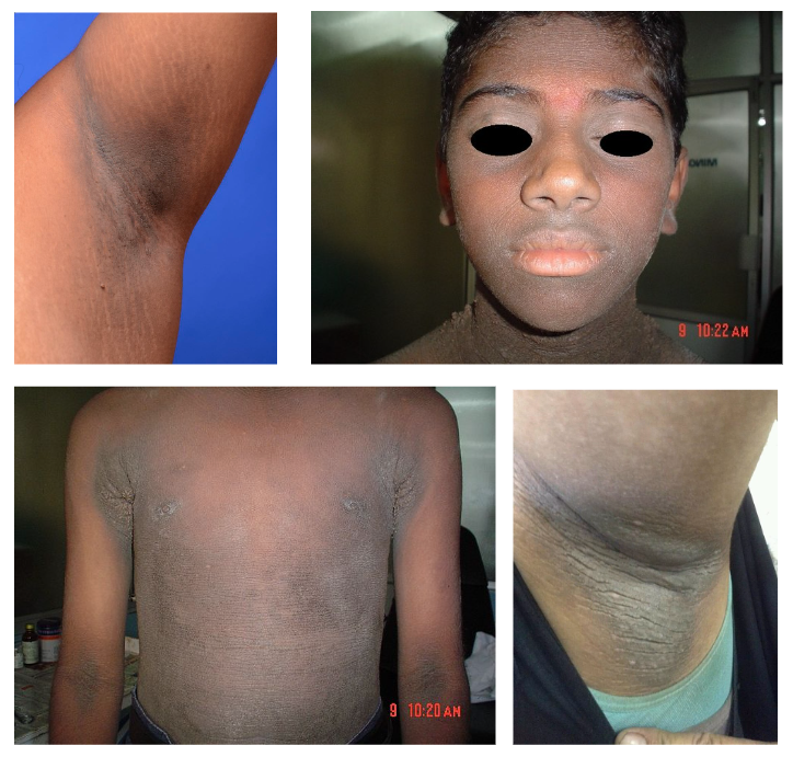
Acanthosis nigricans (underarm)
Acanthosis nigricans (face)
Acanthosis nigricans (body)
Acanthosis nigricans (neck)
Image Sources:
- Wikimedia Commons (2019). Acanthosis nigricans, Licensed under a creative commons licence. Available for free at: https://commons.wikimedia.org/wiki/File:Acanthosis_nigricans.jpg (Accessed on: 9 February 2023).
- Wikimedia Commons (2010). Familial acanthosis nigricans, Licensed under a creative commons licence. Available for free at: https://commons.wikimedia.org/wiki/File:Familial_acanthosis_nigricans.jpg (Accessed on: 9 February 2023).
- Wikimedia Commons (2010). Familial acanthosis nigricans5, Licensed under a creative commons licence. Available for free at: https://commons.wikimedia.org/wiki/File:Familial_acanthosis_nigricans5.jpg (Accessed on: 9 February 2023).
- Wikimedia Commons (2017). Acanthious nigricans new photo for diagnosis, pigmentation appears in neck ,arm,elbow,it be related often to insulin it related to obesity and some times called pseudonigricans, Licensed under a creative commons licence. Available for free at: https://commons.wikimedia.org/wiki/File:Acanthious_nigricans_new_photo_for_diagnosis,_pigmentation_appears_in_neck_,arm,elbow,it_be_related_often_to_insulin_it_related_to_obesity_and_some_times_called_pseudonigricans.jpg (Accessed on: 9 February 2023).
Its aetiology is of increased levels of insulin or insulin-like growth factor (IGF1), and therefore usually found in obese patients with insulin resistance. In the absence of these factors, a malignant cause needs to be explored. Paraneoplastic AN is most commonly associated with gastric adenocarcinomas, comprising ~60% of all malignant causes of AN, due to the ectopic secretion of IGF1. Other malignancies of the upper GI tract are the next most implicated, although the syndrome has been associated with several different solid tumours and Haematological malignancies. The paraneoplastic syndrome is indistinguishable from the benign condition, although in paraneoplastic AN there is an increased incidence of non-pigmented oro-mucosal lesions (15-25%). The management is directed at the treatment of the underlying malignancy.
Dermatomyositis
Dermatomyositis (or dermatopolymyositis), as the name suggests, is a condition of immunological aetiology characterised by myositis along with distinctive skin lesions. Like polymyositis, the exact pathophysiology is unclear, but thought to be via T cell infiltration and subsequent immune-mediated damage of the muscle and cutaneous tissues.

Dermatomyositis (hands)
Wikimedia Commons (2010). Dermatomyositis, Licensed under a creative commons licence. Available for free at: https://commons.wikimedia.org/wiki/File:Dermatomyositis.jpg (Accessed on: 9 February 2023).
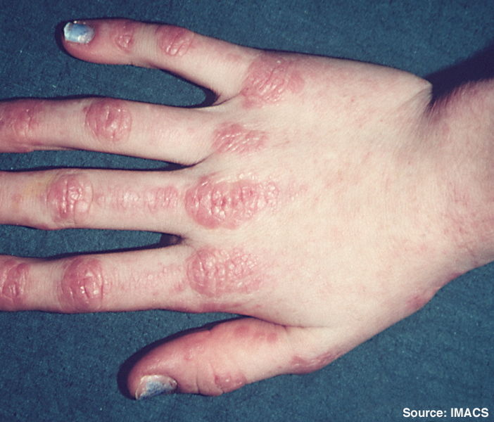
Dermatomyositis (face)
Wikimedia Commons (2010). Dermatomyositis14, Licensed under a creative commons licence. Available for free at: https://commons.wikimedia.org/wiki/File:Dermatomyositis14.jpg (Accessed on: 9 February 2023).
In dermatomyositis, the skin lesions often present first, with a dusky heliotrope rash around the eyes, across the upper back, shoulders, and upper neck in a “shawl” distribution, and on the extensor surfaces of the knees and elbows, as well as the presence of Gottron’s papules (dusky erythematous or violet lesions across the backs of the carpal joints). The myositis can present later or be absent, characterised by symmetrical muscle weakness and pain particularly affecting the girdle muscles. A diagnosis of dermatomyositis is supported by raised creatine kinase and myoglobinuria, and confirmed by muscle and/or skin biopsy.
Dermatomyositis is frequently associated with underlying malignancy and is most associated with ovarian, lung, pancreatic, gastric, colorectal, and non-Hodgkin lymphoma. As with other immune mediated paraneoplastic syndromes, treatment is directed at the underlying malignancy, although steroids and immunosuppression can be used in severe cases, often whereby the morbidity of the syndrome is precluding treatment of the cancer.
Clubbing
Clubbing is the common descriptive term for swelling of the soft tissue of the distal phalanx along with loss of the angle between the nail and nail bed, resulting in characteristic deformity of the ends of the digits, as pictured. This can be further assessed using the Schamroth test: holding two opposing fingers back-to-back against each other. In unaffected fingers, there is usually a small diamond shaped gap at the nail beds, which is lost when clubbing is present.
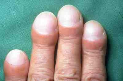
Finger clubbing.
Wikimedia Commons (2013). Clubbing of fingers in IPF, Licensed under a creative commons licence. Available for free at: https://commons.wikimedia.org/wiki/File:Clubbing_of_fingers_in_IPF.jpg (Accessed on: 9 February 2023).

Schamroth test for finger clubbing.
Wikimedia Commons (2021). Schamroths, Licensed under a creative commons licence. Available for free at: https://commons.wikimedia.org/wiki/File:Schamroths.jpg (Accessed on: 9 February 2023).
The pathophysiology of clubbing is not fully understood but is thought to be related to an increase in platelet-derived growth factor (PDGF) and associated factors involved in tissue proliferation. As clubbing is generally asymptomatic and non-morbid, it usually heralds the diagnosis of an underlying condition rather than requiring treatment itself.
Clubbing is typically associated with chronic or inflammatory disease, predominantly cardiac, respiratory, or gastrointestinal in origin, however, there is also an association with underlying malignancy, which needs to be investigated if other common causes have been excluded. Cancers particularly associated with clubbing are primary or metastatic lung tumours, mesothelioma, and Hodgkin lymphoma, although the co-existence of chronic lung or cardiac disease alongside malignancy makes paraneoplastic causality of the clubbing difficult to assess in many cases.
Erythema Gyratum Repens
Erythema Gyratum Repens is a cutaneous condition with a strong association with malignancy, with underlying cancer in ~80% of cases, and as such is considered paraneoplastic until otherwise proven. As with a number of other cutaneous paraneoplastic syndromes, the underlying mechanism is immunological, with similarities between tumour and cutaneous antigens causing cross-reactivity between tumour-directed and cutaneous-directed antibodies.
It is characterised by a classic concentric erythematous plaque-like rash resulting in a “wood grain” effect, typically affecting the trunk and is often itchy. The diagnosis is made on this classic morphology and the diagnosis of an underlying malignancy, as skin biopsy findings can share histological features with other conditions. Lung cancers are the predominant underlying malignancy, or less commonly oesophageal, breast or gastric cancer. Treatment is of the underlying malignancy, although a loss of skin integrity can cause significant morbidity, requiring treatment of rash with steroids or immunosuppression prior to any cancer-directed treatment.
Migratory thrombophlebitis
Thrombophlebitis is a common condition with many different (and usually benign) aetiologies, characterised by a superficial venous thrombus with an associated inflammatory reaction causing pain and erythema around the site. Migratory thrombophlebitis (also known as Trousseau sign of malignancy) is a condition whereby areas of thrombophlebitis resolve and re-present in different sites, often affecting the lower limbs, including moving from one leg to the other. Its pathophysiology, whilst not fully understood, lies in the hypercoagulable states associated with malignancy, though why this condition should affect the superficial veins and predominate in certain malignancies is not known. It is strongly associated with malignancy, particularly adenocarcinoma of the lung, the upper GI tract (including pancreas) and the urogenital tract, and can pre-date a cancer diagnosis by months or even years. A diagnosis of malignancy should be presumed until otherwise proven. The condition is not life-threatening, nor particularly morbid, and so treatment is directed at the underlying malignancy, with topical anti-inflammatories and compression stockings for symptom relief, and thromboprophylaxis if considered clinically appropriate.
Complete Guide to Clinical Genetics ST3 Application and Recruitment (2027)
Written by Medibuddy. Last updated: October 2026. Clinical Genetics is

Complete Guide to Clinical Radiology ST3 Application and Recruitment (2027)
Recruitment paused: NHS England is not running Clinical Radiology ST3+ recru

Complete Guide to Paediatric Surgery ST3 Application and Recruitment in United Kingdom (2026)
Written by Medibuddy. Last updated: October 2026. If you’re planning


Take your subscriptions with you
Our mobile app allows you to access your interview and exam question banks wherever you are.

