


 Close
Close
 22nd June 2023
Nephron
The nephron is the main functional unit of the kidney; there are approximately 1 million nephrons in each kidney, however, with renal disease or normal ageing, the number of nephrons decreases. Each nephron is composed of a renal corpuscle and a renal tubule.
Renal corpuscle
The renal corpuscle consists of the glomerulus and Bowman’s capsule; it is the site of filtration of the blood. Filtration of fluid from the glomerulus to the Bowman’s capsule is based on Starling forces:
- Membrane permeability
- Hydrostatic pressure gradient
- Oncotic pressure gradient
The glomerulus is a bundle of glomerular capillaries covered with a semi-permeable membrane formed of glomerular endothelial cells, the glomerular basement membrane (GBM), and podocytes; these form the filtration barrier. The glomerular capillary endothelium contains pores, or ‘fenestrae’, which are approximately 70 nm in diameter and prevent the filtration of blood cells. The GBM is composed of type IV collagen, heparan sulfate proteoglycans and laminin, and contains three layers: lamina rara externa, lamina densa and lamina rara interna. These layers limit the filtration of larger solutes, such as proteins. Surrounding the GBM is a layer of epithelial cells with foot-like processes called podocytes, separated by small ‘slit pores’ through which flows the filtrate; the slit pores prevent large molecules, e.g. proteins, from crossing. In addition to these physical barriers, the glycocalyx (surrounding the endothelium), the GBM, and the podocytes all contain, or are covered by, negatively charged glycoproteins which restrict the filtration of negatively charged anions and proteins by repelling like charges.
The glomerulus receives its blood supply from the afferent arteriole, and blood leaves the glomerulus via the efferent arteriole. These arterioles are responsible for generating pressure in the glomerulus by constriction of the efferent arteriole and dilatation of the afferent arteriole. The pressure in the glomerulus is ~50mmHg (compared to 35mmgHg in other capillaries). This high hydrostatic pressure generated within the glomerular capillaries drives the ultrafiltration of water and solutes out of the plasma.
Small molecules such as glucose can readily cross the filtration barrier, however, larger molecules such as albumin, and negatively charged anions, are not readily filtered; this creates a colloid pressure due to the presence of proteins in the blood and absence of proteins in the filtrate, forcing water and solutes into the renal tubules.
The glomerulus is encased in Bowman’s capsule, which receives filtrate from the glomerulus and passes it to the proximal tubule of the nephron.

The renal corpuscle.
Image Source: Wikimedia Commons (2019). Renal corpuscle-en, Licensed under a creative commons licence. Available for free at: https://commons.wikimedia.org/wiki/File:Renal_corpuscle-en.svg (Accessed on: 11 January 2023).
Renal tubule
The renal tubule comprises the proximal convoluted tubule, the descending and ascending limbs of the loop of Henle, the distal convoluted tubule, and the collecting duct. The function of the renal tubule is the reabsorption of certain constituents from the filtrate and secretion of molecules from the peritubular capillaries into the distal convoluted tubule.
Urinary excretion = Glomerular filtration – Tubular reabsorption + Tubular secretion
Nephrons which have glomeruli located in the cortex of the kidney are called cortical nephrons; these tend to have shorter loops of Henle and, therefore, only penetrate a small distance into the renal medulla. Nephrons with glomeruli which lie deep in the cortex, on the border of the medulla, are called juxtamedullary nephrons; these represent almost a third of nephrons and tend to have much longer loops of Henle, penetrating deeply into the medulla. The vascular supply of the juxtamedullary nephrons is via the vasa recta, specialised peritubular capillaries which originate from the efferent arterioles.
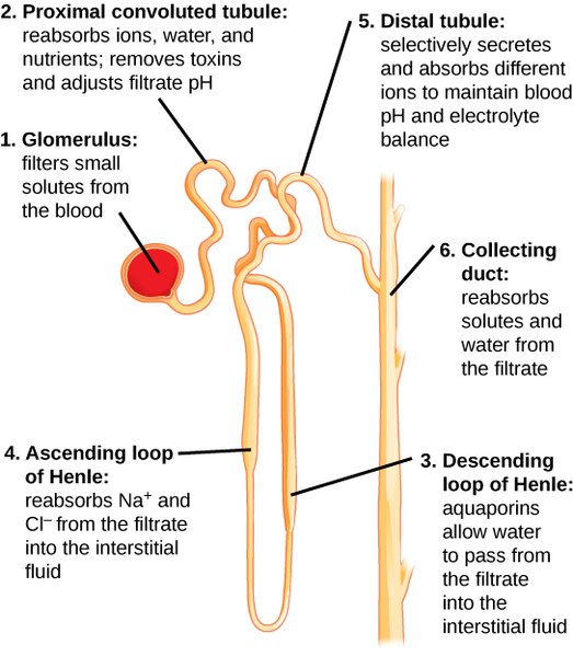
Components of the renal tubule.
Image Source: Wikimedia Commons (2016). Figure 41 03 04, Licensed under a creative commons licence. Available for free at: https://commons.wikimedia.org/wiki/File:Figure_41_03_04.jpg (Accessed on: 9 February 2023).
Complete Guide to Clinical Genetics ST3 Application and Recruitment (2027)
Written by Medibuddy. Last updated: October 2026. Clinical Genetics is

Complete Guide to Clinical Radiology ST3 Application and Recruitment (2027)
Recruitment paused: NHS England is not running Clinical Radiology ST3+ recru

Complete Guide to Paediatric Surgery ST3 Application and Recruitment in United Kingdom (2026)
Written by Medibuddy. Last updated: October 2026. If you’re planning


Take your subscriptions with you
Our mobile app allows you to access your interview and exam question banks wherever you are.

