


 Close
Close
 1st July 2023
Photosensitivity
Background
Photosensitivity describes a group of symptoms, diseases and conditions that are exacerbated by sunlight. Photodermatosis is a rash caused by photosensitivity, whereas it is called photodermatitis if the rash is eczematous. There are many different classifications of photosensitive rashes:
- Primary – exposure to sun causes a specific disease, e.g. polymorphic light eruption, solar urticaria, juvenile spring eruption
- Exogenous – photodermatoses rash is caused by an external photosensitiser such as food, drug or chemical that increases sensitivity to light, e.g. thiazide, tetracycline, NSAID, quinine
- Metabolic – the photodermatitis is caused by a metabolic defect, e.g. porphyria cutanea tarda
- Genetic – associated with a genetic disorder, e.g. xeroderma pigmentosum
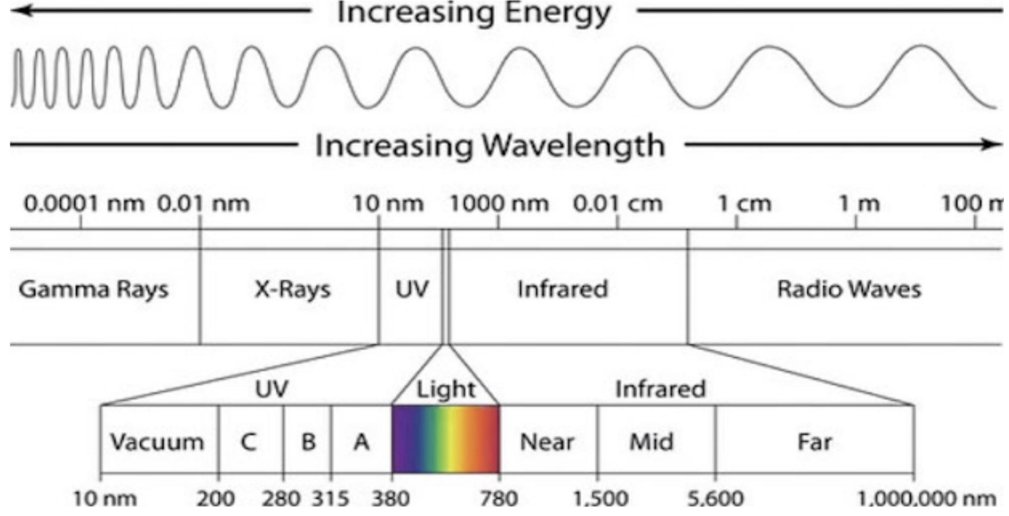
Light Spectrum.
Image Source: Flickr (2020). Light spectrum, Licensed under a creative commons licence. Available for free at: https://www.flickr.com/photos/rosenfeldmedia/49781640696 (Accessed on: 1 April 2022).
Clinical Features
In photodermatosis, the affected areas are those that are exposed to the sun. There is often a sharp line of demarcation between exposed and non-sun exposed areas of the skin. Skin folds and furrows are usually spared. Sometimes, it can only affect certain areas, e.g., photodermatosis on top of the ears in juvenile spring eruption.
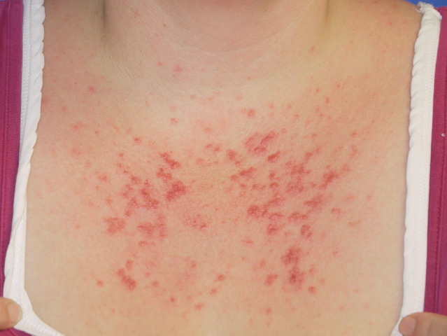
Polymorphic Light Eruption on the Chest.
Image Source: Wikimedia Commons (2013). Polymorphic Light Eruption on the chest, Licensed under a creative commons licence. Available for free at: https://commons.wikimedia.org/wiki/File:Polymorphic_Light_Eruption_on_the_chest.jpg (Accessed on: 1 April 2022).

Investigations
Photosensitivity is usually a clinical diagnosis, but investigations can be done to try and identify the underlying cause. This includes blood tests (full blood count, renal profile, liver function tests, iron studies, autoimmune screen and porphyrias) and stool and urine samples. Autoimmune screens are to look for connective tissue conditions and stool/urine samples are used to check for porphyrins if suspecting porphyria. Rarely, contact photosensitivity tests can be carried out where a photosensitising patch is applied to the skin and checked 2 days after the light is shone to the area.
Management
This mainly involves ensuring adequate sun protection, e.g. sun cream, long sleeved clothing, wide brimmed hat.
Complete Guide to Clinical Genetics ST3 Application and Recruitment (2027)
Written by Medibuddy. Last updated: October 2026. Clinical Genetics is

Complete Guide to Clinical Radiology ST3 Application and Recruitment (2027)
Recruitment paused: NHS England is not running Clinical Radiology ST3+ recru

Complete Guide to Paediatric Surgery ST3 Application and Recruitment in United Kingdom (2026)
Written by Medibuddy. Last updated: October 2026. If you’re planning


Take your subscriptions with you
Our mobile app allows you to access your interview and exam question banks wherever you are.

