


 Close
Close
 6th July 2023
Viral Skin Infections
Herpes Simplex Virus (HSV)
A very common and contagious blistering viral infection. There are two types of HSV:
- HSV-1: usually causes oral and facial infection. Now also the most common cause of genital herpes in the UK.
- HSV-2: Most likely to cause recurrent anogenital disease. Previously the most common cause of anogenital herpes in the UK.
Both variants can affect any area of skin or mucosal membrane, and any age. Transmission is via direct or indirect contact with an individual actively shedding the virus (this may be asymptomatic). The incubation period is 2-12 days. After primary infection, the latent non-replicating HSV resides dormant in the dorsal root ganglion, where it can reactivate. Reactivation can be triggered by excessive UV exposure, illness, stress and immunosuppression.
HSV causes irregular vesicular eruptions on an erythematous base, commonly affecting the lips, mouth, fingers (herpetic whitlow) and anogenital area. Vesicles on mucous membranes quickly develop into painful erosions. Eruptions may be preceded by a few hours by a tingling or burning sensation, fever, malaise and local lymphadenopathy. Eruptions heal within 7-14 days.
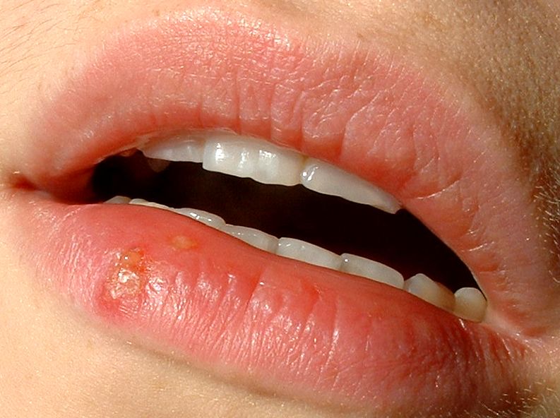
Herpes labialis
Image Source: Wikimedia Commons (2005). Herpes labialis, Licensed under a creative commons licence. Available for free at: https://commons.wikimedia.org/wiki/File:Herpes_labialis.jpg (Accessed on: 10 March 2022).

Diagnosis is usually clinical and can be confirmed on a viral PCR swab. Oral HSV infection can be considered for oral antivirals (e.g. aciclovir or valaciclovir). Patients with anogenital HSV infection should be referred to GUM and managed with 5/7 of oral antiviral within 5 days of the start of an episode or while new lesions are still forming. Suppressive antiviral treatment should be considered in patients having 6 or more attacks a year.
Eczema Herpeticum
A widespread and severe HSV infection of the skin that is mostly associated with atopic eczema. This is a dermatological emergency. Eczema herpeticum usually presents during the first episode of HSV infection but can result from recurrent infection. It has also been seen in non-eczematous conditions such as pemphigus vulgaris, burns and Darier disease.
Eczema herpeticum manifests as clusters of painful and itchy monomorphic vesicles. These rupture leaving small punched-out erosions and ulcerations overlying an erythematous base. Patients often present with extensive vesicles over the face, neck or trunk with associated fever, malaise and lymphadenopathy. Lesions can take up to 6 weeks to heal.
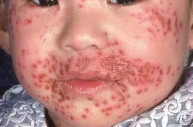
Eczema herpeticum
Image Source: Wikimedia Commons (2018). Eczema herpitcum, Licensed under a creative commons licence. Available for free at: https://commons.wikimedia.org/wiki/File:Eczema_herpitcum.jpg (Accessed on: 15 March 2023).

Diagnosis is usually clinical, however, a viral PCR swab can be taken for HSV. A bacterial swab should also be taken to exclude secondary bacterial infection. Prompt treatment is with oral aciclovir for 10-14 days or until lesions heal. IV aciclovir may be required if the patient is too unwell/deteriorating. An ophthalmologist should be consulted in suspected cases of eye involvement.
Herpes Zoster (Shingles)
A self-limiting blistering rash caused by the reactivation of the varicella zoster virus (VZV), also known as human herpesvirus type 3 (HHV3). Chickenpox is the acute, primary infection which produces widespread lesions at different stages of development. The virus remains dormant in the dorsal root ganglion in the spinal cord and, when reactivated, migrates down cutaneous sensory nerves to induce lesions.

Herpes zoster (shingles)
Image Source: Wikimedia Commons (2007). Herpes zoster chest, Licensed under a creative commons licence. Available for free at: https://commons.wikimedia.org/wiki/File:Herpes_zoster_chest.png (Accessed on: 10 March 2022).

Prior to the skin changes, there may be localised pain associated with fever and tender lymphadenopathy. Red papules then erupt into vesicles in a unilateral dermatomal distribution and are confined to the distribution of one or two sensory nerves. Commonly affects the face, neck and trunk. Involvement of the ophthalmic division of the trigeminal nerve is termed herpes zoster ophthalmicus, and involvement of the VII nerve is termed Ramsay Hunt syndrome. Lesions can take up to a month to resolve.
Herpes zoster is self-limiting and management can include analgesia and emollients. Oral antivirals can be commenced in more vulnerable patients presenting <72 hours from onset or severe cases. Complications include corneal scarring, Ramsay Hunt syndrome and post-herpetic neuralgia. The preventive shingles vaccine is offered in the UK to individuals over 70 years old.
Molluscum Contagiosum
This is caused by a pox virus and presents mostly in children and young adults. Transmitted via close and indirect contact (e.g. towels).
It presents as clusters of pearly umbilicated papules that occur on the face, trunk and extremities. Surrounding dermatitis is frequent. Larger and more widespread molluscum contagiosum occur in immunosuppressed patients.

Molluscum contagiosum
Image Source: Wikimedia Commons (2005). Molluscaklein, Licensed under a creative commons licence. Available for free at: https://commons.wikimedia.org/wiki/File:Molluscaklein.jpg (Accessed on: 10 March 2022).

Molluscum contagiosum is a clinical diagnosis. It is self-limiting and typically resolves without intervention within 18 months. Treatment with cryotherapy, imiquimod, benzoyl peroxide or curettage can also be used.
Viral Warts
This is caused by infection of the epidermal cells with human papillomavirus (HPV). It is transmitted through direct and indirect contact. The most common HPV types infecting the skin are 1, 2, 3, 4, 10, 27, 29 and 57. Anogenital warts are commonly caused by types 6 and 11.
Viral warts are highly contagious and most frequently present in children, immunosuppressed patients (e.g. long-term immunosuppressants, HIV) and in conditions where the skin barrier is defective. Cutaneous warts are papules or nodules with a papillomatous and hyperkeratotic surface. Plantar warts (verruca) are usually painful with dark punctate centres with hyperkeratotic surrounding skin. It is a clinical diagnosis and warts can resolve spontaneously. Other treatment options include podophyllin, imiquimod, cryotherapy or curettage and cautery.
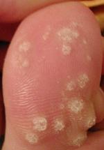
Viral warts
Image Source: Wikimedia Commons (2008). Dornwarzen, Licensed under a creative commons licence. Available for free at: https://commons.wikimedia.org/wiki/File:Dornwarzen.jpg (Accessed on: 15 March 2023).

Complete Guide to Clinical Genetics ST3 Application and Recruitment (2027)
Written by Medibuddy. Last updated: October 2026. Clinical Genetics is

Complete Guide to Clinical Radiology ST3 Application and Recruitment (2027)
Recruitment paused: NHS England is not running Clinical Radiology ST3+ recru

Complete Guide to Paediatric Surgery ST3 Application and Recruitment in United Kingdom (2026)
Written by Medibuddy. Last updated: October 2026. If you’re planning


Take your subscriptions with you
Our mobile app allows you to access your interview and exam question banks wherever you are.

