


 Close
Close
 22nd July 2023
Alcohol-related Liver Disease (ALD)
Background
In contrast to many other major diseases, the mortality associated with liver disease has been increasing in recent decades, as demonstrated in the image below. Alcohol is the leading cause of liver disease in the UK. It can cause either acute or chronic liver disease, and the latter may eventually result in cirrhosis.
ALD is associated with chronic and excessive alcohol intake. However, not all those with a significant alcohol history will develop ALD. Other risk factors associated with ALD are smoking and being overweight.
Pathophysiology
There are multiple pathways through which alcohol can damage the liver, including the production of free radicals and oxidative stress.
The histological stages of ALD include: steatosis, alcohol-related steatohepatitis, fibrosis and cirrhosis, and hepatocellular carcinoma (HCC) as a further complication. Similar stages are seen in non-alcohol related fatty liver disease. However, there are a few key histological findings which are more typical of ALD, such as hyaline sclerosing necrosis and fibro-obliterative damage in the hepatic veins, which can be used to distinguish the two.
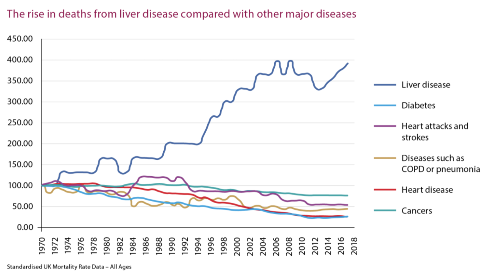
British Liver Trust – Standardised UK Mortality Rate Data.
Image Source: British Liver Trust (no date). Standardised UK Mortality Rate Data – All Ages. Available at: https://britishlivertrust.org.uk/about-us/media-centre/statistics/ (Accessed: 28 June 2023).
Assessment and diagnosis
Taking a thorough alcohol history is key when there is a suspicion of ALD. There are a number of validated tools to screen for alcohol misuse, such as AUDIT, which consists of 10 questions. A link to the AUDIT tool can be found in the References section.
Clinical features
ALD may be asymptomatic or may present with a variety of symptoms, including fatigue, weight loss, anorexia, abdominal discomfort or distension, fever, nausea, vomiting and jaundice.
Findings on examination may be in keeping with chronic liver disease or excess alcohol intake, such as palmar erythema, Dupuytren’s contracture and parotid gland enlargement.
Investigations
Initial investigations should include a full set of liver biochemistry tests (AST, ALT, ALP, GGT, bilirubin and albumin) – i.e. LFTs. AST is often the most raised, and more so than ALT. However, the transaminases can be normal in some people with severe ALD or even cirrhosis, so normal LFTs do not rule out the diagnosis.
Further basic blood tests include an FBC to assess for anaemia and thrombocytopenia, U&Es with magnesium and phosphate levels, and a coagulation screen.
Additional tests may be appropriate to exclude alternative causes of liver dysfunction; for example, a viral hepatitis screen, iron studies and auto-antibodies. This is also known as a ‘non-invasive liver screen’ (NILS).
Ultrasound is the first line imaging investigation, as a non-invasive and cheap way of assessing for pathological markers such as steatosis, and to exclude other pathology, e.g., gallstone disease.
Transient elastography is better for imaging the liver if there is a suspicion of severe fibrosis and cirrhosis, and should be offered to those with a history of chronic and heavy alcohol use. CT and MRI scans may show evidence of liver disease, but are not able to determine aetiology, and offer little additional extra information compared to ultrasound and elastography, so are generally not indicated.
Liver biopsy can be used to help determine the cause of the liver disease but is not necessary in every patient.
Further screening
Hepatocellular carcinoma (HCC) is an important potential complication of ALD. If suspected, α-fetoprotein (AFP) should be measured, as a serum marker strongly associated with HCC. First line imaging remains ultrasound, but CT or MRI may be needed as further investigations if HCC is suspected.
In addition to assessment of the liver, it is important to screen for other alcohol-related organ damage, for example, cardiomyopathy and chronic pancreatitis.
It is also important to include assessment of disease severity.
NICE recommends using the Model for End-Stage Liver Disease (MELD) score, the components of which consist of:
- Dialysis at least twice in the past week or continuous veno-venous haemodialysis for more than 24 hours
- Creatinine level
- Bilirubin level
- INR
- Sodium level
Another commonly used tool is the Child-Pugh score – which uses bilirubin, albumin, INR, the presence of ascites and encephalopathy as its assessment metrics.
Management
The first line treatment – and most important intervention – in the management of ALD is complete abstinence from alcohol.
In the earlier stages of the disease, abstaining from alcohol has the potential to reverse some of the damage that has occurred. For those with more advanced disease, including cirrhosis, there are survival benefits associated with abstinence. It is important to support people in their plans to stop drinking, as some may be at risk of alcohol withdrawal syndrome – see Alcohol withdrawal syndrome section.
Other recommended lifestyle changes include smoking cessation and weight loss.
A multidisciplinary team approach to management is recommended, ideally with input from mental health services and a nutrition expert. People with ALD are highly likely to be malnourished and may require supplementation. This cohort of patients is at high risk of refeeding syndrome and should be monitored closely for this.
Patients should also be monitored and treated for other known complications of alcohol-related cirrhosis. This may include endoscopic surveillance for oesophageal varices and 6-monthly ultrasound scans – with or without AFP serum measurement – to screen for HCC.
Oesophageal varices may be managed with non-selective beta-blockers or endoscopic banding.
Ultimately, those with advanced disease should be considered for liver transplantation. There are criteria when considering if someone may be suitable for a transplant; this often includes a requirement for a period of abstinence and are generally specific to units.
Decompensated liver disease
Cirrhosis secondary to ALD, or any other cause of liver disease, can either be compensated or decompensated. In compensated liver disease, the liver is still able to function, and the patient is relatively well. Decompensation refers to a sudden deterioration in liver function and presents with new or worsened symptoms. Decompensated liver disease is associated with a high mortality rate.
The classic features of decompensation include:
- Jaundice
- Ascites
- Hepatic encephalopathy
- Variceal bleeding
Other indicators of decompensation include hepatorenal syndrome, recurrent infections, and cachexia.
The British Society of Gastroenterology care bundle for decompensated cirrhosis highlights the key areas of management.
The key steps include assessment of liver biochemistry and synthetic function, screening for and treating infection (including an ascitic tap in all patients with ascites to assess for spontaneous bacterial peritonitis), establishing alcohol intake, monitoring and managing alcohol withdrawal, supporting nutrition, and identifying and managing acute kidney injury.
It is important to identify any potential underlying cause for decompensation and treat appropriately.
Additionally, certain features of decompensation have specific management, such as gastrointestinal bleeding, hepatic encephalopathy, and ascites.
Alcohol withdrawal syndrome
Clinical features
People with alcohol dependence are at risk of developing alcohol withdrawal syndrome when they stop or suddenly reduce their alcohol intake.
Alcohol interacts with both GABA and NMDA receptors; chronic use can lead to dysregulation of these receptors. If alcohol consumption is drastically reduced or ceased, this leads to an excess of glutamatergic activity, with reduced GABA activity, which results in overstimulation of the sympathetic nervous system.
Common clinical features of withdrawal include sweating, anxiety, nausea, vomiting, tachycardia and palpitations.
Severe alcohol withdrawal can manifest as ‘delirium tremens’. This is an acute confusional state associated with visual hallucinations, delusions and a coarse tremor. It typically develops 48 hours after the patient’s last drink. This is a medical emergency and can be life-threatening.
Additionally, severe alcohol withdrawal can also cause seizures, which typically manifest 72 hours after the last drink.
Assessment and diagnosis
It is crucial to identify patients with alcohol dependence so that alcohol withdrawal can be monitored for and treatment preempted.
When alcohol withdrawal syndrome is suspected, the severity of symptoms should be assessed in order to guide treatment. The Clinical Institute Withdrawal Assessment for Alcohol Revised (CIWA-Ar) is a validated scoring system to assess severity and is based on key clinical features associated with alcohol withdrawal. (See the References section for more information.
Although alcohol withdrawal is a clinical diagnosis, other investigations should be carried out to support the diagnosis, but also exclude other causes of the presentation.
Management
The mainstay of management of alcohol withdrawal is benzodiazepines. Emergency treatment for seizures is intravenous lorazepam, and delirium tremens can be treated with oral lorazepam or diazepam. Antipsychotics may be used if there is a poor response to benzodiazepines.
For those without seizures or delirium tremens, a benzodiazepine regimen should be initiated. In the majority of people, this would be with chlordiazepoxide, which is a longer-acting benzodiazepine than diazepam and lorazepam.
In addition, all patients should be given thiamine to prevent or treat Wernicke’s encephalopathy. Initially, a high dose parental form is recommended, commonly known as the brand name Pabrinex. Local protocols differ in their recommendations on how long intravenous thiamine should be continued; the patient should then subsequently be established on an oral formulation long term.
Complete Guide to Clinical Genetics ST3 Application and Recruitment (2027)
Written by Medibuddy. Last updated: October 2026. Clinical Genetics is

Complete Guide to Clinical Radiology ST3 Application and Recruitment (2027)
Recruitment paused: NHS England is not running Clinical Radiology ST3+ recru

Complete Guide to Paediatric Surgery ST3 Application and Recruitment in United Kingdom (2026)
Written by Medibuddy. Last updated: October 2026. If you’re planning


Take your subscriptions with you
Our mobile app allows you to access your interview and exam question banks wherever you are.

