


 Close
Close
 8th December 2023
Acute Angle Closure (AAC)
Angle closure is an ophthalmological emergency where a sudden occlusion of aqueous outflow leads to an acute rise in intraocular pressure, which in turn can cause irreversible damage to the optic nerve (glaucoma).
Overview
In angle closure, the trabecular meshwork is occluded by the peripheral iris (irido-trabecular contact). This obstructs the outflow of aqueous humour through the iridocorneal angle, leading to an acute rise in intraocular pressure. These patients will have a normal optic disc. When there is evidence of optic nerve damaged, it is then termed angle closure glaucoma. Angle closure can be classified as primary or secondary. Primary angle closure occurs in an eye with a shallow angle configuration. Secondary angle closure due to a local or systemic factor occurs in an eye with a normal angle configuration. Acute angle closure is sudden onset occlusion of the angle.
Pathophysiology
The mechanisms by which angle closure arises includes relative pupillary block, non-pupillary block and lens-induced angle closure. In relative pupillary block, decreased outflow through the pupil creates a pressure gradient/accumulation of aqueous in the posterior chamber, resulting in the anterior bowing of the iris, which then acutely occludes the angle. In non-pupillary block, there is an abnormal iris configuration. In the lens-induced form, a bulkier lens or a lens that has moved anteriorly decreases aqueous outflow. There is often a mixture of mechanisms.

Image Source: Wikimedia Commons (2006). UBM Angle closure glaucoma, Licensed under a creative commons licence. Available for free at: https://commons.wikimedia.org/wiki/File:UBM_Angle_closure_glaucoma.jpg (Accessed on: 5 May 2022).
Risk factors
- Age (commonest around ages 55-65)
- Female preponderance
- Ethnicity – commoner in people of Asian heritage, particularly far eastern
- Shorter eyes
- Hypermetropia
Presentation
It is typically precipitated by events causing pupil dilatation or ciliary body effusion, such as watching television in a dark room or waking up at night and turning on the lights, iatrogenic (pharmacological) or emotional stress. Common symptoms and signs of acute angle closure are shown below.
Adapted from ‘Angle-closure Glaucoma Straight to the point of care’, the following are common presenting features of acute angle closure:
- Intermittent or constant:
- Unilateral severe ocular or periocular pain with headache
- Nausea and vomiting
- Blurred vision
- Seeing haloes around bright objects such as lights due to corneal oedema
- Decreased VA due to corneal oedema
- Unilateral red eye
- Hazy cornea due to cornea oedema
- Fixed mid-dilated pupil
- Unilateral raised intraocular pressure (usually in the 50s)
- Shallow anterior chamber
- The contralateral eye usually has an occludable angle
Patients typically present acutely and may have a history of episodes of intermittent angle closure, which spontaneously resolves.
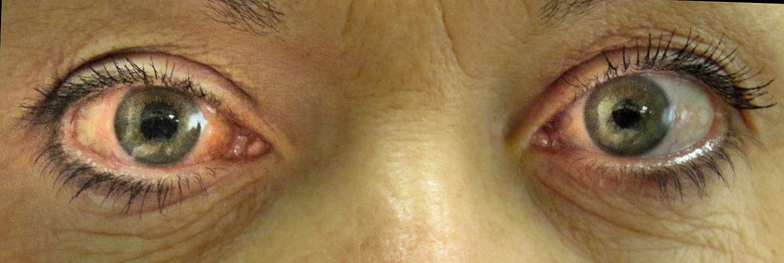
Image Source: Wikimedia Commons (2020). Acute angle closure glaucoma, Licensed under a creative commons licence. Available for free at: https://commons.wikimedia.org/wiki/File:Acute_angle_closure_glaucoma.JPG (Accessed on: 5 May 2022).
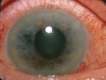
Image Source: Wikimedia Commons (2011). Acute Angle Closure-glaucoma, Licensed under a creative commons licence. Available for free at: https://commons.wikimedia.org/wiki/File:Acute_Angle_Closure-glaucoma.jpg (Accessed on: 5 May 2022).
Diagnosis
Acute angle closure is diagnosed clinically. Angle closure is diagnosed through examination using a set criteria and clinically as set out below.
- Presence of at least 2 of the following symptoms: ocular or periocular pain, nausea and/or vomiting, an antecedent history of intermittent blurring of vision with haloes.
- Presenting intra-ocular pressure (IOP) >21 mmHg.
- Presence of at least 3 of the following signs: conjunctival injection, corneal epithelial oedema, mid- dilated unreactive pupil and shallow anterior chamber.
Ultrasound or OCT of the anterior segment may be utilised in patients suspected to have angle closure or with abnormal iris configuration to confirm diagnosis. Imaging to assess the health of the optic nerve head will also be utilised.
Management
Suspected acute angle closure should be referred urgently same day to ophthalmology or urgent eye service. Acutely, treatments aim to lower the intraocular pressure. Patients should be encouraged to lie in a supine position. First line treatment will include topical treatment such as prostanglandin analogues, oral or IV carbonic anhydrase inhibitors, miotics, and beta blockers. Second-line options in refractory cases can involve medical treatment such as IV mannitol or surgical treatment such as anterior chamber paracentesis. In patients with a clear cornea after successful control of intraocular pressure, laser peripheral iridotomy can be performed as soon as possible. Clear lens extraction is a procedure also considered in primary angle closure. Long-term treatment will include preventative measures such as a laser or surgical peripheral iridotomy.
Complete Guide to Clinical Genetics ST3 Application and Recruitment (2027)
Written by Medibuddy. Last updated: October 2026. Clinical Genetics is

Complete Guide to Clinical Radiology ST3 Application and Recruitment (2027)
Recruitment paused: NHS England is not running Clinical Radiology ST3+ recru

Complete Guide to Paediatric Surgery ST3 Application and Recruitment in United Kingdom (2026)
Written by Medibuddy. Last updated: October 2026. If you’re planning


Take your subscriptions with you
Our mobile app allows you to access your interview and exam question banks wherever you are.

