


 Close
Close
 8th December 2023
Blepharitis
Blepharitis is inflammation of the eyelid margin. It is commonly a chronic condition with acute episodes of marked inflammation. Blepharitis is common; up to 37% of patients seen by ophthalmologists have been reported to have blepharitis. Blepharitis is a multifactorial disease and various interacting mechanisms such as chronic low-grade bacterial infection, inflamed skin from conditions such as eczema or acne rosacea and parasitic infestations. Blepharitis can be classified anatomically, although patients may have an overlap of different types.
The following is a classification system for blepharitis, adapted from ‘Blepharitis’:
- Anterior blepharitis
- Staphylococcal blepharitis
- Seborrheic blepharitis
- Posterior blepharitis/ Meibomian Gland Dysfunction
- Mixed blepharitis with elements of anterior and posterior disease
Anterior blepharitis occurs when there is disease around the eyelashes. It can be staphylococcal or seborrheic. In staphylococcal blepharitis, there is chronic low-grade infection around the base of the eyelashes with an immune response to the bacteria. It is more likely to occur in patients with a history of atopic dermatitis; clinically, there are often hard scales and collarettes around the base of the eyelashes. In seborrheic blepharitis, there is less inflammation than in staphylococcal cases, and there are soft, greasy scales with surrounding skin around the eyes and nasolabial folds greasy, in keeping with seborrheic dermatitis.
Posterior blepharitis can occur in patients with acne and seborrheic dermatitis but can also occur in patients without evidence of a skin condition. It is primarily a disease of the meibomian glands with meibomian gland dysfunction leading to dilatation and obstruction of the glands. The sebum normally acts as a surfactant, but with obstructed glands and decreased expression, the tears evaporate and tear film breaks up easily. Some patients also develop an immune response to the mite Demodex commonly found in hair follicles and sebaceous glands, and this causes blepharitis.
Symptoms are due to irritation of the lid and dry eyes. They include itching of the eyelids, burning sensation, foreign body sensation, watering, intermittent blurred vision, mild photophobia and crusting, particularly in the morning. Patients may also describe episodes of eyelid swelling and redness or give a history of chalazions and styes. Symptoms are commonly bilateral, although one eye may be more affected than the other and present in a relapsing and remitting course.
Examination may reveal scaling of the eyelashes, telangiectasia in the lid margin, eyelid redness and/or swelling and dilated and/or obstructed meibomian glands. Mild conjunctival hypereamia is common in flares. Tear break-up time may be decreased, and the corneal may have evidence of dryness and inflammation, such as punctate epithelial erosions. The patient may have a chalazion, and in those with long-standing disease, there may be notching and scarring of the lid margin and disorders of the eyelashes, such as depigmentation and misdirection.

Image Source: Wikimedia Commons (2017). Anterior Blepharitis, Licensed under a creative commons licence. Available for free at: https://commons.wikimedia.org/wiki/File:Anterior_Blepharitis.jpg (Accessed on: 5 May 2022).

Image Source: Wikimedia Commons (2012). Blepharitis, Licensed under a creative commons licence. Available for free at: https://commons.wikimedia.org/wiki/File:Blepharitis.JPG (Accessed on: 5 May 2022).
Blepharitis is diagnosed clinically based on symptoms and slit lamp examination. It is a chronic condition, and as such, patient education is important. Treatment of blepharitis is usually lid therapy. Patients are advised to apply warm compresses (a microwaveable bag may be purchased) for 5-10 mins and then wash the eyelid gently with a cotton tip applicator with water or an additive such as sodium bicarbonate or tea tree oil if mite infestation. Patients with meibomian gland dysfunction or chalazions should massage the eyelids to express clogged ducts. Lid therapy can be practised a few times a week when stable and more frequently (a few times daily) in exacerbation of disease. Artificial tears can be used to treat dry eye symptoms. Topical antibiotics such as Fusidic acid or Erythromycin may be used to treat active folliculitis in anterior blepharitis. Patients, particularly those with meibomitis or rosacea, will benefit from a course of oral antibiotics (macrolides or tetracylines) to clear bacteria from the lid margin. In marked ocular inflammation, a short course of steroids will decrease inflammation and prevent complications related to this. Refer patients for same-day ophthalmology assessment if there are symptoms of corneal disease, there is rapid onset visual loss, orbital or pre-septal cellulitis is suspected and if the eye is red with red flag features. Chalazions are a common result of meibomian gland dysfunction, and treatment is as above. In patients with chalazions lasting several months despite optimal lid therapy, incision and curettage may be indicated.

Image Source: Wikimedia Commons (2010). Chalazion sup 02, Licensed under a creative commons licence. Available for free at: https://commons.wikimedia.org/wiki/File:Chalazion_sup_02.jpg (Accessed on: 5 May 2022).
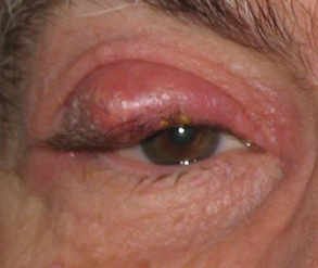
Image Source: Wikimedia Commons (2016). Chalazion2 01242005 (cropped), Licensed under a creative commons licence. Available for free at: https://commons.wikimedia.org/wiki/File:Chalazion2_01242005_(cropped).jpg (Accessed on: 5 May 2022).
Complete Guide to Clinical Genetics ST3 Application and Recruitment (2027)
Written by Medibuddy. Last updated: October 2026. Clinical Genetics is

Complete Guide to Clinical Radiology ST3 Application and Recruitment (2027)
Recruitment paused: NHS England is not running Clinical Radiology ST3+ recru

Complete Guide to Paediatric Surgery ST3 Application and Recruitment in United Kingdom (2026)
Written by Medibuddy. Last updated: October 2026. If you’re planning


Take your subscriptions with you
Our mobile app allows you to access your interview and exam question banks wherever you are.

