


 Close
Close
 13th December 2023
Loss of vision
Assessment of vision loss
Vision loss may occur from an abnormality anywhere in the visual system, from the tear film to the occipital cortex. Initial evaluation should focus on determining the onset and severity of vision loss, associated systemic symptoms, and monocular or binocular involvement. It is important to recognise that patients with homonymous visual field defects may perceive their vision loss as monocular.
Any significant vision loss should be discussed with the local ophthalmology team following initial assessment.
Onset
- Acute vision loss over minutes to hours requires urgent ophthalmic opinion.
- Common causes of acute vision loss include acute angle-closure glaucoma, retinal vascular occlusion, ischaemic optic neuropathy and trauma.
- Sub-acute or chronic vision loss (weeks to months) should also be discussed to arrange formal ophthalmic assessment.
Associated symptoms
- Flashes, floaters and trauma increase the likelihood of retinal detachment.
- Painful eye movements in younger patients may indicate optic neuritis.
- Headaches may be a feature of giant cell arteritis, raised intracranial pressure or migraine.
Past medical history
- Diabetes and hypertension are associated with ocular vascular disease.
- Diabetes and corticosteroid use are associated with accelerated cataract formation.
Ophthalmic history
- It is important to ask about glasses and contact lens use.
- Myopia (short-sightedness) is associated with increased risk of retinal detachment.
- Hypermetropia (long-sightedness) is a risk factor for acute angle closure glaucoma.
- A red eye in a contact lens wearer requires careful ophthalmic examination.
- Previous ocular surgery and its temporal relation to visual loss should be enquired about.
Medication history
- Digoxin is associated with yellowing of vision.
- Phosphodiesterase type-5 (PDE-5) inhibitors (sildenafil, tadalafil, vardenafil) may cause transient blue tints.
- The use of PDE-5 inhibitors has been associated with an increased risk of non-arteritic anterior ischaemic optic neuropathy.
- Amiodarone may cause progressive optic neuropathy.
- Tetracyclines and vitamin A derivatives are associated with raised intracranial pressure.
| Visual acuity | Baseline visual acuity should be documented in all cases, preferably with and without pinhole
Improvement with pinhole suggests a refractive error or ocular media opacity (corneal/lenticular) |
| Visual fields to confrontation | This may detect a hemianopia unnoticed by the patient |
| Pupillary Examination | Fixed and semi-dilated (angle closure glaucoma)
Small, irregular and sluggish (anterior uveitis) |
| Relative afferent pupillary defect | Optic neuropathy even in the presence of a normal-appearing optic nerve head |
| Extraocular eye movements | Painful eye movements (optic neuritis)
Gaze evoked visual loss (orbital lesion) 6TH nerve palsy (raised intracranial pressure) |
| Eyelids | Swollen (orbital cellulitis)
Retraction (thyroid eye disease) |
| Proptosis | Thyroid eye disease
Orbital cellulitis Carotid-cavernous fistula Orbital tumour |
| Conjunctiva/sclera | Redness and injection (orbital or anterior segment disease) |
| Intra-ocular pressure | Significantly raised in acute angle closure glaucoma (>50mmHg) |
| Fundoscopy | Cherry red spot, cotton wool spots, haemorrhages, venous dilation (Retinal vascular occlusion)
Dot/blot haemorrhages (diabetic; hypertensive retinopathy) Macula exudates (diabetic; hypertensive retinopathy) Drusen and macula haemorrhages (age-related macular degeneration) Optic nerve swelling: unilateral vs bilateral (papilloedema) |
| General Examination | Temporal arteries: tenderness, nodules, loss of pulsation
Cardiovascular: atrial fibrillation; heart murmur Neurological: hemiparesis BMI: Consider idiopathic intracranial hypertension in overweight females of child-bearing age Temperature: if elevated, consider orbital cellulitis or infective emboli Blood pressure: uncontrolled hypertension Glucose: undiagnosed diabetes mellitus Urine dipstick for proteinuria if pregnant (pre-eclampsia) |
Source: Denniston, A., & Murray, P. (2018). Oxford Handbook of Ophthalmology (4th ed.) Oxford University Press
Anterior segment disease
Vision loss attributed to anterior segment pathology can be divided into:
- Cornea
- Anterior chamber-related
- Lens
Cornea
A corneal ulcer is a defect of the corneal epithelium and underlying stroma. This is a potentially vision-threatening ocular emergency. Even with prompt treatment, patients can suffer significant morbidity from corneal perforation, scarring, secondary glaucoma and permanent vision loss.
Aetiology:
- Infectious
- Bacterial infection (most common). Organisms include Staphylococcus species (eyelids) and Pseudomonas aeruginosa (contact lens users).
- Viral: Herpes viruses are a common cause of viral keratitis. There may be a history of previous herpetic keratitis, recent cold sore (herpes simplex), or recent/concurrent shingles (herpes zoster) affecting the ophthalmic branch (V1) of the trigeminal nerve.
- Parasitic: Acanthamoeba is a free-living protozoan found in freshwater and soil that can cause corneal ulcers in contact lens wearers. It characteristically causes pain out of proportion to clinical signs due to infiltration of corneal nerves. For this reason, it is often missed in the early stages.
- Fungal: Less than 10% of all corneal infections. They are most often precipitated by corneal trauma or exposure to organic matter.
- Non-infectious
- Exposure keratopathy: incomplete eyelid closure (facial nerve palsy; stroke) results in exposure of the inferior third of the cornea, with drying of the tear film and subsequent corneal surface breakdown.
- Neurotrophic cornea: Corneal sensation is supplied by the trigeminal branch (V1) of the trigeminal nerve. Loss of sensation is a high-risk factor for corneal breakdown. Causes may be local (prolonged contact lens use; herpes virus), systemic neuropathy (diabetes; amyloidosis) or lesion anywhere along the path of V1.
- Autoimmune: vasculitic corneal ulcers often affect the peripheral cornea and cause rapid and circumferential corneal changes.
Risk Factors
- Contact lens wear
- Severe dry eyes
- Trauma/abrasion
- Nutritional deficiency (malabsorption, liver cirrhosis, alcohol excess)
- Systemic collagen vascular disease
- Corneal exposure secondary to incomplete lid closure (facial nerve palsy)
- Loss of corneal sensation
Clinical Presentation
Slit-lamp examination demonstrates an epithelial defect with an underlying yellow/grey infiltrate. A dendritic appearance is characteristic for herpes simplex keratitis. The presence of a hypopyon (white fluid level within the anterior chamber) indicates sight-threatening disease.
Management
All patients presenting with a red eye and a history of contact lens use should be discussed with the local ophthalmic team. Active corneal ulcers require a corneal scrape for gram staining, microbial culture and sensitivity, viral and acanthamoeba PCR to determine underlying aetiology. Definitive management is directed against the underlying cause.

Image Source: Wikimedia Commons (2015). Parasite140120-fig1 Acanthamoeba keratitis, Licensed under a creative commons licence. Available for free at: https://commons.wikimedia.org/wiki/File:Parasite140120-fig1_Acanthamoeba_keratitis.png (Accessed on: 15 May 2022).

Image Source: Wikimedia Commons (2017). Dendritic corneal ulcer, Licensed under a creative commons licence. Available for free at: https://commons.wikimedia.org/wiki/File:Dendritic_corneal_ulcer.jpg (Accessed on: 15 May 2022).

Image Source: Wikimedia Commons (2017). Hypopyon 1, Licensed under a creative commons licence. Available for free at: https://commons.wikimedia.org/wiki/File:Hypopyon_1.jpg (Accessed on: 15 May 2022).
Anterior chamber
Acute angle-closure glaucoma (AACG) is an ophthalmic emergency. The normal range for intraocular pressure is 10 to 22 mmHg. Aqueous humor production begins in the ciliary body, flows through the pupil, and exits the eye through the trabecular meshwork and Schlemm’s canal in the anterior chamber angle (between the iris and the cornea). In acute angle-closure glaucoma, the intraocular pressure increases rapidly due to obstruction of the drainage angle and blockage of aqueous humor outflow. Predisposing factors include an anatomically narrow anterior chamber angle, increasing lens size and an anteriorly located iris-lens diaphragm. AACG is often precipitated by pupillary dilatation. As the pupil enlarges, the peripheral iris contacts the lens, preventing the flow of aqueous through the pupil (pupillary block). This leads to bulging of the iris (iris bombe), crowding of the anterior chamber angle by the iris and obstruction of aqueous humor outflow. The intraocular pressure rises acutely, leading to symptomology.
Risk Factors
- Age: The average age at presentation is 60. This is felt to be due to the increasing size of the lens with age
- Gender: F:M ratio 4 to 1
- Ethnicity: more common in Southeast Asians, Chinese, and Inuit heritage
- Family history: An anatomically narrow anterior chamber angle is usually inherited
Clinical Presentation
- Reduced vision associated with haloes
- Severe headache
- Nausea & vomiting
- Abdominal pain
- Red, injected eye
- Cloudy cornea
- Fixed, semi-dilated pupil (iris ischaemia)
- Intraocular pressures commonly >50mmHg (normal 11-2mmHg)
- Glaucomatous or swollen optic disc
Management
The medical treatment for AACG aims to:
- Decrease production of aqueous humor
- Restore aqueous outflow
- Relieve pupillary block
Initial medical therapy includes a combination of the following medications:
- Intravenous acetazolamide 500 mg, topical beta-blocker (timolol 0.5%) and alpha 2-agonist (apraclonidine 1%) to block the production of aqueous humor.
- Topical pilocarpine 1% to 2% to constrict the pupil and increase the outflow of aqueous humor.
- If the intraocular pressure remains elevated after an hour, intravenous mannitol 1 gram/kg can be given (if there is no contraindication) to rapidly reduce the volume of aqueous humor.
Definitive treatment is by laser peripheral iridectomy (PI) after the acute episode subsides. Iridectomy relieves the pupillary block by allowing the flow of aqueous humor through the iris. The contralateral eye usually has the same predisposing anatomy and carries up to 80% risk of developing an attack of AACG if untreated. Hence, PI is usually performed in both eyes following a first presentation.
Prognosis
The outcome for patients with AACG is good following treatment if recognised early. In the early stages, corneal clouding may be subtle and overlooked. Patients may be mistakenly worked up for intracranial or abdominal symptoms. Delay in treatment can lead to damage to the optic nerve and vision loss.

Image Source: Wikimedia Commons (2011). Acute Angle Closure-glaucoma, Licensed under a creative commons licence. Available for free at: https://commons.wikimedia.org/wiki/File:Acute_Angle_Closure-glaucoma.jpg (Accessed on: 15 May 2022).
Lens
Cataracts cause a painless, slow deterioration of vision. There may be difficulty with focusing and glare experienced with bright lights. Some patients experience a ‘second sight’ – whereby the increased refractive power of an enlarging cataract produces an improvement with near-tasks such as reading. Inevitably, over time, vision progressively declines as the cataract becomes cloudy. Patients are more likely to present to their local optician than an acute practitioner. Certain conditions predispose to accelerated and/or early onset cataract formation:
- Diabetes mellitus
- Corticosteroid use
- Myotonic dystrophy
Cataract extraction is the management of choice.
References
- Ahram DF, Alward WL, Kuehn MH. The genetic mechanisms of primary angle closure glaucoma. Eye (Lond). 2015 Oct;29(10):1251-9.
- Collignon NJ. Emergencies in glaucoma: a review. Bull Soc Belge Ophtalmol. 2005;(296):71-81.
- Shahid H, Salmon JF. Malignant glaucoma: a review of the modern literature. J Ophthalmol. 2012;2012:852659.
Posterior segment disease
Posterior segment disease can be divided into:
- Vitreous haemorrhage
- Retinal detachment
- Macula disease
- Vascular disease
- Optic nerve disease
Vitreous haemorrhage
Vitreous haemorrhage is the presence of blood in the vitreous cavity. The source may be from retinal neovascularisation, rupture of healthy retinal vessels or trauma. Causes include:
- Proliferative diabetic retinopathy (PDR)
- Retinal vein occlusion (RVO)
- Sickle cell retinopathy
- Trauma: blunt or penetrating
- Posterior vitreous detachment
- Blood dyscrasias (Leukaemia, anaemia, thrombocytopenia, haemophilia)
- Iatrogenic: following intra-ocular procedures
Clinical presentation
Patients experience a sudden onset or a shower of floaters, which may or may not obscure the vision. Visual acuity is moderately to severely reduced. The presence of a relative afferent pupillary defect (RAPD) should prompt concern for an associated retinal detachment. Vitreous haemorrhage that is obscuring fundus examination mandates a B-ultrasound scan. This will confirm vitreous echoes and allow visualisation for an associated retinal detachment or posterior wall masses. Ocular ultrasound should not be performed in traumatic cases where there is suspicion of a globe rupture.
Management
All suspected vitreous haemorrhages should be referred to Ophthalmology for full assessment and monitoring. Treatment is aimed at the underlying cause. Surgical intervention (vitrectomy) may be required for non-clearing or repeated vitreous haemorrhage.
Retinal detachment
Retinal detachment (RD) refers to an abnormal separation of the neurosensory retina from the underlying retinal pigment epithelium (RPE). The photoreceptors are susceptible to ischaemia because of separation from the choroidal blood supply, with potentially irreversible vision loss. Urgent surgery is warranted if the macula remains attached to preserve central vision. In macula-off detachments vision may remain poor despite surgical intervention.
Classification
There are three classifications of retinal detachments: rhegmatogenous retinal detachment (RRD), tractional retinal detachment (TRD), and exudative retinal detachment (ERD).
- Rhegmatogenous retinal detachment: A tear, break, or hole occurs in the retina. Vitreous enters the space underneath the neurosensory retina, causing a detachment from the RPE. This detachment can occur over hours to days.
- Tractional retinal detachment: Retinal traction is exerted from the proliferation of fibrotic membranes in the vitreo-retinal interface. When the force is strong enough, the neurosensory retina may be detached from the underlying RPE. Often associated with advanced diabetic eye disease.
- Serous retinal detachment: This is a result of a local exudative process, such as a tumour or local inflammation.
Risk factors for rhegmetogenous retinal detachment:
- Lattice degeneration
- High/pathological myopia
- Previous intraocular surgery
- Trauma
- Previous retinal detachment in the contralateral eye
- Family history
Clinical presentation
Vision loss is often painless and progressive over hours to days. The main characteristics of a retinal detachment are summarised by the four ‘F’s:
- Floaters
- Flashes
- Field defect
- Fall in acuity
Management
The primary treatment of RRD is surgical. If the macula is detached, surgery may be delayed by a few days. If the macula is preserved, urgent surgery is warranted to preserve this. Surgical options include retinopexy (pneumatic or silicone oil) and scleral buckling. A retinal tear in the absence of an RD may be sealed with laser retinopexy.
Differential diagnoses
The main differential diagnosis for new onset floaters and flashes is a posterior vitreous detachment. Posterior vitreous detachment is the most common cause of primary symptomatic floaters. A rapid increase in the number of floaters with sudden onset of photopsia (flashes) needs urgent ophthalmic assessment.
Summary
Any patient complaining of flashes and floaters should be referred for urgent ophthalmological assessment to exclude a retinal detachment.
| Posterior vitreous detachment | Retinal detachment | Vitreous haemorrhage |
|---|---|---|
| Flashes and floaters in the peripheral field of view | Dark, progressive shadow moving across the field of view Decreased VA +/- central vision loss |
Sudden onset shower of floaters |
Macula disease
Age-related macular degeneration (AMD)
AMD is a leading cause of irreversible blindness worldwide. AMD reflects a complex interaction between genes and environmental factors, which have not yet been fully elucidated. The hallmark of AMD is drusen, which are yellow deposits that develop within the outer retina. Drusen disrupt the homeostasis of photoreceptor outer segments, causing a slow, gradual loss of vision. Drusen also predispose to breaks within the outer retina. New vessels may then grow from the choroidal circulation (choroidal new vessels) to the retina, leading to neovascular disease. Choroidal neovascularisation predisposes to macula bleeding, oedema and potentially rapid reduction of vision.
Classification:
Traditionally, two forms of AMD are described: ‘dry’ AMD, characterised by drusen and ‘wet’ AMD by the presence of CNV and retinal haemorrhages. More recently, AMD is now recognised as a spectrum of disease ranging from early and intermediate AMD (drusen and altered RPE) to late AMD (neovascularisation; exudative disease).
Risk Factors:
- Age: most patients are above 60. If below 50 years of age, a secondary cause for choroidal neovascularisation must be sought
- Family history
- Smoking
- Caucasian
- Female
- High cumulative sunlight exposure
- Obesity
- Hypercholesterolaemia
Clinical Presentation:
- Decreased VA: May be sudden reduction in vision with a central scotoma if there has been break-through bleeding from CNV
- Blurred or distorted vision (metamorphopsia)
- Sudden visual loss: Break-through bleeding from CNV; patients often complain of a dense central scotoma
- Fundoscopy: drusen; pigmentary changes; retinal haemorrhages
Management:
There are no proven interventions for early or intermediate AMD. Patients should be advised to stop smoking. Vision should be monitored by an Amsler grid, which allows patients to detect new metamorphopsia. The Age-Related Eye Disease Study (AREDS) and AREDS2 studies investigated the role of high-dose vitamins on the progression of AMD. The results indicated that supplementation may slow the progression from intermediate to advanced disease. The original AREDS formulation contained vitamin C, vitamin E, beta-carotene, zinc oxide and cupric oxide. The AREDS2 formulation replaced beta-carotene with lutein, zeaxanthin and omega-3 fatty acids due to the possible increased risk of lung cancer in former smokers with beta-carotene. The formulation doses are in excess of recommended daily allowances, and no long-term data is available regarding adverse risks associated with excessive use. Decisions to initiate AMD supplements are best taken in conjunction with a medical retina specialist.
Treatment of neovascular AMD has been revolutionised by the introduction of anti-VEGF agents (ranibizumab; bevacizumab; aflibercept), which restore macula architecture, halt progression of neovascularisation and improve visual acuity in approximately 1/3ʳᵈ of patients.
Central serous chorioretinopathy (CSCR)
CSCR is an idiopathic macula disease characterised by the accumulation of subretinal fluid. It typically affects males between the ages of 25-50 and is associated with a ‘type A’ personality. Affected individuals may have a history of systemic or topical corticosteroid use.
Clinical features:
- Blurred/decreased vision
- Metamorphopsia
- Micropsia (minification of objects)
- Central scotoma
Examination usually detects a shallow serous detachment of the macula, best appreciated on slit-lamp examination. Management is usually conservative, with spontaneous resolution of 1-6 months. If persistent or recurrent disease, focal laser photocoagulation may be considered.
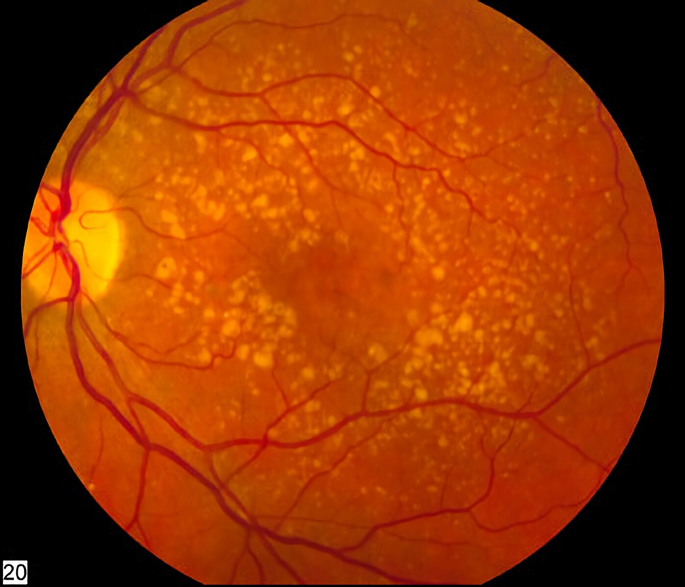
Image Source: Wikimedia Commons (2012). Intermediate age related macular degeneration, Licensed under a creative commons licence. Available for free at: https://commons.wikimedia.org/wiki/File:Intermediate_age_related_macular_degeneration.jpg (Accessed on: 15 May 2022).
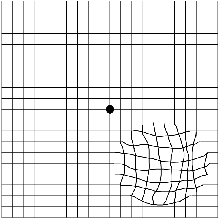
Image Source: Wikimedia Commons (2020). Amsler grid – distorted vision (metamorphopsia), Licensed under a creative commons licence. Available for free at: https://commons.wikimedia.org/wiki/File:Amsler_grid_-_distorted_vision_(metamorphopsia).png (Accessed on: 15 May 2022).
Vascular disease
This may be divided into retinal artery occlusion (central/branch retinal artery occlusion) and retinal vein occlusion (central/branch retinal vein occlusion).
Central retinal artery occlusion (CRAO):
CRAO is the sudden blockage of the central retinal artery, resulting in retinal ischaemia and rapid vision loss. The central retinal artery is the first intra-orbital branch of the ophthalmic artery. Occlusion of the central retinal artery results in retinal ischemia, necrosis and vision loss. Sources of occlusion include:
Embolic:
- Cholesterol (Hollenhorst) plaque: carotid vascular disease
- Platelet-fibrin: thrombo-embolism
- Calcific: cardiac valvular disease
Thrombotic:
- Atherosclerotic disease
- Collagen vascular disease
- Vasculitis: giant cell arteritis
Associations of CRAO:
- Hypertension
- Diabetes mellitus
- Carotid artery disease
- Collagen vascular disease
- Vasculitis
- Trauma
- Coagulopathy
Clinical Presentation:
CRAO typically presents with sudden, painless, monocular vision loss that occurs over seconds. Patients may report an antecedent transient visual loss (amaurosis fugax). Visual acuity is often drastically reduced (VA <6/60). A relative afferent pupillary defect (RAPD) will be present. Fundus examination reveals retinal pallor with a ‘cherry red’ spot at the fovea. At 4-6 weeks, the pale optic disc appears pale, and retinal arterioles are attenuated.
Investigations:
Investigations are aimed at identifying the underlying cause. Inflammatory markers (FBC, CRP, ESR) are mandatory to exclude giant cell arteritis (GCA). Further specific testing, including thrombophilia, autoimmune and infective screening, is directed by concomitant symptoms. A fast-track referral to stroke clinic is recommended to identify thrombo-embolic sources (ECG, ECHO, carotid Doppler).
Management:
There are no established treatments to restore vision following CRAO. Various treatments have been described to restore perfusion to the retina if within 24 hours of onset:
- Ocular massage to dislodge emboli
- Reduction of intraocular pressure with intravenous acetazolamide or anterior chamber paracentesis
- Supplemental oxygen
Fibrinolysis may be beneficial within 4.5 hours of symptom onset. A CT head should be obtained to rule out intracranial haemorrhage.
In cases of suspected GCA, immediate corticosteroids are required to protect the contralateral eye (see section below on GCA). If thrombo-embolic disease is suspected, secondary prevention should be started with clopidogrel 300mg OD followed by 75mg to continue, and atorvastatin 80mg OD pending referral to stroke/TIA clinic.
Prognosis:
Vision loss is usually irreversible. There is a small risk of neovascular disease in the long-term, although patients often do not require long-term surveillance.
Branch retinal artery occlusion
Branch retinal artery occlusion is usually the result of an embolus that lodges at the bifurcation of a retinal arteriole. The retinal and visual changes relate only to the part of the artery occluded. Vasculitis (GCA), in this instance, is extremely rare, and in the absence of associated symptoms, no workup for giant cell arteritis is required. Patients should be referred for further investigations as per TIA. There are no treatments.
Retinal vein occlusion
There are two types of RVO: Central retinal vein occlusion (CRVO) and Branch retinal vein occlusion (BRVO). In CRVO, structural changes in the central retinal artery and the lamina cribrosa leads to compression of the pre-laminar central retinal vein. Subsequent turbulent flow in the central retinal vein predisposes to thrombosis. In BRVO, occlusion of a tributary retinal vein by an adjacent retinal arteriole occurs at the site of an AV crossing (both retinal arterioles and veins share a common adventitial sheath). Underlying structural changes occur as a consequence of arteriosclerosis, hypertension, diabetes mellitus and glaucoma. Thrombi may also be the result of various hyperviscosity syndromes.
The mechanism of visual loss is due to the development of retinal ischaemia and macula oedema.
Associations of CRVO
- Hypertension
- Diabetes
- Smoking
- Obesity
- Protein S, protein C, anti-thrombin III deficiency
- Myeloma
- Antiphospholipid syndrome
- Vasculitis
- Glaucoma
CRVO can be classified into non-ischaemic and ischaemic forms, and the following clinical features can be seen in each:
Non-ischaemic:
- 80% of CRVO
- VA usually >6/60
- May be no RAPD
- Less extensive haemorrhages +/- CWS; swollen optic nerve head; macula oedema
- Better prognosis
Ischaemic:
- 20% of CRVO
- VA <6/60
- Marked RAPD
- Extensive haemorrhages and CWS; swollen optic disc
- Worse prognosis
Investigations:
The following investigations are aimed at identifying associations which require urgent action, such as severe hypertension, uncontrolled diabetes or rarely blood dyscrasias.
- Blood pressure
- FBC
- ESR & CRP
- Glucose
Further routine investigations are not recommended. However, in patients below 40 years of age or those without identifiable risk factors or unusual presentations (bilateral CRVO), further tests may be performed to exclude an underlying systemic prothrombotic disorder
- ACE
- Anti-cardioplin antibodies
- ANA
- ds-DNA
- ANCA
- Thrombophilia screen
- CXR
Management:
Anti-VEGF agents (bevacizumab) can treat macula oedema but may be of little benefit in ischaemic CRVO. Patients with retinal vein occlusions are at risk of retinal, iris and anterior chamber angle neovascularisation and may develop neovascular glaucoma at 3 months (‘100-day glaucoma’). Neovascular disease can be treated by laser photocoagulation to reduce the ischaemic drive.

Image Source: Wikimedia Commons (2019). Cherry red spot in patient with central retinal artery occlusion (CRAO), Licensed under a creative commons licence. Available for free at: https://commons.wikimedia.org/wiki/File:Cherry_red_spot_in_patient_with_central_retinal_artery_occlusion_(CRAO).jpg (Accessed on: 15 May 2022).

Image Source: Wikimedia Commons (2019). Fondo de ojo Ostrucción Vena central, Licensed under a creative commons licence. Available for free at: https://commons.wikimedia.org/wiki/File:Fondo_de_ojo_Ostrucci%C3%B3n_Vena_central.png (Accessed on: 15 May 2022).
Optic nerve disease
Optic neuropathy is damage to the optic nerve. The causes may be divided into:
Acquired
- Demyelinating/idiopathic optic neuritis
- Inflammatory/autoimmune
- Ischaemic optic neuropathy
Congenital
- Leber’s Hereditary Optic Neuropathy (LHON)
Optic neuritis (ON)
Inflammation of the optic nerve may be classified as typical or atypical. Typical ON often refers to either idiopathic or multiple sclerosis-associated disease. This most commonly occurs in young Caucasian women in temperate zones. Vision loss is progressive over hours-to-days, reaching a nadir by 2 weeks. The degree of vision loss is variable, ranging from moderate (6/18) to severe (<6/60). There is associated retrobulbar discomfort with eye movements and disturbance in colour vision. The optic disc may be mildly swollen or normal (retrobulbar ON). Uhthoff’ phenomenon describes a transient worsening of neurological symptoms associated with a rise in body temperature (exercise; bathing) and is associated with both multiple sclerosis (MS) and aquaporin-4 (AQP-4) optic neuritis.
Features of typical ON:
- Ages 20-50 years
- Unilateral
- Progression over hours-to-days
- Retrobulbar discomfort
- Colour vision disturbance
- Relative afferent pupillary defect
- Improvement begins within 2 weeks of onset
Investigations are centred on investigating for multiple sclerosis-associated ON:
- Magnetic resonance imaging (MRI) of the brain and orbits with contrast to look for demyelinating disease.
- In the active phase, the affected optic nerve will enhance.
- Evidence of infratentorial, periventricular and juxtacortical white matter lesions may suggest multiple sclerosis.
Management:
The optic neuritis treatment trial (ONTT) demonstrated that a combination of intravenous methylprednisolone (IVMP) followed by oral Prednisolone may accelerate visual recovery but did not affect final VA for a first episode of optic neuritis. Low-dose oral corticosteroids (1mg/kg) alone were associated with increased frequency of recurrence. For recurrent, idiopathic ON, initiation of treatment within 2 days of symptom onset may provide a better recovery of VA, although further multi-centre studies are warranted to confirm this. Oral corticosteroids in a bioequivalent dose may be as effective as IVMP. The decision to treat typical ON with corticosteroids is often taken on a case-by-case basis.
Prognosis:
In idiopathic or multiple sclerosis-associated optic neuritis, over 90% recover to a visual acuity of 6/12 or better without treatment. However, there may be permanent abnormalities of colour perception, contrast sensitivity or visual field defects. Following a first episode of optic neuritis, the following characteristics have a lower risk of developing multiple sclerosis:
- No demyelinating lesions on MRI
- Optic disc swelling
- Male gender
Atypical ON:
This describes clinical features not consistent with typical optic neuritis. A secondary cause for optic nerve inflammation should be sought. Traditionally, it was acceptable to observe the natural evolution of an ON event over 4-6 weeks. Increased recognition of AQP-4 and myelin oligodendrocyte glycoprotein (MOG) disease has broadened the spectrum of optic neuritis-related autoimmune disorders. AQP-4 and MOG-associated optic neuritis carry significant visual morbidity; prompt initiation of high-dose corticosteroid therapy may prevent a poor outcome. Early clinical suspicion is important because confirmatory antibody testing (aquaporin-4 and MOG antibodies) and neuro-radiological imaging may take weeks. Features which are suggestive of non-MS, autoimmune ON include:
- Bilateral disease
- Moderate-to-severe optic disc swelling
- Non-Caucasian ethnicity
- Age not in keeping with typical optic neuritis
Other investigations for atypical optic neuritis include FBC, ESR, CRP, vitamin B12, folate, HBA1c, LFTs, ANCA, ANA, ACE syphilis, genetic testing for Leber’s hereditary optic neuropathy, syphilis serology and lumbar puncture.
| Idiopathic or MS-related optic neuritis | AQP-4 optic neuritis | MOG-optic neuritis | |
|---|---|---|---|
| History | |||
| Age | 18-40 | 40+ | Any age |
| Ethnicity | Caucasian | Asian; Afro-Caribbean | Southeast Asia; Indian subcontinent |
| Retrobulbar pain | Yes | May be painless | Yes |
| Associated symptoms | May be a previous history or relapse-remitting symptoms | Nausea; vomiting; hiccups (area postrema syndrome) | |
| Examination | |||
| Visual acuity | Usually >6/60 | <6/60 | <6/60 |
| Laterality | Unilateral | Uni-or bilateral ON | Uni- or bilateral ON |
| Optic disc appearance | Normal Mild-to-moderate swelling |
Normal Mild-to-moderate swelling |
Moderate-to-severe disc swelling (disc or peripapillary haemorrhages; subretinal fluid) |
| Radiological findings | |||
| MRI Orbit | Distal optic nerve enhancement | Proximal optic nerve enhancement Chiasmitis |
Distal and proximal optic nerve enhancement Chiasm is spared Often bilateral May have perineural enhancement |
| MRI Head | Lesions in the corpus callosum, preventricular white matter, brainstem and cerebellum | Lesions around the periaqueduct and hypothalamus | Lesions of the cortical grey/juxtacortical white matter |
Source: Cooper SA, Leddy SG, Skipper NT, et al. Optic neuritis with potential for poor outcome. Practical Neurology 2022;22:190-200.
Ischaemic optic neuropathy
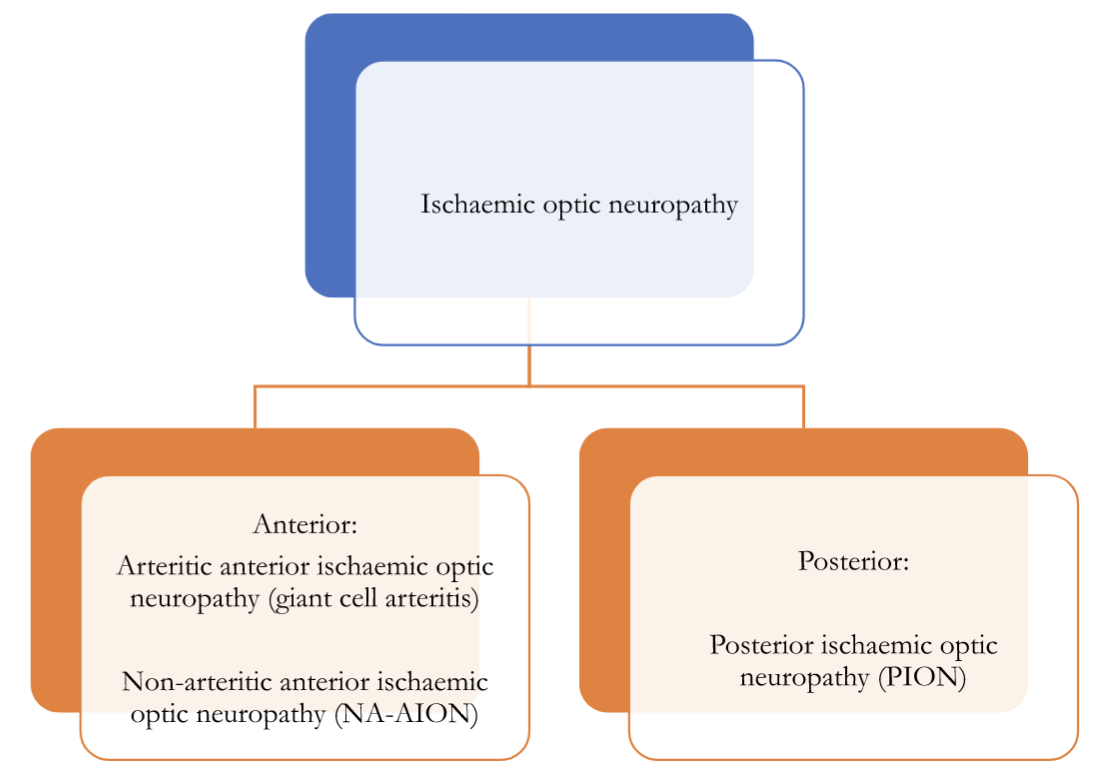
Ischaemic optic neuropathy is a significant cause of acute visual loss in those aged 50 years and over, affecting 2-10/100 000 people per year. It may be divided into anterior and posterior forms.
Giant Cell Arteritis (GCA)
GCA is a chronic granulomatous inflammation of medium-large-sized arteries originating from the arch of the aorta. Ocular involvement occurs in nearly 50%. Vision loss can be attributed to vasculitis affecting the posterior ciliary arteries, central retinal and/or the cilioretinal artery, and the ophthalmic artery respectively. It is important to understand the distinction between arteritic anterior ischaemic optic neuropathy and GCA: the latter is a systemic vasculitis, and ocular involvement is defined by the vision loss described above. Non-GCA causes of arteritic ischaemic optic neuropathy include ANCA-associated vasculitides.
Histopathology:
Early in the disease, the histopathological changes are confined to the region of the internal or external elastic lamina or adventitia. As the disease progresses, the wall of the artery of the artery becomes thickened by significant lymphocytic infiltrate. This reaction is directed against the internal elastic lamina. The vessel lumen may become obliterated by a marked fibroblastic proliferation in the intima. Multinucleated giant cells are present in only 50% of biopsies and are not a requirement for the diagnosis. Inflammation is often segmental, leading to skip lesions. Fibrinoid necrosis is not typical and indicates alternate diagnoses such as ANCA-associated vasculitis.
Clinical presentation:
Approximately 1/3ʳᵈ of patients with GCA will experience vision loss. This is instantaneous and profound (VA<6/60). Patients may experience a prior episode of amaurosis fugax. A brisk RAPD is evident. Fundus changes depend on the vascular territory involved:
Anterior ischaemic optic neuropathy (70-90%)
- Vasculitis of the short posterior ciliary arteries supplying the optic nerve head
- The optic disc displays a characteristic pallid swelling
- Swelling and pallor of the posterior pole: in approximately 1/3ʳᵈ of the population, the posterior ciliary arteries provide a cilioretinal artery branch which perfuses the posterior pole.
Central retinal artery occlusion (15%)
- Inner retinal ischaemia produces a swollen, pale macula areas +/ a cherry-red spot
Posterior ischaemic optic neuropathy (5%)
- Vasculitis of the pial vessels and ophthalmic artery
- The optic nerve head appears normal during the early stages of disease
- Optic disc pallor manifests 6-8 weeks after the initial insult
Other key symptoms in GCA include constitutional symptoms (weight loss, fever, fatigue, anorexia, malaise), headache, scalp tenderness, and jaw claudication. The temporal arteries are often thick, nodular, pulseless and tender to palpation. GCA may also occur in a minority of patients with polymyalgia rheumatica, characterised by pain and stiffness of the shoulder and, less commonly, hip girdles. Approximately 20% of patients with vision loss will have no systemic symptoms.
Investigations:
Supportive laboratory investigations include marked elevation in acute inflammatory markers (ESR; CRP); normocytic, normochromic anaemia and mild thrombocytosis. ANCA and MP3 may be useful in excluding GPA and PAN. Patients with suspected GCA should have a prompt, confirmatory diagnostic test. This could be either:
- Temporal artery biopsy. This should be performed within 2 weeks to maximise diagnostic yield. A sample of at least 2cm is recommended to minimise a false-negative result due to skip lesions.
- Ultrasound. A hyperechoic ‘halo’ may be seen in the affected temporal artery. Ultrasound is user-dependent, with a sensitivity of ∼ 70% and specificity ∼87%. However, this drops markedly within one week of initiating corticosteroids.
Management:
Vision loss in giant cell arteritis is irreversible. Corticosteroids are the mainstay of treatment and should not be delayed pending a confirmatory test. A dose of 1mg/kg/day is continued until symptom resolution. There is usually a marked response within a week. Inflammatory markers should be monitored frequently; once normalised, corticosteroids may be cautiously tapered down. Most patients require at least a year of treatment. The role of intravenous corticosteroids (methylprednisolone 1g daily for three days) is uncertain. With established unilateral visual loss, many centres recommend intravenous treatment to protect the contralateral eye. However, there are no established studies demonstrating superiority of IVMP in preventing contralateral vision loss.
Tocilizumab (Subcutaneous injection 162 mg once weekly) is licensed for relapsed or remitting disease or cases intolerant of corticosteroids. IL-6 levels are significantly elevated in patients with GCA, and IL-6 is thought to play a central role in the pathogenesis of GCA by activation and recruitment of T-cells. There is no role for tocilizumab during the acute phase of disease.

Image Source: Wikimedia Commons (2017). Arteria temporalis superficialis, ramus frontalis, Licensed under a creative commons licence. Available for free at: https://commons.wikimedia.org/wiki/File:Arteria_temporalis_superficialis,_ramus_frontalis.jpg (Accessed on: 15 May 2022).

Image Source: Wikimedia Commons (2014). Giant cell arteritis — low mag, Licensed under a creative commons licence. Available for free at: https://commons.wikimedia.org/wiki/File:Giant_cell_arteritis_–_low_mag.jpg (Accessed on: 15 May 2022).
Non- Arteritic Ischaemic Optic Neuropathy (NA-AION)
NA-AION is attributed to relative hypoperfusion of the short posterior ciliary arteries supplying the optic nerve head. This results in oedema, ischaemia and infarction of the optic nerve fibres. Changes are often sectorial, affecting either the superior or inferior half of the optic nerve head.
Risk Factors:
Small optic nerve cup (‘disc at risk’) is the strongest risk factor for developing NA-ION. Cup-to-disc ratio is typically 0.2 or less. Optic nerve head oedema induced by ischemic injury is thought to cause axonal compression within a crowded scleral tunnel. Other notable associations include:
- optic disc drusen
- systemic hypertension
- diabetes mellitus
- obstructive sleep apnoea
- use of phosphodiesterase-5 inhibitors
Clinical presentation:
Vision loss is often painless and instantaneous. The following examination findings are present:
- VA: may be moderately-to-severely reduced (6/18 -(<6/60)
- Visual field: altitudinal visual field defect
- Colour vision: may or may not be affected
- Pupil examination: RAPD
- Optic nerve head: swollen; often sectorial and hyperaemic
Investigations:
All patients should have inflammatory markers (FBC, CRP, ESR) to exclude giant cell arteritis. Additionally, blood pressure should be documented.
Management:
Patients should have a full review of their medication history, with specific attention to the use of PDE-5 inhibitors and anti-hypertensive medications, especially the use of these medications at night. There is no accepted treatment for NA-AION. Corticosteroids are associated with an increased risk of second eye involvement. Aspirin does not aid in visual recovery nor reduce the risk to the contralateral eye. The Ischemic Optic Neuropathy Decompression Trial (IONDT) demonstrated optic nerve fenestration surgery is ineffective and potentially harmful.
Prognosis:
The Ischemic Optic Neuropathy Decompression Trial demonstrated a 40% improvement in visual acuity. The risk of second eye involvement is about 15% over 5 years. Poor baseline visual acuity and diabetes were risk factors for second eye involvement.
Posterior ischaemic optic neuropathy
Posterior ischaemic optic neuropathy affects the plial plexus, serving the retrolaminar optic nerve. The major risk factors are peri-operative hypotension and GCA. There may be unilateral or bilateral involvement. Visual acuity is often dramatically reduced. Initial examination reveals a RAPD but otherwise normal-appearing optic nerve head and fundus. Over 4-6 weeks, profound optic atrophy is revealed. All peri-operative patients complaining of visual loss should have a careful pupil examination. FBC, CRP and ESR should be performed in all patients >50 years of age. There are no treatment options.
Leber’s hereditary optic neuropathy
Leber’s hereditary optic neuropathy is a rare optic neuropathy of maternal inheritance arising from point mutations in mitochondrial DNA. Vision loss is attributed to free radical oxidate damage of the retinal ganglion cell layer. Patients may have concomitant cardiac abnormalities. There is often a family history of visual loss. The presentation is often a young male <50 years old with subacute bilateral, sequential vision loss (<6/60). Pupillary reactions are normal. Fundus examination may reveal peripapillary telangiectasia, arteriolar narrowing, optic disc oedema, or atrophy. Mitochondrial DNA analysis of a serum sample is required to confirm the diagnosis. An atypical optic neuropathy screen (see above) is recommended to exclude compressive and inflammatory optic neuropathies. An ECG should be performed for cardiac conduction defects associated with mitochondrial cytopathies. Patients should be advised to avoid smoking and avoid excess alcohol. Idebenone, a co-enzyme Q10 derivative, may be offered to maintain good residual vision. However, no clear benefit in final visual acuity has been demonstrated in randomised controlled trials.
References
- Age-Related Eye Disease Study Research Group. A randomised, placebo-controlled, clinical trial of high-dose supplementation with vitamins C and E and beta carotene for age-related cataract and vision loss: AREDS report no. 9. Arch Ophthalmol. 2001 Oct;119(10):1439-52.
- Aldrich EM, Lee AW, Chen CS, et al. Local intraarterial fibrinolysis administered in aliquots for the treatment of central retinal artery occlusion: the Johns Hopkins Hospital experience. Stroke 2008;39:1746–50.
- Beck RW, Cleary PA, Anderson MM, Keltner JL, Shults WT, Kaufman DI, Buckley EG, Corbett JJ, Kupersmith MJ, Miller NR. A randomised, controlled trial of corticosteroids in the treatment of acute optic neuritis. The Optic Neuritis Study Group. N Engl J Med. 1992 Feb 27;326(9):581-8.
- Beck RW, Cleary PA, Backlund JC. The course of visual recovery after optic neuritis. Experience of the Optic Neuritis Treatment Trial. Ophthalmology. 1994 Nov;101(11):1771-8.
- Beck RW, Cleary PA. Optic neuritis treatment trial. One-year follow-up results. Arch Ophthalmol. 1993 Jun;111(6):773-5.
- CATT Research Group. Martin DF, Maguire MG, Ying GS, Grunwald JE, Fine SL, Jaffe GJ. Ranibizumab and bevacizumab for neovascular age-related macular degeneration. N Engl J Med. 2011 19 May;364(20):1897-908.
- Cooper SA, Leddy SG, Skipper NT, et al. Optic neuritis with potential for poor outcome. Practical Neurology 2022;22:190-200.
- Hayreh SS, Podhajsky PA, Zimmerman B. Ocular manifestations of giant cell arteritis. Am J Ophthalmol 1998;125:509–20.
- Kertes PJ, Galic IJ, Greve M, Williams G, Baker J, Lahaie M, Sheidow T. Efficacy of a Treat-and-Extend Regimen With Ranibizumab in Patients With Neovascular Age-Related Macular Disease: A Randomised Clinical Trial. JAMA Ophthalmol. 2020 01 March;138(3):244-250.
- Klopstock T, Wai-Man Y, Dimitriadis K, et al. A randomised placebo-controlled trial of Idebenone in Leber’s Hereditary Optic Neuropathy. Brain 2011;134(Pt 9):2677-86.
- Nakamura M, Nakazawa T, Doi H, Hariya T, Omodaka K, Misu T, Takahashi T, Fujihara K, Nishida K. Early high-dose intravenous methylprednisolone is effective in preserving retinal nerve fiber layer thickness in patients with neuromyelitis optica. Graefes Arch Clin Exp Ophthalmol. 2010 Dec;248(12):1777-85.
- Park K, Tanaka K, Tanaka M. Uhthoff’s phenomenon in multiple sclerosis and neuromyelitis optica. Eur Neurol. 2014;72(3-4):153-6. doi: 10.1159/000361045. Epub 2014 28 August. PMID: 25195501.
- Petzold A, Braithwaite T, van Oosten BW, et al. Case for a new corticosteroid treatment trial in optic neuritis: review of updated evidence. J Neurol Neurosurg Psychiatry 2020;91:9–14.
- Schmidt-Erfurth U, Eldem B, Guymer R, Korobelnik JF, Schlingemann RO, Axer-Siegel R, Wiedemann P, Simader C, Gekkieva M, Weichselberger A., EXCITE Study Group. Efficacy and safety of monthly versus quarterly ranibizumab treatment in neovascular age-related macular degeneration: the EXCITE study. Ophthalmology. 2011 May;118(5):831-9.
- Schmidt-Erfurth U, Kaiser PK, Korobelnik JF, Brown DM, Chong V, Nguyen QD, Ho AC, Ogura Y, Simader C, Jaffe GJ, Slakter JS, Yancopoulos GD, Stahl N, Vitti R, Berliner AJ, Soo Y, Anderesi M, Sowade O, Zeitz O, Norenberg C, Sandbrink R, Heier JS. Intravitreal aflibercept injection for neovascular age-related macular degeneration: ninety-six-week results of the VIEW studies. Ophthalmology. 2014 Jan;121(1):193-201.
- Sivaprasad, S., Amoaku, W., Hykin, P. et al. The Royal College of Ophthalmologists Guidelines on retinal vein occlusions: executive summary. Eye 29, 1633–1638 (2015).
- Snodderly DM. Evidence for protection against age-related macular degeneration by carotenoids and antioxidant vitamins. Am J Clin Nutr. 1995 Dec;62(6 Suppl):1448S-1461S.
- Stiebel-Kalish H, Hellmann MA, Mimouni M, et al. Does time equal vision in the acute treatment of a cohort of AQP4 and MOG optic neuritis? Neurol Neuroimmunol Neuroinflamm Jul 2019, 6 (4) e572.
- Varma DD, Cugati S, Lee AW, et al. A review of central retinal artery occlusion: clinical presentation and management. Eye 2013;27:688–97.
Disorders of the visual pathway
Most intracranial disorders are associated with significant neurological morbidity; however, occipital pathology may be clinically silent. Patients often describe vague or unilateral problems with vision. A dense homonymous hemianopia may be missed unless visual fields are adequately assessed.
Anatomical considerations
The optic chiasm permits the hemi-decussation of the visual field information, such that the information from the left visual field (left temporal and right nasal field) and right visual field (right temporal and left nasal field) is processed in the contralateral visual cortex. Lesions at the optic chiasm produce bi-temporal hemianopia. Retro-chiasmal lesions produce homonymous hemianopia. Congruity describes the degree of similarity between the visual field defects in both eyes. The more posterior the lesion in retro-chiasmal visual pathway, the more congruous the visual field defect is.
Causes of neurological visual field defects
- Tumours
- Pituitary adenoma
- Craniopharyngioma
- Meningioma
- Lymphoma
- metastases
- Infection
- Intracerebral abscess
- Inflammation/infiltration
- Sarcoidosis
- Histiocytosis
- Lymphocytic hypophysitis
- Vascular
- Stroke
- Pituitary apoplexy
- Arteriovenous malformation
- Intracranial aneurysm
- Trauma

Image Source: Wikimedia Commons (2017). Hemianopsio eo, Licensed under a creative commons licence. Available for free at: https://commons.wikimedia.org/wiki/File:Hemianopsio_eo.png (Accessed on: 15 May 2022).
1 – right monocular vision loss 2 – bi-temporal homonymous hemianopia 3. Left complete homonymous hemianopia 4. Left superior homonymous quadrantopia 5 – Left inferior homonymous quadrantopia with macula sparing 6 – left superior quadrantopia with macula sparing.
Visual aspects of stroke
Visual impairment is a common consequence of stroke. There is significant overlap between the arterial circulations. The lateral geniculate nucleus may be affected by compromise to the internal carotid artery (anterior choroidal artery branch), middle or posterior cerebral artery. The optic radiations are served by both the anterior choroidal and middle cerebral arteries. The occipital cortex is supplied by the middle and posterior cerebral arteries. A posterior cerebral artery stroke may produce a complete homonymous hemianopia with macular sparing owing to an intact blood supply from the middle cerebral artery.
| ANTERIOR CIRCULATION | POSTERIOR CIRCULATION | ||||
|---|---|---|---|---|---|
| Visual field defect | Localisation | Visual field defect | Localisation | ||
| Internal carotid artery | Monocular visual field loss (transient or permanent) | Retina (central/retinal artery branch occlusion) | Posterior cerebral artery | Incongruous homonymous hemianopia | Lateral geniculate body (posterior choroidal artery) |
| Anterior choroidal artery | Incongruous homonymous hemianopia | Optic tract Optic radiation |
Homonymous sectoranopia | Lateral geniculate body | |
| Homonymous sectoranopia | Lateral geniculate body | Congruous homonymous hemianopia with macula sparing | Anterior visual cortex | ||
| Middle cerebral artery | Homonymous sectoranopia | Lateral geniculate body | – | – | – |
| Incongruous homonymous hemianopia | Optic radiation | – | – | – | |
| Congruous homonymous central field defect (macula VF defect) | Posterior visual cortex | – | – | – | |
Exceptional visual field defects
Rarely, intracranial insults may give rise to unusual visual field defects that may mimic an intra-ocular or pre-chiasmal insult. There are two circumstances of note:
- Bilateral occipital lobe lesions: will give rise to bilateral altitudinal visual field defect mimicking retinal or optic nerve head lesion. Full dilated fundus examination is recommended to exclude local causes. Neuroimaging is diagnostic. Patients are at risk of cortical blindness syndrome.
- Unilateral temporal crescenteric visual field defect: The nasal visual field extends to 60 degrees from fixation, whereas the temporal field extends to 90%. This leaves an area of the temporal visual field that has no nasal correlate in the contralateral eye, represented within the parieto-occipital sulcus. Damage in this area of the visual cortex leads to a monocular, contralateral temporal crescent defect.
| Cortical blindness | decreased visual acuity secondary to bilateral retro-chiasmal lesions |
| Anton syndrome | Cortical blindness with denial of blindness |
| Riddoch phenomenon | Moving targets are visualised, however stationary targets are missed |
| Benson syndrome (posterior cortical atrophy) | A form of atypical Alzheimer’s disease with visuo-spatial and visuo-perceptual deficits |
| Posterior reversible encephalopathy syndrome (PRES) | Acute headaches, seizures, altered consciousness, visual disturbance. Associated with hypertensive emergency and eclampsia |
| Balint syndrome | Ocular apraxia (cannot move eyes on command but normal spontaneous movements)
Visual inattention |
References
- Ali K The temporal crescent syndrome Practical Neurology 2015;15:53-55.
- File [modified]: Hemianopsio eo.png. Wikimedia Commons [Internet]. Commons.wikimedia.org. 2021 [cited 15 May 2022]. Available from: https://commons.wikimedia.org/wiki/File:Hemianopsio_eo.png
- Newman RP, Kinkel WR, Jacobs L. Altitudinal Hemianopia Caused by Occipital Infarctions: Clinical and Computerised Tomographic Correlations. Arch Neurol. 1984;41(4):413–418.

Transient visual loss
Transient loss of vision is a fully reversible episode of visual loss lasting less than 24 hours. It often causes concern to the clinician for fear as a harbinger of imminent stroke. Patients often present after resolution of the episode and may have a normal ocular and systemic examination.
Differential diagnoses
The most important distinction in the assessment of TVL is vascular (amaurosis fugax) vs non-vascular aetiology.
| Acute angle-closure glaucoma
Uncontrolled diabetes Optic nerve dysfunction
|
Vascular causes
- Emboli
- TVL can occur due to embolic occlusion of the retinal, optic disc, and choroidal blood vessels.
- Often, the emboli may be visible on ophthalmoscopic examination.
- Ocular ischaemic syndrome
- Ocular ischemic syndrome (OIS) is a disorder of ocular function that occurs subsequent to the stenosis of the carotid artery.
- TVL occurs over seconds to minutes.
- The eye may be red from dilated conjunctival and episcleral vessels.
- There is usually a slow, progressive decline in visual acuity over weeks to months.
- Characteristic features include episodes precipitated by bright light and dull ocular pain often relieved by lying down (‘ocular angina’).
- Migraine
- May cause transient monocular visual loss due to retinal vasospasm.
- Episodes may last minutes to hours, and there may be a history of stereotyped events.
- An associated headache is common.
- Mostly, the visual loss is transient, lasting 5 to 20 minutes, and may recur several times during the day.
- Other visual disturbances include monocular positive visual phenomena (flashing lights or scintillating scotoma).
- Haematological abnormalities
- Severe anaemia, sickle cell disease, and hypercoagulable states may cause episodes of transient visual loss.
- Vasculitis
- vision loss in GCA may be heralded by TVL.
- Examination reveals a swollen optic disc.
- There may be concomitant systemic symptoms (See above section on GCA)
Investigations
All patients should undergo routine haematological testing. In patients under the age of 50 without atherosclerotic risk factors, further investigation is not required.
- Haematological evaluation: FBC, CRP, ESR, cholesterol and lipid profile. Thrombophilia screening should be guided by history and concomitant symptoms.
- Cardiac investigations: ECG +/- 24-hour Holter monitor; ECHO; carotid Doppler scanning.
- Neuroimaging to exclude a haemorrhagic lesion in the brain. Angiographic imaging (CTA/ MRA) may also be utilised in imaging the head and neck.
Management
Management of TVL is directed at the underlying cause.
The North American Symptomatic Carotid Endarterectomy Trial (NASCET) demonstrated that patients with ocular TIA had a 2-year stroke incidence of 17%. This risk may be reduced in patients who have carotid stenosis greater than 70%, reducing future risks of stroke. Individuals with <75% ipsilateral internal carotid artery stenosis or complete carotid occlusion are treated with anti-platelet agents. Those with >75% stenosis should be referred to a vascular surgeon for consideration of endarterectomy or carotid artery stenting.
Ischaemic heart disease is the most common cause of death following an episode of amaurosis fugax. Vascular risk factors, such as hypertension, diabetes, and hyperlipidaemia should be addressed.
References
- Ahmed R, Foroozan R. Transient monocular vision loss. Neurol Clin 2010;28:619-629.
- Barnett HJ, Taylor W, Eliasziw M et al. Benefit of Carotid Endarterectomy in Patients with Symptomatic Moderate or Severe Stenosis. N Engl J Med 1998; 339:1415-1425
- Poole CJ, Ross Russell RW Mortality and stroke after amaurosis fugax. Journal of Neurology, Neurosurgery & Psychiatry 1985;48:902-905.
Complete Guide to Clinical Genetics ST3 Application and Recruitment (2027)
Written by Medibuddy. Last updated: October 2026. Clinical Genetics is

Complete Guide to Clinical Radiology ST3 Application and Recruitment (2027)
Recruitment paused: NHS England is not running Clinical Radiology ST3+ recru

Complete Guide to Paediatric Surgery ST3 Application and Recruitment in United Kingdom (2026)
Written by Medibuddy. Last updated: October 2026. If you’re planning


Take your subscriptions with you
Our mobile app allows you to access your interview and exam question banks wherever you are.

