


 Close
Close
 27th December 2023
Diagnosis
As with many presentations in medicine, an accurate history and examination is crucial and can reveal the cause of the diplopia in the majority of cases. There are two types of diplopia, monocular and binocular, and differentiating between these is a crucial first step. Refractive errors or lens diseases should be suspected in monocular diplopia when it can be resolved with a pinhole, whereas binocular diplopia requires the clinician to determine the underlying cause of the ocular misalignment.
Monocular or binocular assessment
Monocular diplopia is diagnosed when the double vision does not resolve upon closing or covering either eye and, therefore present with monocular viewing. Monocular diplopia can be bilateral, which is not the same as binocular diplopia, which requires both eyes to be open and have the symptom of double vision. In binocular diplopia, covering either eye will eliminate the diplopia.
Use of a pinhole device
Where monocular diplopia is confirmed, a pinhole device should be utilised to identify whether the cause may be related to anterior structures such as the cornea or the lens. The pinhole will block peripheral light rays, only allowing central light to pass through the cornea and pupil, improving acuity and eliminating diplopia secondary to an optical cause. If the diplopia persists, one should consider issues with the macula, such as oedema or problems with the cortex. The symptom of multiple images (> 2) is known as cerebral polyopia and is a rare occurrence of monocular diplopia secondary to structural disorders of the cerebral cortex.
Assess the brainstem
Diplopia can be a symptom seen in stroke patients where structures in the brainstem are affected. Therefore, when monocular diplopia is excluded, the logical next step would be to assess the brainstem and organise urgent imaging where indicated. Structures which may be affected include:
- The diencephalon, a primary relay and processing centre for sensory (including visual) information
- Nuclei or fascicles of the 3ʳᵈ, 5ᵗʰ, or 6ᵗʰ cranial nerves, each responsible for extraocular muscle function
- The medial longitudinal fasciculus – a specialised nerve bundle involved in extraocular function
- The vestibulo-ocular pathway – important in the gaze-stabilising reflex
Assess the diplopia axis
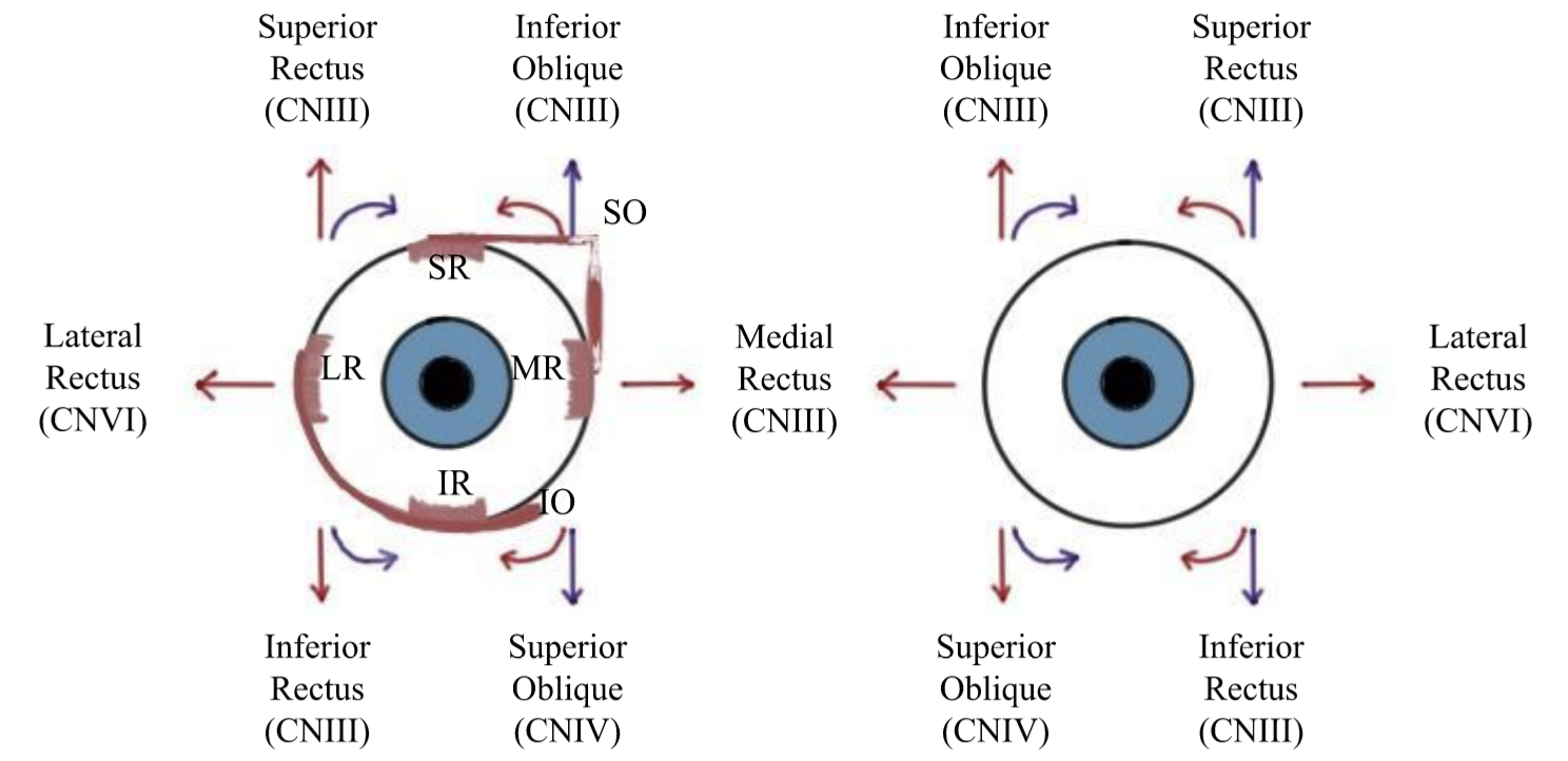
Purely vertical diplopia is caused by 3ʳᵈ or 4ᵗʰ nerve palsy, myasthenia gravis, skew deviation (acquired vertical misalignment not due to any single muscle or ocular motor nerve) or processes related to the orbital tissues such as thyroid eye disease. Purely horizontal diplopia is usually related to abduction (6ᵗʰ cranial nerve) and adduction (third cranial nerve) deficits. Identifying problems with extraocular motility can be difficult and requires a detailed examination, observing each eye separately. Whilst assessing the axis, it is important to observe any signs of thyroid eye disease, such as proptosis or lid lag and for signs of myasthenia, such as fatigable ptosis.
Assess eye misalignment
Cover and alternate cover testing is an efficient and quick way to assess the alignment of the eyes and establish if a diplopia may be due to strabismus. Cover tests can help identify and quantify ocular misalignment, and there are three types of cover test, two of which can be performed at the bedside with little equipment.
Begin with the monocular cover-uncover test, which differentiates between tropias, where a deviation is always present, and phorias, where a deviation is present when one eye is closed or covered. Ask the patient to fixate on a point ahead, roughly 50cm or further away, and observe for any deviations. If placing an occluder over the non-affected eye causes the affected eye to fixate, this is known as a tropia. A phoria would be seen when the covered eye deviates on occlusion and returns to fixation on removal of the occluder.
The alternate cover test involves the patient fixating on a point ahead of them whilst the examiner moves an occluder from one eye to the other whilst focusing on the movement of the uncovered eye. This test allows the examiner to note whether the deviation is worse in specific gazes, i.e. when looking to the left, right, up, down or on a diagonal plane. A deviation which remains the same in all positions of gaze is classed as concomitant and incomitant if it is affected. Incomitant squints should raise concern for neurological, myogenic or neuromuscular junction disorders and may involve specialist input from neurology, whereas concomitant squint should be referred to ophthalmology and may not require further investigation.
Investigations
Blood pressure, blood glucose, urine analysis and erythrocyte sedimentation rate should be performed in cases where a microvascular pathology is suspected. Where thyroid or myasthenia causes are suspected, thyroid function tests, anti-acetylcholine receptor antibodies and single fibre electromyography (EMG) of the orbicularis muscles should be considered. The ice test is a simple bedside investigation which is highly sensitive and specific to myasthenia. The test is interpreted as positive when a ptotic lid is improved after ice application for 2-5 minutes. Acute CT or MRI scans should be obtained when the onset of diplopia is acute and/or present with neurological signs or papilloedema.
Complete Guide to Clinical Genetics ST3 Application and Recruitment (2027)
Written by Medibuddy. Last updated: October 2026. Clinical Genetics is

Complete Guide to Clinical Radiology ST3 Application and Recruitment (2027)
Recruitment paused: NHS England is not running Clinical Radiology ST3+ recru

Complete Guide to Paediatric Surgery ST3 Application and Recruitment in United Kingdom (2026)
Written by Medibuddy. Last updated: October 2026. If you’re planning


Take your subscriptions with you
Our mobile app allows you to access your interview and exam question banks wherever you are.

