


 Close
Close
 29th December 2023
Retinal artery occlusion
Background & Epidemiology
Retinal artery occlusion (RAO) occurs due to blockage of blood flow through a retinal artery, either from a thrombus, embolus, or vascular inflammation or spasm.
RAOs tend to occur in the elderly population and are predominantly due to atherosclerotic emboli from the carotid bifurcation, with giant cell arteritis comprising a smaller proportion of these cases (<5%). Multiple studies have reported an association between RAO and concurrent silent ischaemic stroke and/or subsequent stroke risk. Both share similar risk factors and patient demographics, including older age, male gender, smoking, systemic hypertension, diabetes, obesity, and dyslipidaemia.
Systemic associations should be considered in younger patients (particularly those aged <50 years), including migraine, trauma, hypercoagulable states, exogenous emboli such as talc in intravenous drug users, cardiac abnormalities producing emboli (endocarditis, valvular disease, cardiac myxoma), infections, and autoimmune disorders (e.g. systemic lupus erythematosus, polyarteritis nodosa, Susac syndrome),
Any artery supplying the eye may be affected. Depending on the affected artery(s), RAOs can be classified into central retinal artery occlusion (CRAO), branch retinal artery occlusion (BRAO), cilioretinal artery occlusion (CLRAO), or ophthalmic artery occlusion. The central retinal artery supplies the inner two-thirds of the retina, while the ophthalmic artery supplies the retina and choroid.
Clinical Features
CRAOs present with acute onset painless loss of vision. A relative afferent pupillary defect may be present. A ‘cherry red spot’ may be seen on fundoscopic examination – this is the fovea, which is the thinnest part of the retina and retains relative transparency, in contrast to the surrounding retina, which develops oedema and opacification of the inner retinal layers and therefore appears pale. The arteries are attenuated and demonstrate segmental blood flow (‘box-carring’ or ‘cattle-trucking’ of the blood column within it). Up to 25% of patients may retain preserved central vision due to the presence of a cilioretinal artery supplying the papillomacular bundle; this is supplied by the posterior ciliary artery and, therefore, unaffected by CRAO.
BRAO presents with acute onset visual defect corresponding to the area of retina supplied by the affected artery, which tends to appear pale due to intracellular oedema in the acute phase. The affected artery may be attenuated with ‘box-carring’ or ‘cattle-trucking’ of the blood column within it. Emboli may be visible within the relevant retinal arteries in up to two-thirds of cases, appearing as yellow refractile plaques (Hollenhorst plaques), whitish non-refractile calcified particles, or greyish elongated fibrin-platelet aggregates. Cotton wool spots may be present.
Ophthalmic artery occlusion should be suspected in patients with profoundly reduced vision (no perception of light) because it affects both the inner and outer retina as well as choroidal perfusion. There is, therefore, retinal pallor, albeit without a ‘cherry red spot’ due to absence of choroidal perfusion. A relative afferent pupillary defect may be present.
Management
Acute management:
RAOs represent one of the few ophthalmic emergencies, particularly as there is a short treatment window before irreversible vision loss sets in. Experiments on animal models have demonstrated irreversible retinal damage after 100 minutes of retinal arterial occlusion. Ocular massage is often undertaken in an attempt to dislodge the thrombus/ embolus, as are measures to reduce the intraocular pressure, such as intravenous Acetazolamide, anterior chamber paracentesis, and paper bag rebreathing to produce hypercarbia. Thrombolysis is performed in some centres as well. However, none of these options have been proven to reliably treat RAO.
Management of associated risk factors:
Clinical features of giant cell arteritis (GCA) (new onset headache, amaurosis fugax, temporal tenderness and absent pulse, jaw claudication, anterior ischaemic optic neuropathy, etc.) should be evaluated, and blood tests such as FBC, ESR, CRP should be requested as a matter of urgency, with high-dose systemic steroids started to protect the fellow eye if the degree of suspicion is high. Temporal artery ultrasound and/or biopsy should then be considered as appropriate. GCA is more relevant in CRAO and CLRAO, in contrast to BRAO, which only occurs very rarely in the context of GCA.
Patients should then be referred to a specialist stroke clinic for further workup due to the high risk of stroke (2.2% risk of symptomatic stroke in the 15 days preceding and following the RAO). Underlying risk factors such as hypertension and atrial fibrillation should be sought and addressed. Carotid Doppler ultrasound is often undertaken, and carotid endarterectomy may be indicated in patients with severe (70-99%) carotid stenosis.
Monitoring for complications:
Close monitoring for the sequelae of RAO (retinal and anterior segment neovascularisation) should follow. This occurs less commonly than in patients with RVOs.
Differentials
Differentials of a cherry-red spot in the macula include metabolic storage disorders such as Tay-Sachs disease and Niemann-Pick disease and drug toxicity from methanol poisoning, carbon monoxide poisoning, or intravitreal administration of Gentamicin.
Differentials of retinal pallor include infective retinitis, such as progressive outer retinal necrosis or commotio retinae from blunt trauma.
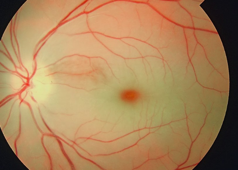
Image Source: Wikimedia Commons (2019). Cherry red spot in patient with central retinal artery occlusion (CRAO), Licensed under a creative commons licence. Available for free at: https://commons.wikimedia.org/wiki/File:Cherry_red_spot_in_patient_with_central_retinal_artery_occlusion_%28CRAO%29.jpg (Accessed on: 27 May 2022).
Complete Guide to Clinical Genetics ST3 Application and Recruitment (2027)
Written by Medibuddy. Last updated: October 2026. Clinical Genetics is

Complete Guide to Clinical Radiology ST3 Application and Recruitment (2027)
Recruitment paused: NHS England is not running Clinical Radiology ST3+ recru

Complete Guide to Paediatric Surgery ST3 Application and Recruitment in United Kingdom (2026)
Written by Medibuddy. Last updated: October 2026. If you’re planning


Take your subscriptions with you
Our mobile app allows you to access your interview and exam question banks wherever you are.

