


 Close
Close
 10th January 2024
Diagnosis
A diagnosis of pleural effusion should be considered in any patient presenting to hospital with shortness of breath. The combination of history and thorough physical examination revealing findings, as detailed above, should alert the clinician to the possibility of an underlying pleural effusion.
Chest x-ray
Plain posteroanterior (PA) chest x-rays are the first-line imaging modality used in suspected pleural effusion and become abnormal with around 200mL of pleural fluid accumulation. The fluid results in blunting of the costophrenic angle with associated development of the meniscus. If there is air associated with the pleural effusion, called a hydropneumothorax, a straight line at the air-fluid interface will be seen, rather than a meniscus.

Large L-sided unilateral pleural effusion in a female patient.
Image Source: Wikimedia Commons (2022). Pleural Effusion, Licensed under a creative commons licence. Available for free at: https://commons.wikimedia.org/wiki/Category:Pleural_effusion (Accessed on: 17 May 2022).
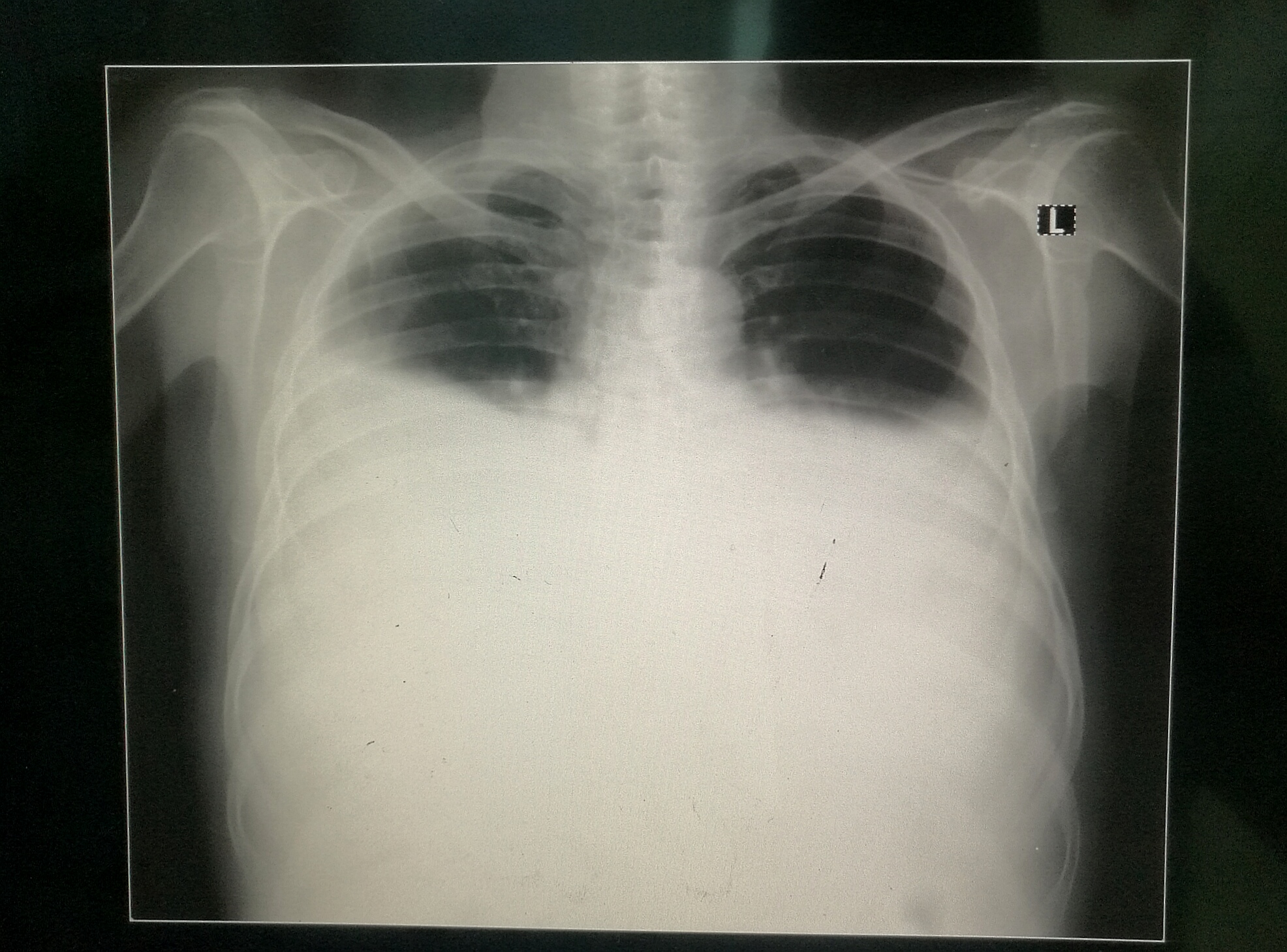
Large bilateral pleural effusions.
Image Source: Wikimedia Commons (2017). Bilateral Pleural Effusion, Licensed under a creative commons licence. Available for free at: https://upload.wikimedia.org/wikipedia/commons/3/34/Bilateral_Pleural_Effusion.jpg (Accessed on: 17 May 2022).
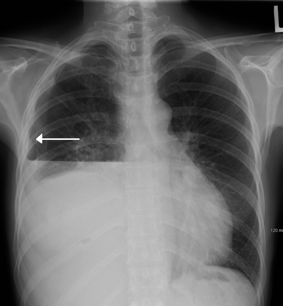
R-sided hydropneumothorax in a male patient. Key difference to note compared to a pleural effusion without associated pneumothorax is the straight line caused by the air-fluid interface.
Image Source: Wikimedia Commons (2017). Hydropneumothorax, Licensed under a creative commons licence. Available for free at: https://upload.wikimedia.org/wikipedia/commons/7/7d/HydropneumoX.png (Accessed on: 17 May 2022).
Ultrasound
Bedside pleural ultrasound can be used to identify pleural effusions and be utilised to identify a site for diagnostic aspiration of pleural fluid. It is particularly useful in the diagnosis of small pleural effusions, which may not be evident on a plain radiograph. In addition to its uses for ultrasound-guided diagnostic aspiration of pleural fluid, ultrasound can be used to determine additional characteristics of the fluid, e.g. septations, which may be suggestive of an underlying malignant or infective effusion.
Diagnostic Aspiration
Diagnostic aspiration is the key diagnostic test in evaluating a unilateral pleural effusion. British Thoracic Guidelines (BTS) state this should be a bedside direct ultrasound-guided aspiration of fluid in the most lateral position possible to reduce associated complications. A 50mL sample of fluid is obtained and sent for:
- Protein
- Lactate dehydrogenase
- Gram staining
- Microbiology culture
- Cytology
- pH (if an infected pleural effusion is suspected)
On aspiration, pleural fluid can take on many appearances. “Normal” pleural fluid is straw-coloured and inodorous. However, in the setting of pathology, many different appearances of pleural fluid can be noted, including blood-strained, frank blood, purulent (suggesting empyema), milky (suggesting chylothorax) or bile-stained.

Figure 4: Diagnostic algorithm for pleural effusion once identified.
Flowchart adapted from guidance available at – Maskell N. British Thoracic Society pleural disease guidelines – 2010 update. Thorax. 2010;65(8):667–9.
Pleural Effusion Fluid Analysis
The key distinction pleural fluid analysis attempts to achieve is exudative vs. transudative pleural effusions. This distinction allows a narrowing of the differential diagnosis (see below).
Pleural fluid protein (g/L) measurement is the key to differentiating between transudates and exudates. Pleural fluid protein >30g/L is considered exudative and protein <30g/L is considered transudative. However, the closer the level is to 30g/L, the less accurate this distinction becomes. In order to achieve a distinction between transudate and exudate in this setting we employ Light’s Criteria:
- Pleural fluid protein to serum protein ratio >0.5
- Pleural fluid LDH to serum LDH ratio >0.6
- Pleural LDH >0.67 x upper limit of normal for serum LDH
If any one of these criteria are met, an exudative pleural effusion diagnosis can be made. Lactate dehydrogenase is utilised in Light’s Criteria as it is a non-specific marker of inflammation – a key component of exudative pleural effusion pathology.
Microscopy, Culture and Sensitivity
Microscopy, culture and sensitivity (M, C + S) is the gold-standard test that is sent with all diagnostic pleural aspirations as per BTS guidelines. TB cultures can be sent in addition if this is suspected in order to look for acid-fast bacilli.
Cell differential is widely reported to be of limited diagnostic benefit as, in the majority of cases, the differential diagnosis remains wide. Pleural fluid can be predominantly lymphocytic, neutrophilic or eosinophilic.
- Lymphocytic. Defined as >50% of pleural fluid cells being lymphocytes. In this instance differentials to consider would be tuberculosis (TB) and malignancy. If a significantly raised lymphocyte count is noted (>80%), this should prompt the clinician to think about TB, as well as rheumatological conditions: rheumatoid arthritis (RA) and sarcoidosis.
- Neutrophilic. This is seen in acute processes and, most commonly, in parapneumonic effusions.
- Eosinophilic. Defined as >10% of pleural fluid cells, and the differential remains wide, but the most common cause is pneumothorax (air in the pleural space) or haemothorax (blood in the pleural space). Rarer conditions, such as eosinophilic granulomatosis with polyangiitis (EGPA), can cause eosinophil-predominant pleural effusions.
Specific Diagnostic Tests of Pleural Effusion
- Amylase. A raised amylase within pleural fluid is highly sensitive for pancreatic disease, oesophageal rupture or malignancy. The presence of a raised pleural fluid amylase (defined as any pleural amylase level above the limit of normal) narrows the differential significantly to these conditions.
- Triglycerides. High levels of pleural fluid triglyceride concentrations >110mg/dL are diagnostic of chylothorax.
- Cholesterol. Significantly raised cholesterol (>250mg/dL) aids with diagnosis of a pseudochylothorax.
- pH. pH can be utilised in suspected non-purulent pleural infections (empyema). Normal pleural pH is around 7.60, and pleural fluid acidosis is defined as a pH <7.30 and can be seen in a range of causes: malignancy, TB and connective tissue disease. A pleural fluid pH of <7.2 though is highly suggestive of an empyema and should be treated as per pleural infection guidelines (see “Management” below).
- Cytology. Cytological analysis can be utilised in patients with pleural effusions suspected to be secondary to malignancy and has a sensitivity of around 60%. The presence of malignant cells within a pleural fluid sample is the gold standard diagnostic test for malignant pleural effusions.
Complete Guide to Clinical Genetics ST3 Application and Recruitment (2027)
Written by Medibuddy. Last updated: October 2026. Clinical Genetics is

Complete Guide to Clinical Radiology ST3 Application and Recruitment (2027)
Recruitment paused: NHS England is not running Clinical Radiology ST3+ recru

Complete Guide to Paediatric Surgery ST3 Application and Recruitment in United Kingdom (2026)
Written by Medibuddy. Last updated: October 2026. If you’re planning


Take your subscriptions with you
Our mobile app allows you to access your interview and exam question banks wherever you are.

