


 Close
Close
 10th January 2024
Management
Several pieces of information are key to the management of a spontaneous pneumothorax. The first is the question of haemodynamic stability. If haemodynamic instability is noted, urgent management is required (see Management: Tension Pneumothorax). If the patient, like in the majority of cases, is haemodynamically stable, then an important distinction between primary and secondary pneumothorax needs to be made. These distinctions are important as secondary pneumothoraces are often tolerated much less well and, therefore, may require more significant management, e.g. intercostal drainage. The third question to ask is how symptomatic is that patient’s pneumothorax? Patients who are significantly dyspnoeic are likely to need more active intervention than those who are asymptomatic. The fourth and final question to consider is the size of the pneumothorax, which can be assessed on the PA chest x-ray or CT Thorax.
What defines a secondary pneumothorax? ‘Secondary’ simply means there is either a known history of underlying lung disease or there is evidence to suggest this:
- Age >50 with significant smoking history.
- Clinical examination consistent with evidence of chronic lung disease.
- Radiological evidence of chronic lung disease.
Tension Pneumothorax
Tension pneumothorax is a life-threatening medical emergency that requires prompt treatment to avoid cardiovascular collapse. If identified either clinically or radiologically, it should be treated with high-flow oxygen and urgent needle decompression with a large-bore needle into the 2nd intercostal space, midclavicular line. This initial life-saving treatment releases some air to decrease risk of progress to cardiovascular collapse but is not definitive management in itself and should be followed by prompt insertion of an intercostal drain for drainage of the pneumothorax. The needle decompression should remain in place until the full procedure for intercostal drain insertion is completed.
Primary Spontaneous Pneumothorax
In small (<2cm) primary spontaneous pneumothoraces which are asymptomatic, a conservative approach to management can be utilised, providing the patient is able to seek medical attention again should they become more unwell at home. Lifestyle advice on smoking cessation should be provided to reduce the risk of recurrence with early review in clinic to ensure resolution of the pneumothorax.
In PSP that is symptomatic with dyspnoea or considered large (>2cm using British measurements), an initial trial of needle aspiration should be utilised alongside high-flow oxygen. Needle aspiration, in the context of PSP, has been shown to have equal success in management of the pneumothorax as insertion of chest drain. It involves the use of a needle and 50mL syringe to drain up to 2.5L of air from the chest. If this fails to improve the patient symptomatically or radiologically (considered to be pneumothorax <2cm), then proceeding to chest drain is the recommendation.
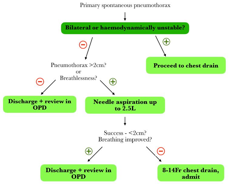
Flowchart adapted from guidance available at – Maskell N. British Thoracic Society pleural disease guidelines – 2010 update. Thorax. 2010;65(8):667–9.
Secondary Spontaneous Pneumothorax
Patients with secondary spontaneous pneumothorax (SSP) tolerate pneumothoraces less well due to their underlying chronic lung disease, which significantly alters the management strategy for this set of patients. In addition to increased symptomatology, SSPs are less likely to spontaneously resolve, meaning active management with insertion of intercostal drainage is more frequently needed.
Given the above, in any SSP that is symptomatic of their pneumothorax, you should proceed immediately to intercostal drainage with a small-bore intercostal drain (8-14Fr), with admission and early input from the respiratory specialist team. In the setting where the patient is asymptomatic, the size of the pneumothorax determines the ongoing management. Pneumothoraces 1-2cm can undergo a trial of needle aspiration up to 2.5L of air with response assessed afterwards. If the pneumothorax is subsequently <1cm, then no further intervention is required other than high-flow oxygen (with caution in those with known hypercapnic respiratory failure) and admission to hospital for at least a 24-hour monitoring period. If needle aspiration fails to create a pneumothorax <1cm in size, then proceeding to drainage is the most appropriate next step.
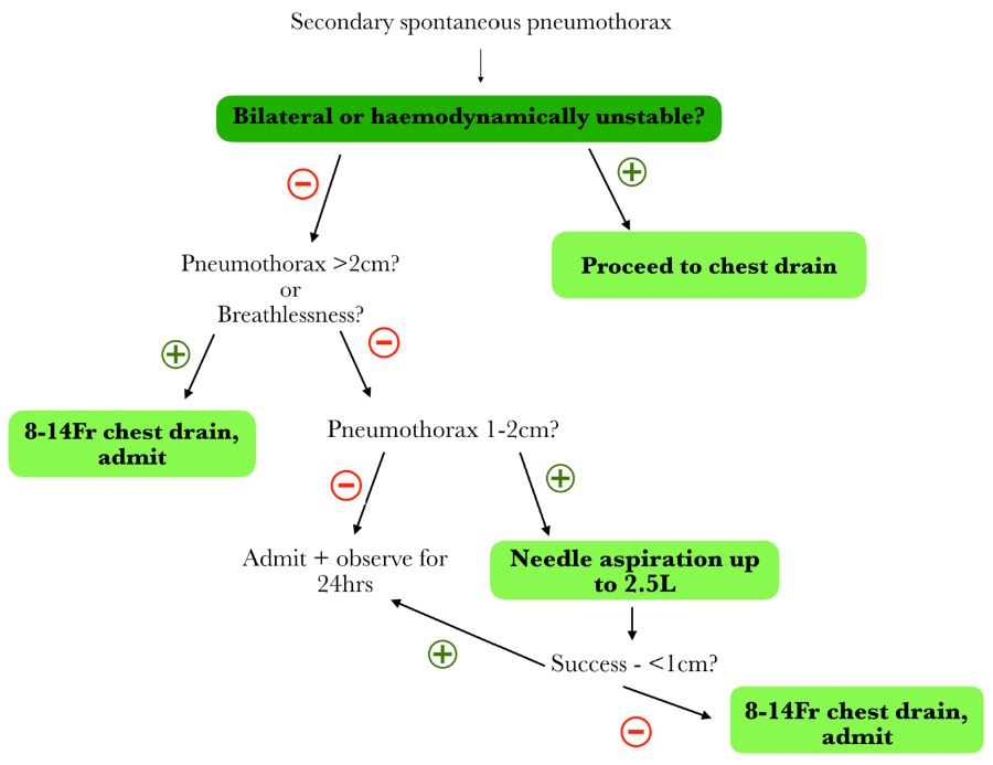
Flowchart adapted from guidance available at – Maskell N. British Thoracic Society pleural disease guidelines – 2010 update. Thorax. 2010;65(8):667–9.
Persistent Air Leak Management in Primary and Secondary Spontaneous Pneumothorax
In some cases, pneumothoraces fail to resolve despite initial management as guided by the algorithms above. This is seen more frequently in secondary spontaneous pneumothorax than primary but is possible in both subtypes. A persistent air leak is usually defined as a failure for the pneumothorax to fully resolve despite 24-48 hours of appropriate management. There are some management options available:
- Suction. Low-grade suction can be considered in patients with a persistent leak at 24-48 hours to help with re-expansion of the lung. The recommendation is to start with low rates of suction, approximately -10cm H20, which can be increased to -20cmH20 dependent on resolution and how well the patient tolerates the added pressure.
- Referral to cardiothoracic surgeons for consideration of surgical management. Consideration of early referral to cardiothoracic surgeons (with persistent air leak at 3-5 days) for surgical management is important in patients fit for surgical procedure and can provide patients with definitive management. This is especially important in patients with SSP.
- Chest drainage with ambulatory drainage device. In selected patients with persistent air leak, a chest drain can remain in situ with a drainage device attached. This drainage device can allow ongoing drainage of air in the community. Decisions to utilise this lie with specialised respiratory colleagues.
- Pleurodesis.
Preventing Recurrence
Patients who present with their first episode of PSP do not typically require definitive treatment, e.g. surgical management unless they have a persistent air leak at the initial presentation or they have a repeat episode.
On the other hand, patients who present with SSP are at high risk of recurrence. As such, in SSP, consideration of definitive management during their initial admission should be completed. In patients considered fit for surgery, surgical methods for definitive management are the preferred option. In those where surgery is not an option or not the patient’s preferred course of action, then chemical pleurodesis with sterile talc pleurodesis or tetracycline pleurodesis can be used (BTS recommends tetracycline pleurodesis over talc if available). Talc or tetracycline pleurodesis results in pleural inflammation and formation of adhesions to cause pleural effusion and prevention of further episodes of pneumothorax.
Management of Recurrent Pneumothorax Episodes
Recurrent pneumothoraces are a common clinical event with an estimated recurrence rate of around 30% in PSP and 50% in SSP, although data on this varies. In PSP, recurrence is thought to be related to some risk factors: being tall, low BMI, smoking and anatomical changes identified on CT (small blebs or bullae). The key risk factor for a further pneumothorax, though, is having had one previously.
In the initial setting, recurrent pneumothoraces should be managed with insertion of an intercostal drain. Recurrent pneumothoraces should prompt the clinician to consider early discussion with cardiothoracic surgical colleagues for definitive management. The agreed criteria for referral, as per BTS guidelines, are the following:
- Second ipsilateral pneumothorax.
- First contralateral pneumothorax.
- Synchronous bilateral spontaneous pneumothorax.
- Persistent air leak at 5-7 days despite intervention, or failure of lung re-expansion.
- Spontaneous haemothorax.
- At risk Profession, e.g. pilots, divers
- Pregnancy.
In patients not suitable for surgery, chemical pleurodesis can be completed (see Management: Preventing Recurrence).
Special Circumstances
Air Travel following Pneumothorax
The British Thoracic Society provided guidance on air travel and history of pneumothorax in 2022. It recommends that any patient with a current, unresolved pneumothorax should not travel by air. In those who have a resolved pneumothorax, they should wait for 7 days after full resolution is confirmed on a chest x-ray before travelling.
Diving following Pneumothorax
The British Thoracic Society published specific recommendations for diving and respiratory disease in 2003. It recommends that anyone with evidence of bullae on CT imaging should not dive due to the increased risk of barotrauma during the activity. In addition, any previous spontaneous pneumothorax is a contraindication to diving (unless specifically treated with bilateral pleurectomy and subsequent normal lung function and imaging following the procedure).
If a patient has had a previous traumatic pneumothorax, this is not a contraindication to diving, providing they have been left with normal lung function and imaging.
Complete Guide to Clinical Genetics ST3 Application and Recruitment (2027)
Written by Medibuddy. Last updated: October 2026. Clinical Genetics is

Complete Guide to Clinical Radiology ST3 Application and Recruitment (2027)
Recruitment paused: NHS England is not running Clinical Radiology ST3+ recru

Complete Guide to Paediatric Surgery ST3 Application and Recruitment in United Kingdom (2026)
Written by Medibuddy. Last updated: October 2026. If you’re planning


Take your subscriptions with you
Our mobile app allows you to access your interview and exam question banks wherever you are.

