


 Close
Close
 13th January 2024
Sleep-Related Breathing Disorders
Introduction
Sleep-related breathing disorders are common and often unrecognised. It is estimated that 9% of women and 24% of men have some form of sleep-disordered breathing. The disorders are associated with an increased risk of all-cause mortality and have been especially linked with cardiovascular and metabolic complications. In addition, these disorders have significant social consequences, with effects on executive function, changes to behaviour and excessive daytime sleepiness.
Physiology of Sleep
Sleep is the natural periodic rest state of our mind and body characterised by reduced consciousness, inhibition of sensory activity and drastically reduced muscle activity. Despite spending roughly a third (or ~8 hours/day) of our lives sleeping, sleep is poorly understood, and its exact purpose is not completely certain. There is evidence to suggest that sleep has roles in:
- Restoration and clearance: sleep has been shown to clear waste products from the brain that build up during the day via the ‘glymphatic system’ (clearance system of the brain reliant on glial cells)
- Growth and repair: sleep has been suggested to help in body repair and growth; growth hormone levels peak during sleep
- Memory and learning: experimental data shows how sleep is critical to brain plasticity, promoting synaptic growth and maintenance while helping consolidate memory.
While the science is not completely clear about the exact functions of sleep, it is readily apparent to any of us who has had a poor night’s sleep that one doesn’t tend to feel clear-headed, energetic, or particularly sharp without a full night of deep restorative sleep!
Sleep is cyclical and goes through various stages with differing physiological activity. Sleep is broadly divided into two categories: Rapid Eye Movement (REM) sleep and Non-Rapid Eye Movement (NREM) sleep. NREM sleep is further subdivided into N1, N2 and N3 substages. Each stage/sub-stage is characterised by changes in muscle tone (as noted on Electromyography or EMG), brain waves (as seen on Electroencephalography or EEG) and changes in response to sensory input.
Stages of Sleep
1) Wakefulness: one is awake, alert, and responsive to outside stimuli. Higher cortical activity (voluntary movement, speech, etc) is readily apparent. As one settles to sleep, one becomes drowsier and response to external stimuli starts to decrease.
EEG tends to show either beta waves (alert) or alpha waves (drowsy).
NREM Sleep:
2) N1 Stage: this is the transition between wakefulness and sleep and where sleep is the lightest. High levels of N1 sleep on sleep studies can suggest that sleep is fragmented and unable to deepen (e.g. due to sleep-related breathing disorders). With a transition to N1 sleep, there is a general decrease in muscle tone and awareness of external stimuli. EEG shows theta waves.
3) N2 Stage: as sleep deepens, there is a transition to N2 sleep. This is where one spends the majority (up to 55%) of total sleep. In N2 sleep, muscle tone further decreases, and there is further loss of awareness (and thus response) to external stimuli. EEG shows sleep spindles and K complexes.
4) N3 Stage (or deep/slow wave sleep): This is the deepest phase of sleep, where the sleeper is least likely to awake in response to external stimuli. It tends to occur more frequently during the first part of the night. EEG characterised by delta waves.
Historically, NREM sleep was divided into four stages. This convention has changed; the previous N4 stage is now classified as part of N3 sleep. In NREM sleep, we can observe release of hormones from the anterior pituitary gland and clinically can observe decreases in temperature, blood pressure, heart rate and respiratory rate.
REM Sleep:
5) Characterised by rapid eye movement, REM sleep is the phase of sleep that is most associated with dreaming. This is the state of sleep where there is complete atonia of the muscles (excluding the extraocular muscles and the diaphragm). This is a result of inhibition of the alpha motor neurones.
REM sleep has been associated with learning, memory consolidation and emotional processing. Observing clinically, we see irregular breathing patterns and periodic rises in blood pressure and heart rate. It has also been seen that there is an increase in O2 consumption from the brain during this stage of sleep. Brain waves on EEG mimic awake states. On average, one spends 20-25% of total sleep in REM sleep. The proportion of time spent in REM sleep decreases as we age.
The Sleep Cycle:
A normal uninterrupted sleep cycle lasts for 70 -110 minutes and progresses in a cyclical fashion (Image 1). We tend to cycle through 4-5 cycles of sleep during an average night’s sleep (Image 2). N3 sleep tends to occur predominantly in the first few cycles of sleep (which thus tend to be when sleep is deepest). As the night progresses, one has longer periods of REM sleep, and sleep tends to lighten. There may be very brief arousals (awakenings) that often aren’t remembered or registered by the sleeper on waking.

Image Source: Wikimedia Commons (2013). Hypro zyklus 1 en 103, Licensed under a creative commons licence. Available for free at: https://upload.wikimedia.org/wikipedia/commons/b/bb/Hypro_zyklus_1_en_103.svg (Accessed: 16 August 2022).

Note that by current convention, N4 is part of N3 Sleep
Image Source: Wikimedia Commons (2011). Sleep Hypnogram, Licensed under a creative commons licence. Available for free at: https://commons.wikimedia.org/wiki/File:Sleep_Hypnogram.svg (Accessed: 16 August 2022).
References
- Douglas K. Stages and architecture of normal sleep [Internet]. Uptodate.com. 2021 [cited 21 July 2022]. Available from: https://www.uptodate.com/contents/stages-and-architecture-of-normal-sleep
- Fuller P, Gooley J, Saper C. Neurobiology of the Sleep-Wake Cycle: Sleep Architecture, Circadian Regulation, and Regulatory Feedback. Journal of Biological Rhythms [Internet]. 2006 [cited 5 August 2022];21(6):482-493. Available from: https://journals.sagepub.com/doi/10.1177/0748730406294627
- Widmaier E, Raff H, Strang K, Vander A. Vander’s Human Physiology: The Mechanisms of Body Function. 12th ed. New York: McGraw-Hill; 2011.
Physiology of Breathing and Sleep
Regulating breathing while asleep differs from regulating wakeful breathing. While awake, ventilation is both a conscious and unconscious process, with a wakefulness drive to breathe augmenting our underlying rhythm of breathing. As we slumber, we lose our conscious stimulation of breathing, leaving our unconscious pathways in control. Therefore, we naturally hypoventilate (in comparison to wakefulness) when asleep. For those with respiratory abnormalities (e.g. smaller airways, underlying lung disease, etc.), the loss of conscious compensatory mechanisms can lead to sleep-disordered breathing.
There are two key components in breathing that are of interest to us: central (or neural) control and muscle tonicity (in the upper airways, the chest wall, and the diaphragm).
Central Control of Respiration
Central control of respiration is via the brain-stem respiratory network in the medulla and pons. There are three components to this complex series of neurons and nuclei:
- Respiratory Neurons: that generate the underlying respiratory rhythm and act as the primary driver of respiratory activity. There are two groups of respiratory neurons: the Dorsal Respiratory Group (DRG) and the Ventral Respiratory Group (VRG).
- Chemosensors: detect changes in blood gases and pH and trigger a physiological response to maintain each parameter within the normal range.
- Respiratory Motor Groups: innervate the respiratory muscles.
The DRG is composed of inspiratory neurons and is closely associated with nucleus of the solitary tract. This area receives important vagal sensory input critical for reflex control of breathing: peripheral chemoreceptors (for pO2 and pCO2 from aorta and carotids), central chemoreceptors (pH in the ventral medulla) and lung vagal afferents (stretch, irritant and pain receptors). The VRG is composed of both inspiratory and expiratory neurons. The Pre-Botzinger Complex (a grouping of inspiratory neurons) within the VRG has some intrinsic pacemaker activity that generates an underlying respiratory rhythm. This complex contains mu-opioid receptors, which slow respiratory pacemaker activity when activated. Thus, we can see how opiates will trigger the mu receptors and slow the pacemaker activity, causing respiratory depression when given at sufficient doses.
The various chemosensors will respond to changes in the pCO2 and pO2 to trigger respiration. One of the primary aims of ventilation is to maintain the pCO2 within a range of 4.5- 6.0 kPa, and respiration is thus very sensitive to even slight changes in pCO2. Hypercarbia will trigger an increase in respiratory drive. In sleep, this can lead to an arousal, pulling patients from deeper sleep and thus disrupting sleep. Hypocapnia, in turn, can lead to a decrease in respiratory drive when, indeed, can lead to periodic central apnoeas (if it crosses the apnoeic threshold). The hypoxic drive alone is relatively insensitive, but when coupled with the hypercarbic drive, it has synergistic effects on increasing arousal and wakefulness and the initiation of some of the conscious waking respiratory drive.
Muscle Tone
As one progresses through the sleep cycle, one’s muscle activity and tone decreases. One loses the wakefulness drive to breathe, and our breathing lightens: the respiratory rate drops, the minute ventilation drops, and muscles become flaccid. This loss of ventilatory motor output is noted in the upper airway muscles, chest wall, and accessory muscles.
Upper Airway
The loss of tonicity in the upper airway manifests itself as a narrowing of the upper airway lumen and increased flaccidity of the upper airway walls. This, in turn, causes increased upper airway resistances and more turbulent airflow. In healthy individuals, this tends not to cause an issue. However, some people are at increased risk of having more substantial upper airway narrowing or even collapse leading. If the upper airway resistance increases enough (i.e. there is sufficient narrowing of the lumen), one can have noisier breathing or snoring (especially if the soft palate vibrates or flaps with inhalation). This can lead to reduced (but not absent) airflow. For some individuals, the increased resistance and the floppiness of their upper airway structures lead to collapse and obstruction, completely stopping airflow and leading to an obstructive apnoeic episode. This, in turn, leads to a rise in pCO2 and increased airway pressure until it triggers an arousal and a surge in catecholamines, leading to increased wakefulness and, therefore, increased drive to the respiratory muscles.
Chest Wall and Diaphragm
The reduction of tonicity in the chest wall muscles increases as our sleep deepens. The diaphragm remains the main respiratory pump during sleep but loses the assistance of the intercostal and accessory respiratory muscles. This leads to a reduction in the minute ventilation. In healthy individuals, the body can manage. However, for certain patient groups, the loss of the supporting respiratory muscles can lead to worsening of their breathing and gas exchange. They develop hypercarbia/hypoxia, which in turn triggers arousals.
References
- Punjabi N, Caffo B, Goodwin J, Gottlieb D, Newman A, O’Connor G et al. Sleep-Disordered Breathing and Mortality: A Prospective Cohort Study. PLoS Medicine. 2009;6(8):e1000132.
- Mandal S, Kent B. Obstructive sleep apnoea and coronary artery disease. Journal of Thoracic Disease. 2018;10(S34):S4212-S4220.
- Beebe D, Groesz L, Wells C, Nichols A, McGee K. The Neuropsychological Effects of Obstructive Sleep Apnea: A Meta-Analysis of Norm-Referenced and Case-Controlled Data. Sleep. 2003;26(3):298-307.
- Sowho M, Amatoury J, Kirkness J, Patil S. Sleep and Respiratory Physiology in Adults. Clinics in Chest Medicine. 2014;35(3):469-481.
- Newton K, Malik V, Lee-Chiong T. Sleep and Breathing. Clinics in Chest Medicine. 2014;35(3):451-456.
- Malhotra A, Horner R. Control of breathing and upper airways during sleep. In: Broaddus V, Mason R, Gotway M, Ernst J, King Jr T, Lazarus S et al., ed. by. Murray & Nadel’s Textbook of Respiratory Medicine. 6th ed. Elsevier; 2015. p. 1511-1526.
Sleep Studies
Sleep studies are required to investigate sleep-related breathing disorders. There are various sleep studies of increasing degrees of complexity dependent on the requirements (and resources available to the particular centre). Tests can be done at home or overnight at the testing centre.
The gold standard is the Polysomnogram (PSG) which has multiple tools to measure different physiologic signals that help stage sleep, monitor respiratory airflow, monitor respiratory effort and monitor oximetry and heart rate and rhythm. Optionally, there are mechanisms to measure ventilation as well.
Components of a full Polysomnogram
A full sleep study will include all the components below and be done overnight in a centre.
Staging Sleep
- Electroencephalography (EEG): to monitor brain waves and help stage sleep.
- Electrooculogram (EOG): to pick up the movement and activity of the eyes
- Electromyogram (EMG): to measure muscle tension and movement
They measure for:
- Arousals: changes in sleep stage to full wakefulness or a lighter stage of sleeping. These are counted and then divided by total sleep time to give an Arousal Index (AI)
Monitor Respiratory Airflow
- Oronasal thermistor and nasal pressure transduction: to monitor for changes to airflow
They measure for:
- Apnoeas: absence or near-complete absence of airflow. These are classified as obstructive (apnoea with evidence of respiratory effort), central (apnoea with absence of any respiratory effort) or mixed (apnoea with a period of absent respiratory effort followed by evidence of some effort).
- Hypopnea: Reduction of airflow, not sufficient to be classified as an apnoea
Respiratory Effort
- Thoracic and abdominal Respiratory Inductance Plethysmography (RIP): bands across the abdomen and thorax to measure chest and abdominal wall movement and thus respiratory effort
Oximetry
- Pulse oximeters: the most basic element to measure the oxygen saturations through sleep
They measure for:
- Hypoxaemia during sleep
Cardiac Rate and rhythm
- Electrocardiogram (ECG): 1-3 lead ECG to measure heart rate and monitor for any cardiac dysrhythmias
Ventilation
Monitoring of ventilation is often only performed if there is a high clinical suspicion of hypoventilation.
- Transcutaneous CO2 monitoring
- End-tidal CO2 monitoring
From the above measures, one can make interpretations on:
- Sleep quality: disturbance/ fragmented sleep will exhibit a higher proportion of N1 or N2 sleep. The timing of apnoeas can also help differentiate central from obstructive apnoeas.
- Apnoea- Hypopnea Index (AHI): Apnoea + hypopneas divided by the total sleep time. This can be subdivided into an obstructive AHI and central AHI.
- Degree of Hypoxaemia
Home studies
Home studies tend to be a cheaper and more available alternative. The portable devices can be taken home and used to assess airflow, respiratory effort, oximetry and cardiac rhythm (or even used just to measure overnight oximetry!). The sleep staging component is the absent measure. Home studies are particularly useful in patients with a high pre-test probability of diagnosis.
Portable or home studies will not measure sleep and stages. From the other data points though we can derive:
- Respiratory Event Index (REI): the number of apnoeas or hypopneas during recorded time. Because this time doesn’t record sleep itself (the recorded time is longer than the total time spent), this can underestimate the ratio of apnoeas/hypopneas to actual time asleep.

Image Source: Wikimedia Commons (2017). Polysomnography tester, Licensed under a creative commons licence. Available for free at: https://upload.wikimedia.org/wikipedia/commons/0/0d/Polysomnography_tester.jpg (Accessed: 9 August 2022).
References:
- Schulman D. Polysomnography in the evaluation of sleep-disordered breathing in adults [Internet]. Uptodate.com. 2022 [cited 8 August 2022]. Available from: https://www.uptodate.com/contents/polysomnography-in-the-evaluation-of-sleep-disordered-breathing-in-adults
- Understanding the Results (Diagnosing OSA) [Internet]. Sleep.hms.harvard.edu. 2021 [cited 8 August 2022]. Available from: https://sleep.hms.harvard.edu/education-training/public-education/sleep-and-health-education-program/sleep-health-education-34
- Obstructive sleep apnoea in adults [Internet]. BMJ Best Practice. 2020 [cited 8 August 2022]. Available from: https://bestpractice.bmj.com/topics/en-gb/215
Sleep Apnoea Syndromes
Background and Presentation
Sleep apnoea syndromes are the commonest sleep-related breathing disorders. They are characterised by pauses in breathing (apnoeas) or periods of especially shallow or light breathing (hypopneas) that occur during sleep. The main syndromes are Obstructive Sleep Apnoea/ Hypopnea Syndrome and Central Sleep Apnoea.
Patients with sleep apnoea typically complain of daytime sleepiness, fatigue or low energy.
Clinical History
The following points can be teased out through a good history:
- Excessive daytime sleepiness: whether patients fall asleep during mundane tasks, including while driving. Indeed, patients’ first presentation may be due to a road traffic accident. This is an especially key symptom to pick up. The Epworth Sleepiness Scale is a useful screening tool for this (see Table 1)
- Partners may describe snoring, choking or gasping during sleep or witness apnoeic episodes.
- Patients may have morning headaches (frontal, bilateral, squeezing pain).
- Irritability/ mood instability
- Loss of libido
- Sleep that doesn’t refresh them
- Whether they wake to pass urine multiple times in the night (arousals are stressful, and the increase of cortisol increases urine output).
- A good drug (including recreational drugs) history can tease out chronic opioid use or benzodiazepine use.
| Situation | Chance of Dozing (0-3) |
|---|---|
|
1) Sitting and reading |
|
|
2) Watching Television |
|
|
3) Sitting inactive in a public place (e.g. theatre) |
|
|
4) As a passenger in a car for an hour without a break |
|
|
5) Lying down to rest in the afternoon |
|
|
6) Sitting and talking to someone |
|
|
7) Sitting quietly after lunch (when you’ve not had alcohol) |
|
|
8) In a car, while stopped in traffic |
Table 1: Epworth Sleepiness Score 0= Would never doze 1=slight chance of dozing 2=moderate chance of dozing 3= high chance of dozing.
Scores of >9 should prompt further assessment for an underlying sleep disorder
Clinical Examination
- When examining the patient, obesity (with a large neck and waist) is often the primarily clinical finding that may highlight an increased risk of obstructive apnoeas.
- Patients can have a crowded oropharynx (macroglossia, large tonsils or uvula), small jaws or narrow or high-arched palates.
- It may be incidentally noticed that patients exhibit significant hypoxaemia while asleep in hospital.
- Patients may exhibit the signs and symptoms of other diseases that predispose them to sleep apnoeas:
- Signs of heart failure
- Stroke
- Concurrent atrial fibrillation
- Signs of chronic lung disease.
- Also, look if the patient has developed complications of sleep apnoea, such as hypertension, erectile dysfunction, mood disorders or ischaemic heart disease.
Obstructive Sleep Apnoea/Hypopnea Syndrome (OSAHS)
Overview and Clinical Significance
OSAHS is a common disorder, with NICE estimating that ~5% of UK adults have undiagnosed OSAHS. It is a syndrome with a spectrum of severity. Patients with the syndrome have obstructed or partially obstructed airways during sleep, stopping ventilation, leading to arousals and disrupting sleep.
This impairment of sleep has been linked to an increased risk of developing cardiovascular disease, pulmonary hypertension, hypertension, weight gain and diabetes. Patients with excessive daytime sleepiness are at an increased risk of road accidents. They can develop mood disorders and may even exhibit some cognitive dysfunction.
Pathophysiology and Risk Factors:
Patients who have OSAHS tend to have upper airways that are more collapsible or narrower than the healthy population. As they sleep, their upper airway collapse and can partially or fully obstruct the airway; this tends to happen in REM sleep. The patient is unable to ventilate effectively, which leads to hypoxaemia and hypercarbia and triggers arousals during sleep. This impairs sleep quality, and patients have unrefreshing sleep.
Risk factors associated with developing OSAHS are:
- Population-based:
- Men > Women
- Increasing age (estimated 15-20% of patients with OSAHS are >70 years old)
- Smoking
- Co-Morbidities based
- Obesity
- T2DM
- Hypertension
- Chronic lung disease (e.g. COPD, Pulmonary Fibrosis)
- Acromegaly
- Hypothyroidism
- Down’s Syndrome
- Prader Willi
- Anatomy:
- Large neck
- Large soft tissues of the mouth and upper airways (tongue, adenoids and tonsils)
- Maxillo-mandibular abnormalities
- Nasal Congestion (polyposis, deviated septum, sinusitis etc)
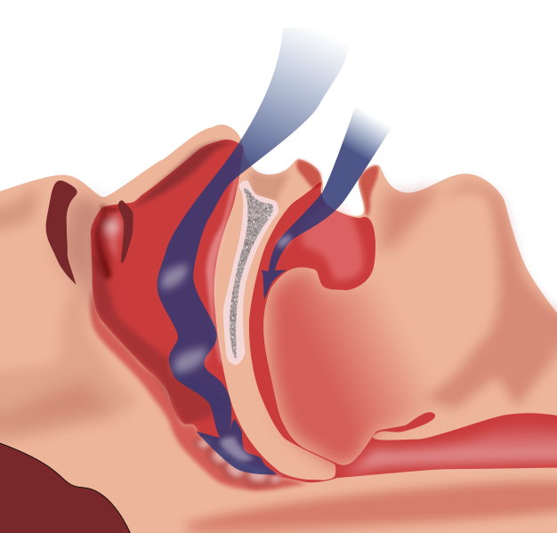
Image Source: Wikimedia Commons (2010). Obstruction ventilation apnee sommeil, Licensed under a creative commons licence. Available for free at: https://commons.wikimedia.org/wiki/File:Obstruction_ventilation_apn%C3%A9e_sommeil.svg (Accessed: 5 August 2022).
Diagnosis
While clinical assessment and history can be strongly suspicious of OSAHS, definitive diagnosis requires polysomnography or home sleep studies. Diagnosis for OSAHS requires either/or:
- Obstructive AHI or REI of ≥15 episodes/hour
- AHI or REI of ≥5 episodes/hour AND EITHER:
- Symptoms: excessive daytime sleepiness, insomnia
- Complications: hypertension, ischaemic heart disease, stroke, mood disorder, cognitive dysfunction
OSAHS is then classified as:
- Mild: mild/absent sleepiness and AHI/REI of 5-15 episode/hour
- Moderate: sleepiness during activities that require some attention and AHI/REI 15-30 episodes/hour
- Severe: sleepiness during active activities (e.g. eating or driving) and AHI/REI of >30episodes/hour
Management
Management of OSAHS is primarily aimed at improving quality of life for patients and reducing symptom burden. In addition, the aim is to stop the development of complications. This can be measured by symptomatic improvement (especially excessive daytime sleepiness) via reduction of the AHI/REI, control of blood pressure and control of hyperglycaemia.
First Line:
Overnight CPAP is the first-line intervention for symptomatic adults (especially in patients with severe OSAHS).
- CPAP helps splint open the upper airway and prevent collapse, thus preventing airway obstruction and apnoea.
- CPAP therapy has a survival benefit and improves sleep quality (reducing daytime sleepiness), mood, and cognition. There is some evidence that it can improve glycaemic control and help control hypertension.
- Improving excessive daytime sleepiness reduces the risk of road traffic accidents (See DVLA requirements)
Adherence to CPAP:
CPAP is often not well tolerated, and adherence is thus low. The mask is often found to be uncomfortable, the pressures not well tolerated or too noisy, and patients can get a dry mouth/nasopharynx. Given that adherence is critical to controlling symptoms, it is critical to help patients with adapting to and accepting CPAP therapy.
- Addition of humidification
- Proper fit and interface:
- The mask should be adjusted to fit the patient, and additional straps may be necessary
- The mask should be nasal or oro-nasal or full face (dependant on patient preference/ presence of oral air leak)
- Reduce nasal resistance
- Nasal surgery (see below under Surgery)
- Intranasal steroid sprays (e.g. with concurrent rhinitis or turbinate hypertrophy)
- Patient education, support, and behavioural therapy
- Behavioural interventions (e.g. Cognitive Behavioural Therapy). There is good evidence that this can significantly improve adherence to therapy
- Supportive therapy (e.g. additional follow-up, phone call check-ins, adherence monitoring equipment, etc.). There is moderate evidence that this can improve adherence
- Patient education around CPAP and/or OSAHS. There is mixed to low evidence that this, in isolation, can help with adherence.
Adjuncts:
Lifestyle advice
- Obese patients are advised to lose weight. Loss of 10% body weight was found to reduce AHI by 26% in one study.
- Advice to avoid alcohol and sedating medications (e.g. benzodiazepines/opiates).
Surgery
- Surgery may be a first-line option in patients with OSAHS caused by distinct anatomy (e.g. bulky palatine tonsils)
- Surgery can improve upper airway flow via reducing upper airway resistance (e.g. nasal polypectomy, nasal septal surgery) or increasing the calibre of the pharyngeal airway (tonsillectomy, adenoidectomy, uvulopalatopharyngoplasty).
- Surgery to help improve nasal airflow may be required to help with CPAP adherence.
- Bariatric surgery may be an option in patients with a BMI >40 or <40 with comorbidities.
Second Line:
Oral Appliances
- For patients with mild to moderate disease who fail to adhere to or tolerate CPAP, oral appliances (e.g. mandibular splints or tongue retainers) can be used to help keep the upper airway open.
DVLA Requirements
Given the dangers of driving while excessively sleepy, there are DVLA requirements for patients who have OSAHS. The rules depend on the severity of OSAHS and the presence of excessive daytime sleepiness. In addition, the guidance for follow-up differs for Group 1 drivers (car and motorcycle) versus Group 2 drivers (bus, taxi or lorry drivers).
The penalties are strict for patients not following the guidance, with fines of up to £1000.00 for not telling the DVLA about medical conditions that affect their driving. They can also be liable to prosecution if involved in an accident.
On diagnosis:
- Mild, Moderate or Severe OSAHS, without symptoms of excessive daytime sleepiness:
- Can drive as usual
- No need to notify the DVLA
- Mild OSAHS with symptoms of excessive daytime sleepiness
- Must NOT drive until symptoms satisfactorily controlled
- If symptoms cannot be controlled within 3 months of initiation of treatment, the DVLA must be informed
- Moderate/ Severe OSAHS with symptoms of excessive daytime sleepiness
- Must NOT drive until sleep clinic has confirmed that OSAHS is under control, sleepiness is no longer excessive, and that patient is compliant with CPAP treatment.
- DVLA must be informed. DVLA require medical confirmation of control of symptoms before allowing driving/restoring driving licences.
- Excessive sleepiness with suspected OSAHS
- Must NOT drive until OSAHS diagnosed and sleep clinic has confirmed excessive sleepiness is controlled by treatment (as per above guidance dependent on severity)
Monitoring:
- Group 1 Drivers: Condition reviewed at least every 3 years
- Group 2 Drivers: Condition review at least annually
Responsibility lies with the driver to contact the sleep services to arrange this review (can be by telephone or in-person, depending on the sleep service).
Central Sleep Apnoea Syndromes (CSAS)
Overview
CSAS is less common than OSAHS and often occurs secondary to another condition; rarely, there is primary or idiopathic cause for the condition. It is more common amongst older adults (especially >65 years old), males and patients with heart failure, renal failure, atrial fibrillation/flutter (AF), neurological disorders and those using medications that cause respiratory depression. It is estimated to affect about 0.9% of patients above 40 years old.
Patients with CSAS most often tend to develop CSA with Cheyne-Stokes breathing. Cheyne-Stokes breathing is a pathological breathing pattern. It is when there are periods of progressively increased hyperpnea (in a crescendo-decrescendo pattern) to compensate for an increased pCO2. This leads to hypocapnia, triggering a central apnoea. This, in turn, causes a rise in pCO2, restarting the cycle again.
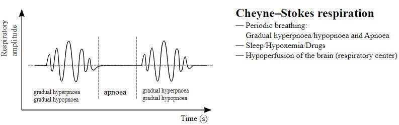
Note original image adapted to only show Cheyne-Stokes Respiration
Image Source: Wikimedia Commons (2012). Breathing abnormalities, Licensed under a creative commons licence. Available for free at: https://commons.wikimedia.org/wiki/File:Breathing_abnormalities.svg (Accessed: 17 August 2022).
Diagnosis
CSA requires formal overnight lab Polysomnography (PSG) to be diagnosed.
Diagnosis of CSA with Cheyne-Stoke breathing requires:
- PSG showing ≥5 central AHI/hour, there are three consecutive central apnoeas/hypopneas in a Cheyne-Stokes breathing pattern, ≥50% of the total apnoeas/hypopneas are central in nature.
- Patient has concurrent AF, heart failure or a neurological disorder
- Patient has clinical features of a sleep apnoea syndrome
- There is no better diagnosis for the symptoms, nor is there any association with medication use
Diagnosis of CSA with medication or substance use requires:
- PSG showing ≥5 central AHI/hour, ≥50% of the total apnoeas/hypopneas are central in nature, there is no evidence of Cheyne-Stokes breathing
- Patient is taking opioids or other respiratory depressant medication
- Patient has clinical features of a sleep apnoea syndrome
- There is no better diagnosis for the symptoms
Diagnosis of Primary CSA
- PSG showing ≥5 central AHI/hour, ≥50% of the total apnoeas/hypopneas are central in nature, there is no evidence of Cheyne-Stokes Breathing
- Patient is not taking opioids or other respiratory depressant medication
- Patient has clinical features of a sleep apnoea syndrome
- There is no better diagnosis for the symptoms
Management
Management of CSA is often rooted in treating the underlying conditions associated with CSA. Any possible drug culprits should be stopped, and the associated medical condition should be optimised. For most patients with mild-moderate complications secondary to the sleep apnoea, this tends to be primary approach to treatment. Often, this, in turn, helps resolve or improve the CSA.
In patients with severe complications, management of the underlying condition occurs in conjunction with targeted treatments for the CSA. This involves:
– Positive Airway Pressure (PAP) therapy: this can be CPAP, BiPAP or even Adaptive Servo -Ventilation (ASV). ASV machines monitor the airflow and deliver PAP dependent on a person’s breathing patterns
– Supplemental oxygen during sleep. This may be in conjunction with PAP therapy or as a second-line option
References
- Obstructive sleep apnoea/hypopnoea syndrome and obesity hypoventilation syndrome in over 16s | Guidance | NICE [Internet]. Nice.org.uk. 2021 [cited 13 August 2022]. Available from: https://www.nice.org.uk/guidance/ng202
- Obstructive sleep apnoea in adults [Internet]. BMJ Best Practice. 2020 [cited 13 August 2022]. Available from: https://bestpractice.bmj.com/topics/en-gb/215
- Badr S. Pathophysiology of upper airway obstruction in obstructive sleep apnea in adults [Internet]. Uptodate.com. 2022 [cited 14 August 2022]. Available from: https://www.uptodate.com/contents/pathophysiology-of-upper-airway-obstruction-in-obstructive-sleep-apnea-in-adults
- Kline L. Clinical presentation and diagnosis of obstructive sleep apnea in adults [Internet]. Uptodate.com. 2022 [cited 14 August 2022]. Available from: https://www.uptodate.com/contents/clinical-presentation-and-diagnosis-of-obstructive-sleep-apnea-in-adults
- Kryger M, Malhotra A. Management of obstructive sleep apnea in adults [Internet]. Uptodate.com. 2022 [cited 14 August 2022]. Available from: https://www.uptodate.com/contents/management-of-obstructive-sleep-apnea-in-adults
- Bourke S, Burns G, Macfarlane J. Ventilatory failure and sleep related breathing disorders. Respiratory Medicine: Lecture Notes. 10th ed. John Wiley & Sons Ltd; 2022. p. 391- 410.
- Askland K, Wright L, Wozniak D, Emmanuel T, Caston J, Smith I. Educational, supportive and behavioural interventions to improve usage of continuous positive airway pressure machines in adults with obstructive sleep apnoea. Cochrane Database of Systematic Reviews. 2020;2020(4).
- Excessive sleepiness and driving [Internet]. GOV.UK. [cited 13 September 2022]. Available from: https://www.gov.uk/excessive-sleepiness-and-driving
- DETAILED DVLA GUIDANCE FOR UK DRIVERS WITH SLEEP APNOEA [Internet]. Sleep Apnoea Trust; 2022 [cited 8 September 2022]. Available from: https://sleep-apnoea-trust.org/driving-and-sleep-apnoea/detailed-guidance-to-uk-drivers-with-sleep-apnoea/
- Central sleep apnoea [Internet]. BMJ Best Practice. 2022 [cited 17 August 2022]. Available from: https://bestpractice.bmj.com/topics/en-gb/648
- Central Sleep Apnea (CSA) [Internet]. American Sleep Association. 2022 [cited 16 August 2022]. Available from: https://www.sleepassociation.org/sleep-apnea/central-sleep-apnea/
- Badr S. Central sleep apnea: Pathogenesis [Internet]. Uptodate.com. 2022 [cited 16 August 2022]. Available from: https://www.uptodate.com/contents/central-sleep-apnea-pathogenesis
- Badr S. Central sleep apnea: Risk factors, clinical presentation, and diagnosis [Internet]. Uptodate.com. 2021 [cited 16 August 2022]. Available from: https://www.uptodate.com/contents/central-sleep-apnea-risk-factors-clinical-presentation-and-diagnosis
- Badr S. Central Sleep Apnoea: Treatment [Internet]. Uptodate.com. 2021 [cited 16 August 2022]. Available from: https://www.uptodate.com/contents/central-sleep-apnea-treatment
Complete Guide to Clinical Genetics ST3 Application and Recruitment (2027)
Written by Medibuddy. Last updated: October 2026. Clinical Genetics is

Complete Guide to Clinical Radiology ST3 Application and Recruitment (2027)
Recruitment paused: NHS England is not running Clinical Radiology ST3+ recru

Complete Guide to Paediatric Surgery ST3 Application and Recruitment in United Kingdom (2026)
Written by Medibuddy. Last updated: October 2026. If you’re planning


Take your subscriptions with you
Our mobile app allows you to access your interview and exam question banks wherever you are.

