


 Close
Close
 16th January 2024
Hypersensitivity pneumonitis (HP)
Hypersensitivity pneumonitis (HP)
Background
Epidemiology
Hypersensitivity pneumonitis (or extrinsic allergic alveolitis) is an immune-mediated respiratory syndrome caused by allergen inhalation and subsequent reaction. This leads to alveolar and bronchiole inflammation via a non-IgE mediated process. The most common allergen is organic dust; however, there are hundreds of known causative agents, so identifying the allergen can be difficult in some cases. Pigeon fancier’s lung and Farmer’s lung are examples of hypersensitivity pneumonitis caused by pigeon exposure (avian protein antigen in faeces) and grain/straw mould (usually due to Aspergillus, Alternaria or Trichosporon fungi) respectively.
Prevalence and incidence of the condition overall are thought to be low; however, mild cases can masquerade as other conditions, such as viral illness and, therefore, remain undetected for a long period of time. Smoking may be a protective factor against developing acute HP; some evidence suggests that smokers exposed to causative allergens form fewer antibodies against antigens present and, therefore, do not manifest a clinical presentation of HP. However, in cases of established disease, smoking may influence a more severe presentation and cause recurrent episodes of exacerbation.

Adapted from: King Jr, T. (2021) Hypersensitivity pneumonitis (extrinsic allergic alveolitis): Epidemiology, causes, and pathogenesis, UpToDate. Edited by K. Flaherty and P. Dieffenbach. UpToDate. Available at: https://www.uptodate.com/contents/hypersensitivity-pneumonitis-extrinsic-allergic-alveolitis-epidemiology-causes-and-pathogenesis?search=hypersensitivity+pneumonitis&source=search_result&selectedTitle=1~150&usage_type=default&display_rank=1 (Accessed: January 15, 2023).
References
- Grammer, L. (2022) Hypersensitivity pneumonitis, Hypersensitivity pneumonitis – Symptoms, diagnosis and treatment | BMJ Best Practice. BMJ Best Practice. Available at: https://bestpractice.bmj.com/topics/en-gb/647 (Accessed: January 15, 2023).
- King Jr, T. (2021) Hypersensitivity pneumonitis (extrinsic allergic alveolitis): Epidemiology, causes, and pathogenesis, UpToDate. Edited by K. Flaherty and P. Dieffenbach. UpToDate. Available at: https://www.uptodate.com/contents/hypersensitivity-pneumonitis-extrinsic-allergic-alveolitis-epidemiology-causes-and-pathogenesis?search=hypersensitivity+pneumonitis&source=search_result&selectedTitle=1~150&usage_type=default&display_rank=1 (Accessed: January 15, 2023).
- Selman, M., Pardo, A. and King, T.E. (2012) “Hypersensitivity Pneumonitis Insights in Diagnosis and Pathobiology,” American Journal of Respiratory and Critical Care Medicine, 186(4), pp. 314–324. Available at: https://doi.org/10.1164/rccm.201203-0513ci.
Pathophysiology
Not every individual exposed to the same allergen will go on to develop HP – indeed, most of those exposed will develop no clinical signs or symptoms post-exposure. In susceptible individuals, it has been proposed that in acute cases, exposure to the antigens in an allergen causes a type 3 hypersensitivity reaction, leading to IgG antibody production for that allergen’s antigens. Then, subsequent exposure to the same allergen results in a type 4 hypersensitivity reaction with increased CD4+ cell activity and cytokine release, leading to granuloma formation and other chronic pathological changes.
A genetic component to the disease has been suggested in a number of reports; however, no definite genetic risk factor appears to have been identified with a consistent link to HP development yet.
References
- Grammer, L. (2022) Hypersensitivity pneumonitis, Hypersensitivity pneumonitis – Symptoms, diagnosis and treatment | BMJ Best Practice. BMJ Best Practice. Available at: https://bestpractice.bmj.com/topics/en-gb/647 (Accessed: January 15, 2023).
- Jacobs, K. and Kligerman, S. (2019) “Immune-mediated lung diseases,” Seminars in Ultrasound, CT and MRI, 40(3), pp. 213–228. Available at: https://doi.org/10.1053/j.sult.2018.11.011.
Selman, M., Pardo, A. and King, T.E. (2012) “Hypersensitivity Pneumonitis Insights in Diagnosis and Pathobiology,” American Journal of Respiratory and Critical Care Medicine, 186(4), pp. 314–324. Available at: https://doi.org/10.1164/rccm.201203-0513ci.
Presentation
The presentation of HP can be broadly classed as acute, subacute and chronic, and the clinical presentation of an individual with HP may differ depending on their disease classification status. The below diagram lists common symptoms experienced in HP.

Adapted from: Grammer, L. (2022) Hypersensitivity pneumonitis, Hypersensitivity pneumonitis – Symptoms, diagnosis and treatment | BMJ Best Practice. BMJ Best Practice. Available at: https://bestpractice.bmj.com/topics/en-gb/647 (Accessed: January 15, 2023).
The above symptoms can begin as soon as four hours after exposure to an allergen. They are usually accompanied by physical signs such as: diffuse crackles, mid-inspiratory noises and clubbing (advanced disease). Wheeze is not normally a prominent feature of HP, and if present, other diagnoses such as severe asthma, should be considered instead.
References
- Grammer, L. (2022) Hypersensitivity pneumonitis, Hypersensitivity pneumonitis – Symptoms, diagnosis and treatment | BMJ Best Practice. BMJ Best Practice. Available at: https://bestpractice.bmj.com/topics/en-gb/647 (Accessed: January 15, 2023).
- Selman, M., Pardo, A. and King, T.E. (2012) “Hypersensitivity Pneumonitis Insights in Diagnosis and Pathobiology,” American Journal of Respiratory and Critical Care Medicine, 186(4), pp. 314–324. Available at: https://doi.org/10.1164/rccm.201203-0513ci.
Diagnosis
Firstly, a detailed history should be taken, particularly in relation to identifying if any confirmed exposure to a causative agent has occurred at any point. In two-thirds of cases, the allergen is never found but known exposure to a known causative agent makes this diagnosis likely alongside relevant clinical signs and symptoms.
Testing for sensitivity to known allergens is not always recommended, as false positive and negative results are common. The diagram below details the typical panel of tests ordered when working up a patient with suspected HP, alongside an explanation of expected findings in someone with this diagnosis.
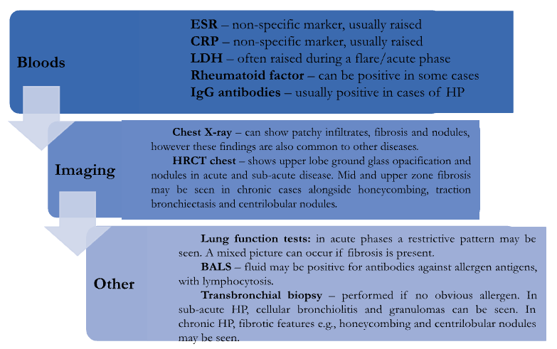
Adapted from: Grammer, L. (2022) Hypersensitivity pneumonitis, Hypersensitivity pneumonitis – Symptoms, diagnosis and treatment | BMJ Best Practice. BMJ Best Practice. Available at: https://bestpractice.bmj.com/topics/en-gb/647 (Accessed: January 15, 2023).
References
- Grammer, L. (2022) Hypersensitivity pneumonitis, Hypersensitivity pneumonitis – Symptoms, diagnosis and treatment | BMJ Best Practice. BMJ Best Practice. Available at: https://bestpractice.bmj.com/topics/en-gb/647 (Accessed: January 15, 2023).
- Hsieh, C. and Kamangar, N. (2022) Hypersensitivity pneumonitis, Background, Pathophysiology, Etiology. Medscape. Available at: https://emedicine.medscape.com/article/299174-overview (Accessed: January 15, 2023).
- King Jr, T. et al. (2022) Hypersensitivity pneumonitis (extrinsic allergic alveolitis): Clinical manifestations and diagnosis, UpToDate. UpToDate. Available at: https://www.uptodate.com/contents/hypersensitivity-pneumonitis-extrinsic-allergic-alveolitis-clinical-manifestations-and-diagnosis?search=hypersensitivity+pneumonitis&source=search_result&selectedTitle=2~150&usage_type=default&display_rank%5D (Accessed: January 15, 2023).
- Selman, M., Pardo, A. and King, T.E. (2012) “Hypersensitivity Pneumonitis Insights in Diagnosis and Pathobiology,” American Journal of Respiratory and Critical Care Medicine, 186(4), pp. 314–324. Available at: https://doi.org/10.1164/rccm.201203-0513ci.
Differential diagnoses
HP should be considered as part of the initial work up for any patient presenting with confirmed exposure to one of the identified causative allergens, alongside clinical findings. Other conditions to consider include:
- Sarcoidosis – this condition usually demonstrates symptoms and signs not limited to the respiratory system, e.g., erythema nodosum. A transbronchial biopsy may be needed in some cases to accurately diagnose HP.
- Idiopathic pulmonary fibrosis (IPF) – in IPF, HRCT chest would show more evidence of abnormalities in the lower zones of the chest rather than the upper zones, and centrilobular nodules would likely not be seen. However, the imaging can look very similar and respiratory opinion may be needed. Additionally, BALS in IPF would not show lymphocytosis.
- Pneumoconiosis – this usually only occurs in patients with an obvious exposure history for this condition, e.g., miners who worked with mineral dust. Chest imaging tends to show signs of asbestosis and silicosis, e.g., calcified hilar lymph nodes.
- Viral pneumonia – the symptoms of viral pneumonia tend to last for some time (at least a week) and would not repeatedly occur once resolved. In HP, symptoms may last only a couple of days but occur repeatedly in an episodic fashion every time the patient is exposed to the allergen. A respiratory virus panel can be helpful to deduce if any viral infection is present.
- Other more common respiratory diseases, e.g., asthma, COPD – these should also be considered if the history does not contain any clear evidence of an allergen and other suspicious factors are present, e.g., history of atopy for asthma, smoking history in COPD, etc., alongside imaging and other initial investigations.
References
- Grammer, L. (2022) Hypersensitivity pneumonitis, Hypersensitivity pneumonitis – Symptoms, diagnosis and treatment | BMJ Best Practice. BMJ Best Practice. Available at: https://bestpractice.bmj.com/topics/en-gb/647 (Accessed: January 15, 2023).
- Jacobs, K. and Kligerman, S. (2019) “Immune-mediated lung diseases,” Seminars in Ultrasound, CT and MRI, 40(3), pp. 213–228. Available at: https://doi.org/10.1053/j.sult.2018.11.011.
- King Jr, T. et al. (2022) Hypersensitivity pneumonitis (extrinsic allergic alveolitis): Clinical manifestations and diagnosis, UpToDate. UpToDate. Available at: https://www.uptodate.com/contents/hypersensitivity-pneumonitis-extrinsic-allergic-alveolitis-clinical-manifestations-and-diagnosis?search=hypersensitivity+pneumonitis&source=search_result&selectedTitle=2~150&usage_type=default&display_rank%5D (Accessed: January 15, 2023).
- Ungprasert, P., Ryu, J.H. and Matteson, E.L. (2019) “Clinical manifestations, diagnosis, and treatment of sarcoidosis,” Mayo Clinic Proceedings: Innovations, Quality & Outcomes, 3(3), pp. 358–375. Available at: https://doi.org/10.1016/j.mayocpiqo.2019.04.006.
Management
The mainstay of treatment for HP revolves around reducing/removing exposure to any identified causative allergens. Patients are advised to avoid either an allergen they are known to be sensitive to, or other potential allergens that may be present in their environment. This can be difficult, especially if the allergen is linked to their livelihood. Total avoidance can result in regression of disease and/or prevent progression to fibrosis, so the importance of avoiding causative allergens must be emphasised and encouraged.
Acute and sub-acute HP
For mild cases of acute HP, steroids are not recommended as the clinical presentation will often improve once the causative allergen is removed. If symptoms are recurrent and persistent, despite allergen avoidance, a short course of oral steroids can be offered if no contraindications are present. These can usually be tapered off unless fibrosis is present – patients in this group normally require long-term low-dose therapy.
Prednisolone is the 1st line oral steroid choice, and dosing is 0.5-1mg/kg/day on average. When symptoms improve, the dose can be tapered by about 5mg every other day over 4-6 weeks, depending on the ongoing clinical presentation.
Chronic HP
For those with chronic HP of a fibrotic nature, smoking avoidance/cessation, seasonal respiratory vaccinations and chest physiotherapy are recommended in addition to the above management strategy. For some, long-term oxygen therapy may eventually also be needed. Use of immunosuppressive agents such as azathioprine and mycophenolate mofetil (MMF) are not currently part of standard care, and further data is needed to support their use in this condition.
For those with very severe disease, lung transplant may be an option, depending on comorbidities, functional status and overall suitability for transplant. There have been several reports of the condition reoccurring in the transplanted organ, likely due to ongoing exposure to the allergen post-procedure.
References
- American Lung Association (2022) How Hypersensitivity Pneumonitis Is Treated, American Lung Association. Available at: https://www.lung.org/lung-health-diseases/lung-disease-lookup/hypersensitivity-pneumonitis/treating-and-managing (Accessed: January 15, 2023).
- Grammer, L. (2022) Hypersensitivity pneumonitis, Hypersensitivity pneumonitis – Symptoms, diagnosis and treatment | BMJ Best Practice. BMJ Best Practice. Available at: https://bestpractice.bmj.com/topics/en-gb/647 (Accessed: January 15, 2023).
- King Jr, T. (2022) Hypersensitivity pneumonitis (extrinsic allergic alveolitis): Treatment, prognosis and prevention, UpToDate. Edited by K. Flaherty and P. Dieffenbach. UpToDate. Available at: https://www.uptodate.com/contents/hypersensitivity-pneumonitis-extrinsic-allergic-alveolitis-treatment-prognosis-and-prevention?search=hypersensitivity%20pneumonitis&source=search_result&selectedTitle=3~150&usage_type=default&display_rank=3#H3199492110
Complete Guide to Clinical Genetics ST3 Application and Recruitment (2027)
Written by Medibuddy. Last updated: October 2026. Clinical Genetics is

Complete Guide to Clinical Radiology ST3 Application and Recruitment (2027)
Recruitment paused: NHS England is not running Clinical Radiology ST3+ recru

Complete Guide to Paediatric Surgery ST3 Application and Recruitment in United Kingdom (2026)
Written by Medibuddy. Last updated: October 2026. If you’re planning


Take your subscriptions with you
Our mobile app allows you to access your interview and exam question banks wherever you are.

