


 Close
Close
 17th January 2024
Sarcoidosis
Background
Epidemiology
Sarcoidosis is a multi-system disorder which can affect most organs in the body. Pulmonary sarcoidosis is the most common presentation, affecting up to 90% of patients with the disease and accounting for a large proportion of morbidity and mortality. Prevalence of sarcoidosis in the UK is around 175 cases per 100,000 population, affecting males and females equally. Most people are diagnosed between the ages of 30 and 60. There is a higher prevalence among people of Irish and Afro-Caribbean descent, and therefore, across the UK, cases are most concentrated in London, Scotland and Northern Ireland due to the prominence of these population groups in those regions (1).
Pathophysiology
The exact aetiology of sarcoidosis is unclear, although it is characterised by the accumulation of non-caseating granulomas in various tissues, including, but not limited to, the lungs. The granulomata are comprised of accumulated macrophages, CD4-positive T-lymphocytes and multinucleated giant cells. There is an associated increased production of cytokines such as tumour necrosis factor alpha, (TNF-α), interferon-gamma (IF-γ), interleukin-2 (IL-2) and transforming growth factor beta (TGF-β), mediating a widespread inflammatory process. These granulomata have a propensity for the thoracic lymph node sites, but can also form in the lung parenchyma (2).
Presentation
Sarcoidosis can have an indolent and non-specific presentation with multi-systemic symptoms and can sometimes be a challenging diagnosis to make. Patients with lymph-node limited disease may be entirely asymptomatic, and the respiratory exam is usually normal in patients with pulmonary sarcoidosis. Below are outlined some of the most common symptoms and signs based on site of disease.
Signs and symptoms
Symptoms and signs can be best appreciated by delineating them by different presentations of disease. Patients often have multiple groups of presenting symptoms depending on their sites of disease.
Lymph node disease:
- Fevers
- Night sweats
- Fatigue
- Weight loss
- Myalgia
Lung parenchymal disease:
- Dyspnoea
- Reduced exercise tolerance
- Cough
- Pleuritic chest pain
- Chest tightness
Skin manifestations:
- Erythema nodosum – tender, erythematous nodules found mostly on the shins
- Erythema multiforme – target lesions
- Lupus pernio – violaceous, plaque-like lesions on the nose and cheeks which, when biopsied, show non-caseating granulomas
- Calcinosis cutis – skin deposition of calcium
- Skin plaques
- Papules and papulonodules
- Subcutaneous nodules
- Erythroderma
- Nummular eczema
- Nail changes such as pitting, scarring, thickening and clubbing (3)
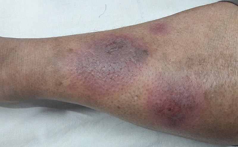
Image Source: Wikimedia Commons (2018). Erythema Nodosum, Licensed under a creative commons licence. Available for free at: https://commons.wikimedia.org/wiki/File:Erythema_Nodosum.jpg (Accessed: 6 June 2022).

Image Source: Wikimedia Commons (2010). Lupus Pernio 01, Licensed under a creative commons licence. Available for free at: https://commons.wikimedia.org/wiki/File:Lupus_pernio_01.jpg (Accessed: 6 June 2022).
Eye manifestations:
- Uveitis – blurred vision, red, watery eyes, photophobia
- Scleritis/episcleritis
- Proptosis
- Iris nodules
- Lacrimal gland involvement, causing sicca syndrome
Cardiac manifestations:
- Arrhythmias, most commonly conduction abnormalities
- Sudden cardiac death from ventricular arrhythmias or complete heart block
- Symptoms of congestive cardiac failure such as shortness of breath, peripheral oedema
- Symptoms and signs of pulmonary hypertension
Neurological manifestations – neurosarcoidosis can affect any part of the nervous symptom, most commonly:
- Cranial nerve palsies, commonly facial nerve palsy, optic nerve dysfunction
- Brain parenchyma can be affected, resulting in a range of neurological symptoms and signs, including cognitive impairment
- Peripheral neuropathy
- Myelopathy – causing leg weakness and sensory changes
- Meningitis
Musculoskeletal manifestations:
- Polyarthritis, often acute
- Dactylitis
Endocrine and metabolic manifestations:
- Parotid gland enlargement
- Hyperprolactinaemia, resulting in amenorrhoea and galactorrhoea in women
- Hypercalcaemia, resulting in renal stones
Gastrointestinal manifestations:
- Asymptomatic liver enzyme derangement
- Hepatomegaly
- Jaundice
- Right upper quadrant pain
Haematological manifestations – can produce non-specific symptoms related to the following blood dyscrasias:
- Lymphadenopathy
- Non-specific symptoms of fatigue, easy bruising and infection can also occur with lymphopenia, anaemia and thrombocytopenia.
Lofgren’s syndrome – this is an acute presentation of sarcoidosis characterised by bilateral hilar lymphadenopathy, erythema nodosum and polyarthralgia, often predominantly affecting the ankles.
Diagnosis
Investigations
- Blood tests are primarily useful for assessing for the complications of sarcoidosis and monitoring on treatment rather than being diagnostic in and of themselves. All patients should have:
- Full blood count
- Bone profile (including serum calcium)
- Liver function tests
- Urea and electrolytes
- Serum angiotensin converting enzyme (ACE) level
- 12-lead ECG – important as a baseline screening test for cardiac sarcoid, which most commonly presents with arrhythmia.
- Chest x-ray – essential for the diagnosis of pulmonary sarcoidosis; disease staging is based on radiographic appearance (see table 5). In cases where the clinical and radiological picture is clear cut, the diagnosis can be made based on chest x-ray alone without need for further imaging. Most commonly, there is a symmetrical hilar lymphadenopathy, and if parenchymal involvement is present, it tends to be mid and upper zone predominant nodular or reticulonodular shadowing. Where there is progression to fibrosis, this is upper lobe predominant. Surveillance chest x-ray imaging is used on a 3-monthly basis.
- High-resolution CT chest – can be helpful to further characterise appearances where there is diagnostic uncertainty. It is also more sensitive than chest x-ray, so can be used to detect disease where there is a clinical suspicion in the context of a normal plain radiograph.
- FDG-PET – occasionally used in scenarios of diagnostic uncertainty – looks for active inflammatory disease and is useful for detecting cardiac involvement.
- Pulmonary function testing – can often be normal, but in the presence of fibrosis will show a restrictive lung defect picture.
- Bronchoscopy and biopsy – where there is any diagnostic uncertainty regarding alternative differentials, such as lymphoma and tuberculosis, biopsy is the only way to reach a confirmed diagnosis. On the biopsy sample, a CD4:CD8 ratio >4 in the absence of other inflammatory cells is highly specific for a diagnosis of sarcoidosis.
- If there are any cutaneous lesions, they can be biopsied in the first instance.
- Endobronchial ultrasound (EBUS) guided biopsy should be undertaken if there is primarily lymph node disease.
- Transbronchial biopsies are used if there is primarily parenchymal disease present (4).
Table 5 – Scadding criteria for radiographic staging of pulmonary sarcoidosis
| Scadding stage | Chest x-ray findings |
|---|---|
| 0 | Normal |
| I | Bilateral hilar lymphadenopathy alone |
| II | Bilateral hilar lymphadenopathy with pulmonary infiltrates |
| III | Pulmonary infiltrates without hilar lymphadenopathy |
| IV | Pulmonary fibrosis |
Table 5 Source: Thillai M, Atkins CP, Crawshaw A, Hart SP, Ho L-P, Kouranos V, et al. BTS Clinical Statement on pulmonary sarcoidosis. Thorax. 2020 Dec 2;76(1):4–20.

Image Source: Wikimedia Commons (2016). HilarAdenopathy, Licensed under a creative commons licence. Available for free at: https://commons.wikimedia.org/wiki/File:HilarAdenopathy.png (Accessed: 6 June 2022).
Differential diagnoses
Lymphoma
For patients presenting with bilateral hilar lymphadenopathy, lymphoma is an important differential. They can also present with “B-symptoms” of weight loss, night sweats and persistent fatigue, all of which can also occur with sarcoidosis. Differentiating features would include painless lymphadenopathy elsewhere and splenomegaly. Full blood count and blood film are minimally invasive investigations which can help with the diagnosis, but if there is a significant diagnostic uncertainty, a lymph node biopsy or bone marrow aspirate should be undertaken.
Tuberculosis
Pulmonary tuberculosis (TB) presents with cough, haemoptysis and shortness of breath in addition to systemic symptoms such as weight loss, fevers and night sweats. Appearance of hilar lymphadenopathy can be similar to sarcoidosis on chest x-ray. TB is characterised by caseating granulomas, whereas sarcoidosis is associated with non-caseating granulomas. Travel and exposure history are important in delineating risk of an infective cause of symptoms, particularly where there are red flag symptoms such as haemoptysis. It is important to rule out TB prior to giving immunosuppressive therapy.
Interstitial lung disease
In patients with fibrosis on imaging, it is important to consider other forms of interstitial lung disease. Diseases of occupational exposure, such as berylliosis and silicosis, can have a similar CT appearance, so a thorough occupational history is key.
Management
The decision to commence pharmacological treatment in sarcoidosis should be considered based on the expected morbidity and mortality for the individual patient, as well as potential impacts on their quality of life. Some patients have minimal symptoms from their sarcoidosis, and introducing immunosuppressant therapy with associated side effects is not without risk. Attention should also be paid to managing patient symptoms such as fatigue with supportive lifestyle measures, such as structured exercise routines and counselling on sleep hygiene.
Steroids
In pulmonary sarcoidosis, when steroids are indicated, a commonly used regime would be 20-40mg prednisolone once daily for 4-6 weeks, followed by a tapering period to a maintenance dose of 5-10mg per day. In life-threatening disease or extrapulmonary cases such as neurosarcoidosis or cardiac sarcoid, high-dose intravenous methylprednisolone can be used as initiation therapy.
Second line immunosuppression
Indications for second-line immunosuppressive therapy:
- Progression of pulmonary disease on steroid treatment
- Unacceptable symptom burden
- Unacceptable steroid side effects or strong patient aversion to steroid treatment
- Inability to taper steroids dose to below 10-15mg prednisolone per day without rebound symptoms
- Significant contraindications to long-term steroid therapy, such as obesity, diabetes mellitus or osteoporosis
There are multiple agents of choice for steroid-sparing agents.
- Methotrexate – an anti-folate medication, introduced as a once weekly regimen of 5-10mg, it can be uptitrated to 20mg as tolerated. Once weekly folic acid 5mg should be given concurrently to reduce risk of myelosuppression. Potential side effects include pneumonitis, hepatic fibrosis and teratogenicity (both male and female patients should be counselled on this risk).
- Azathioprine – an inhibitor of purine synthesis, it causes immunosuppression by interrupting DNA and RNA formation. This affects lymphocyte proliferation in sarcoidosis. Started at 50mg once daily, it can be uptitrated to a therapeutic dose of 2mg/kg/day. Side effects include pancytopenia, increased risk of malignancy, nausea, vomiting, diarrhoea, rash, malaise and fever. Patients should have thiopurine methyltransferase (TPMT) levels tested prior to commencing treatment; those with a pre-existing enzyme deficiency are more at risk of pancytopenia, and an alternative agent should be used.
- Mycophenolate mofetil – an inhibitor of lymphocyte proliferation, this can be started at a dose of 1-1.5g twice a day. Side effects include gastrointestinal disturbances, teratogenicity as well as a small risk of neutropenia.
- Leflunomide – works as an anti-metabolite, similar to methotrexate. Can be used in preference to methotrexate due to a lower gastrointestinal toxicity profile, although it can still cause this. Other side effects include hepatotoxicity, hypertension and peripheral neuropathy.
- Cyclophosphamide – this is an alkylating agent that results in increased cell apoptosis, reducing circulating lymphocyte numbers. Use in sarcoidosis is rare due to the high side effect profile, which includes pancytopenia, haemorrhagic cystitis, nausea, vomiting, diarrhoea and alopecia.
- Hydroxychloroquine – works by increasing lysosomal pH in antigen-presenting cells. It is rarely used as monotherapy in pulmonary fibrosis, but is commonly used in cutaneous and joint disease and to treat fatigue. It can also be used as an adjunctive therapy to help with steroid weaning, at a dose of 200mg twice a day. Side effects include cardiac and retinal toxicity, in addition to gastrointestinal symptoms and headache. Prior to commencing treatment, all patients must have a baseline ECG to ensure there is no pre-existing long QTc interval, as well as a baseline ophthalmic examination.
All second-line immunosuppressive agents require frequent blood test monitoring of FBC, U&Es and LFTs. The following regime is recommended:
- 2-weekly for the first 2-months
- If stable, can reduce frequency to monthly for the next 6-months
- If stable, can reduce frequency to 3-monthly, which should be ongoing whilst the patient is on treatment
Biological agents and antifibrotics
- Infliximab is an anti-tumour necrosis factor (anti-TNF) agent. TNF is one of the cytokines that has a significant pro-inflammatory effect in sarcoidosis, promoting granuloma formation; inhibiting it, therefore, has an important therapeutic effect. Infliximab can be used as a third-line agent in combination therapy with methotrexate or azathioprine, given initially as 2-weekly infusions and then 6-8 weekly for maintenance. Side effect profile includes immunosuppression, with increased susceptibility to mycobacterium and invasive fungal infections, infusion reactions, alopecia, visual field defects and increased risk of pulmonary embolism. Prior to starting treatment, patients should be screened for latent TB infection using Interferon Gamma Release Assay (IGRA) testing. If latest TB is detected, patients should complete anti-TB treatment prior to starting infliximab.
- Antifibrotic medications such as pirfenidone and nintedanib are only licensed in the UK for idiopathic pulmonary fibrosis, but recent studies have shown a positive therapeutic effect in fibrosis secondary to sarcoidosis (4).
Lung transplantation
Bilateral lung transplantation is an option for patients with advanced fibrosis or pulmonary hypertension who have a significant, acute drop in pulmonary function (e.g., a 10% drop in FVC over a 6-month period) (4).
References
- Sarcoidosis statistics | British Lung Foundation [Internet]. statistics.blf.org.uk. Available from: https://statistics.blf.org.uk/sarcoidosis
- UpToDate [Internet]. www.uptodate.com. Available from: https://www.uptodate.com/contents/pathology-and-pathogenesis-of-sarcoidosis
- Sarcoidosis: Symptoms, Causes, and Images – DermNet [Internet]. dermnetnz.org. Available from: https://dermnetnz.org/topics/sarcoidosis
- Thillai M, Atkins CP, Crawshaw A, Hart SP, Ho L-P, Kouranos V, et al. BTS Clinical Statement on pulmonary sarcoidosis. Thorax. 2020 Dec 2;76(1):4–20.
Complete Guide to Clinical Genetics ST3 Application and Recruitment (2027)
Written by Medibuddy. Last updated: October 2026. Clinical Genetics is

Complete Guide to Clinical Radiology ST3 Application and Recruitment (2027)
Recruitment paused: NHS England is not running Clinical Radiology ST3+ recru

Complete Guide to Paediatric Surgery ST3 Application and Recruitment in United Kingdom (2026)
Written by Medibuddy. Last updated: October 2026. If you’re planning


Take your subscriptions with you
Our mobile app allows you to access your interview and exam question banks wherever you are.

