


 Close
Close
 18th January 2024
Investigations
Blood tests
Blood tests are not only useful for making the initial diagnosis of RA but are also used as baseline tests for monitoring disease activity and adverse effects of medications.
- FBC (with differential and platelet count), LFTs, U&Es, serum uric acid
- ESR and CRP are both elevated in RA- useful in initial diagnosis and for monitoring disease activity
- Immunology
- Rheumatoid Factor (RF)- present in 70-80% of patients with RA but poor specificity (also found in healthy individuals, patients with SLE, Sjogren’s syndrome, and those with mixed cryoglobulinemia, as well as in many other inflammatory conditions).
- Anti-CCP – similar sensitivity as RF but much higher specificity (95-98%)
- ANA- a negative ANA is useful in excluding SLE. It may be positive in up to ⅓ of patients with RA. If positive, anti-dsDNA and anti-Smith testing should be performed as these are highly specific for SLE.
- Seronegative RA can be diagnosed in patients lacking both RF and anti-CCP if they have findings otherwise characteristic of RA if appropriate exclusions have been met.
- Infectious screen – especially in patients with a very short history and those who are seronegative for RF and anti-CCP:
- Parvovirus B19
- HBV and HCV
- Borrelia in areas endemic for Lyme disease
- Chikungunya virus in areas where mosquito bite-transmitted viral diseases are prevalent
Imaging
Plain radiography is usually the first mode of imaging performed in patients suspected of RA, albeit not sensitive enough to pick up changes such as bony erosions in early disease. Typical radiological findings associated with RA include osteopenia, joint space narrowing, erosions and subluxation.
Ultrasound (US) may be more useful in allowing earlier diagnosis of RA, particularly in patients with seronegative disease. It can assess soft tissues and identify synovial thickening, presence of fluid in joints and abnormalities in tendons, ligaments and entheses. Doppler US assesses for soft tissue hyperaemia and is, therefore, useful for distinguishing between active and inactive inflammatory tissues.
Magnetic resonance imaging (MRI) is more sensitive still and can evaluate all structures (including soft tissue, cartilage, and bones) which can be affected by RA.
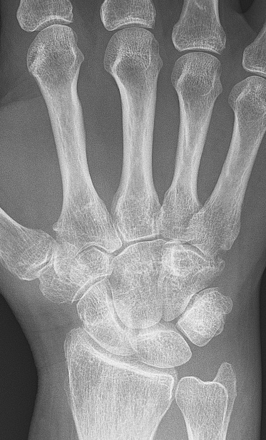
Image Source: Wikimedia Commons (2009). Rheumatoid arthritis with unaffected carpal bones 2009, Licensed under a creative commons licence. Available for free at https://en.wikipedia.org/wiki/Rheumatoid_arthritis#/media/File:Rheumatoid_arthritis_with_unaffected_carpal_bones_2009.jpg (Accessed: 24 November 2021).
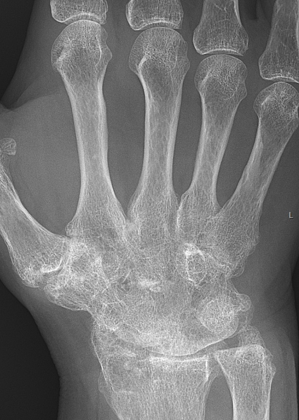
Image Source: Wikimedia Commons (2017) Rheumatoid arthritis with carpal ankylosis 2017, Licensed under a creative commons licence. Available for free at https://en.wikipedia.org/wiki/Rheumatoid_arthritis#/media/File:Rheumatoid_arthritis_with_carpal_ankylosis_2017.jpg (Accessed 24 November 2021)

Image Source: Wikimedia Commons (2006). X-ray of right fourth PIP joint with bone erosions by rheumatoid arthritis, Licensed under a creative commons licence. Available for free at: https://upload.wikimedia.org/wikipedia/commons/6/6c/X-ray_of_right_fourth_PIP_joint_with_bone_erosions_by_rheumatoid_arthritis.jpg (Accessed: 24 November 2021).
Synovial fluid analysis
- For diagnosis or exclusion of gout, pseudogout, or an infectious arthritis if there is a joint effusion
- Analysis should include:
- Cell count and differential
- Crystal search
- Gram stain and culture
Complete Guide to Clinical Genetics ST3 Application and Recruitment (2027)
Written by Medibuddy. Last updated: October 2026. Clinical Genetics is

Complete Guide to Clinical Radiology ST3 Application and Recruitment (2027)
Recruitment paused: NHS England is not running Clinical Radiology ST3+ recru

Complete Guide to Paediatric Surgery ST3 Application and Recruitment in United Kingdom (2026)
Written by Medibuddy. Last updated: October 2026. If you’re planning


Take your subscriptions with you
Our mobile app allows you to access your interview and exam question banks wherever you are.

