


 Close
Close
 19th January 2024
Reactive arthritis
Background
Definition
Reactive arthritis is an inflammatory arthritis which occurs typically following a gastrointestinal or genitourinary infection. The infection may precede symptom onset by around 1 to 4 weeks. The classical triad of arthritis, urethritis and conjunctivitis, also known as Reiter’s syndrome, is frequently described but only occurs in a minority of patients.
Causes and pathophysiology
Reactive arthritis commonly affects young Caucasian adults. The syndrome is often subdivided into two groups depending on the underlying cause:
- Post-enteric – associated with gram-negative bacteria such as Campylobacter (most common), Salmonella, Shigella, and Yersinia.
- Post-venereal – associated with Chlamydia infection, Neisseria gonorrhoeae or human immunodeficiency virus (HIV).
Reactive arthritis associated with sexually transmitted infections is most commonly seen in men, whereas post-enteric arthritis has an equal sex incidence. The most frequent causative organism of reactive arthritis is Chlamydia trachomatis, with Chlamydia pneumoniae also being a known trigger. Bacterial DNA of causative organisms have been identified in the synovial tissue of patients presenting with reactive arthritis. A strong association with HLA B27 has been noted.
Presentation
Reactive arthritis generally develops around 2-4 weeks after an infection; however, in 10% of patients, no symptomatic infection is identified. The onset is usually acute, and systemic features such as fever, weight loss and fatigue may be present.
- Classic triad: urethritis, conjunctivitis, arthritis = Reiter’s syndrome.
- Peripheral arthritis:
- Usually asymmetrical
- Predominantly affects large joints in the lower limbs
- Usually oligoarticular – affects less than 6 joints
- Tends to present as tender, swollen, stiff joints, usually in the morning
- Axial arthritis:
- Sacroiliitis and inflammation in the lumbosacral spine occurs commonly
- May present as lower back pain and stiffness, particularly in the morning and after periods of inactivity – symptoms are generally alleviated with exercise
- Enthesitis
- Achilles tendonitis, which presents as heel pain and plantar fasciitis can occur
- Dactylitis
- Skin manifestations:
- Erythema nodosum (palpable, tender, erythematous subcutaneous nodules)
- Circinate balanitis (painless ulcers and plaque-like lesions on the shaft or glans of the penis)
- Keratoderma blennorrhagicum (vesicles which can develop into plaque-like or pustular lesions, most commonly on the palms and soles of the feet)
- Dystrophic nail changes
- Mucous membrane involvement:
- Mouth ulcers – usually superficial and painless
- Genital ulcers
- Cystitis
- Prostatitis
- Ocular manifestations:
- Conjunctivitis – can present with eye erythema, tearing and purulent discharge
- Anterior uveitis (iritis) – associated with HLA B27 positivity. Can present with pain, erythema and photophobia
- Episcleritis
- Keratitis
- Corneal ulcerations
- Gastrointestinal manifestations:
- Some patients may have intermittent symptoms of abdominal pain and diarrhoea
- Cardiovascular manifestations:
- Aortitis – can occur with or without aortic regurgitation and may precipitate heart failure
- Cardiac conduction defects leading to arrhythmias
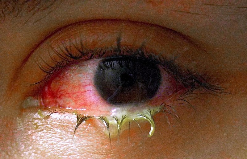
Image Source: Wikimedia Commons (2022). Swollen eye with conjunctivitis, Licensed under a creative commons licence. Available for free at: https://commons.wikimedia.org/wiki/File:Swollen_eye_with_conjunctivitis.jpg (Accessed: 30 June 2021).

Image Source: Wikimedia Commons (2022). Keratoderma blennorrhagic, Licensed under a creative commons licence. Available for free at: https://commons.wikimedia.org/wiki/File:Keratoderma_blennorrhagica.jpg (Accessed: 30 June 2021).
Diagnosis
Investigations
There are no confirmatory tests for reactive arthritis, and diagnosis is based on clinical suspicion in a patient with inflammatory arthritis and a history of a preceding genitourinary or gastrointestinal infection. Investigations are often performed to rule out other diagnoses.
Blood tests:
- FBC: may reveal normocytic normochromic anaemia, mild leucocytosis, and thrombocytosis in the acute stage
- ESR and CRP are usually very high in the acute stage
- HLA B27 is positive in most affected patients; however, is not specific for reactive arthritis
- Rheumatoid factors and antinuclear antibodies are negative
Identifying causative organism:
- Urogenital and stool cultures: positive at the time of infection, but are usually negative post onset of arthritis
- Men: Urine nucleic acid amplification test (NAAT) for C. trachomatis and N. gonorrhoeae and urethral culture for N. gonorrhoeae
- Women: Vulvovaginal NAAT for C. trachomatis and N. gonorrhoeae and endocervical culture for N. gonorrhoeae
- Both sexes: HIV, syphilis, hepatitis B and hepatitis C screening depending on clinical history
- Serology for Yersinia, Campylobacter, Salmonella and Shigella if indicated
Joint aspiration
- Joint aspiration may be needed to rule out septic arthritis or crystal arthropathy.
- Synovial fluid in patients with reactive arthritis would demonstrate a high white cell count (2000-50,000 cells/mm3. ) with a neutrophil count above 50% and may reveal triggering organisms, e.g. gonorrhoea.
Imaging
- X-rays: may be normal in early disease or show non-specific changes such as soft tissue swelling. In advanced disease, joint space narrowing and erosions in the joints of the hands and feet may be seen. X-rays may also reveal bilateral but asymmetrical sacroiliitis or enthesitis as fluffy periosteal new bone formation. Non-marginal syndesmophytes are typically seen.
- MRI: may be more sensitive in detecting sacroiliitis than x-rays in patients with lower back pain
- Ultrasound: may detect synovial hypertrophy and increased vascularity using doppler imaging
Differential diagnoses
- Ankylosing spondylitis – can present similarly to reactive arthritis; however, spinal involvement tends to be symmetrical, and x-rays can reveal the presence of syndesmophytes. AS also tends to also affect peripheral joints less frequently than reactive arthritis.
- Psoriatic arthritis – tends to affect mostly distal interphalangeal joints, which can be identified on x-rays
- Rheumatoid arthritis – usually presents as a symmetrical polyarthritis and does not affect lumbar spine or sacral joints. Rheumatoid factors and anti-CCP are frequently positive.
- Rheumatic fever – associated with an upper respiratory tract infection. Rheumatic fever tends to present as a migratory arthritis in the upper and lower limbs and does not involve the spine. Antistreptolysin-O titre may be positive.
- Adult-onset Still’s disease – may also present with fever and joint pain. Joint pain tends to occur in two spikes throughout the day and is associated with a salmon-coloured rash. Serum LDH and ferritin may be elevated.
- Enteric arthritis – patients will have a history of inflammatory bowel disease and tend to present with bilateral and symmetrical sacroiliitis. X-rays reveal marginal syndesmophytes in contrast to reactive arthritis, where non-marginal syndesmophytes are usually seen.
- Gout – patients usually present with an acutely swollen and tender joint, and distal interphalangeal joint involvement is common. Gout can be differentiated from reactive arthritis by synovial fluid examination, which would reveal crystals in gout.
- Septic arthritis – usually affects a single joint, and presents as a swollen, erythematous and warm joint in the presence of a fever. Synovial fluid analysis and culture would reveal a very high white cell count (>50,000mm ³) and neutrophil count >75%, with identification of the underlying organism. Blood cultures may also be positive in septic arthritis.
- Post-viral arthritis – parvovirus infection can present with a flu-like illness followed by arthralgia. This tends to mostly demonstrate a symmetrical small joint polyarticular pattern, and patients may often have a maculopapular rash. Serology would reveal the presence of parvovirus B19
Management
The main aim of treatment is directed towards symptomatic relief and prevention or slowing down of progressive joint damage. In the acute phase, rest is recommended, but physiotherapy can be used in later stages for symptomatic management.
Pharmacological management
- NSAIDS: NSAIDs are often used first line to treat symptomatic axial and peripheral arthritis, and can improve functional status if used early in the disease process.
- Corticosteroids: In patients unresponsive to NSAIDs, corticosteroids can be trialled. Steroids have been shown to be beneficial in improving symptoms in peripheral joints, but their efficacy in the management of axial disease is limited. Patients with monoarticular or oligoarticular joint involvement may be offered intra-articular steroid injections in acute flares, but if multiple joints are affected or if there are cardiac or ocular manifestations, systemic corticosteroids may be required. Topical corticosteroids can be used to treat skin disease associated with reactive arthritis, i.e. circinate balanitis and keratoderma blennorrhagicum.
- Conventional DMARDs: DMARDs such as Sulfasalazine can be used in patients who do not respond sufficiently to NSAIDs and demonstrate progressive joint damage and functional impairment
- Antibiotics: NICE guidelines recommend treating the initial infection if a causative organism is found, but to not offer long-term treatment to manage the arthritic symptoms following the infection.
References
- Barth WF, Segal K. Reactive arthritis (Reiter’s syndrome). American family physician. 1999 Aug 1;60(2):499.
- BMJ Best Practice [Internet] Bestpractice.BMJ.com [cited April 2022]. Available from: https://bestpractice.bmj.com/topics/en-gb/597/aetiology
- Carlin E, Marzo-Ortega H, Flew S. British Association of Sexual Health and HIV national guideline on the management of sexually acquired reactive arthritis 2021. International journal of STD & AIDS. 2021 Oct;32(11):986-97.
- Carlin E, Marzo-Ortega H, Flew S. British Association of Sexual Health and HIV national guideline on the management of sexually acquired reactive arthritis 2021. International journal of STD & AIDS. 2021 Oct;32(11):986-97.
- Cheeti A, Chakraborty RK, Ramphul K. Reactive Arthritis. 2018
- Martel W, Braunstein EM, Borlaza G, Good AE, Griffin Jr PE. Radiologic features of Reiter disease. Radiology. 1979 Jul;132(1):1-0.BMJ Best Practice [Internet] Bestpractice.BMJ.com [cited April 2022]. Available from: https://bestpractice.bmj.com/topics/en-gb/597/aetiology
- McAllister K, Goodson N, Warburton L, Rogers G. Spondyloarthritis: diagnosis and management: summary of NICE guidance. Bmj. 2017 Mar 1;356.
Complete Guide to Clinical Genetics ST3 Application and Recruitment (2027)
Written by Medibuddy. Last updated: October 2026. Clinical Genetics is

Complete Guide to Clinical Radiology ST3 Application and Recruitment (2027)
Recruitment paused: NHS England is not running Clinical Radiology ST3+ recru

Complete Guide to Paediatric Surgery ST3 Application and Recruitment in United Kingdom (2026)
Written by Medibuddy. Last updated: October 2026. If you’re planning


Take your subscriptions with you
Our mobile app allows you to access your interview and exam question banks wherever you are.

