


 Close
Close
 19th January 2024
Presentation
The clinical presentation of psoriatic arthritis varies greatly, with a diverse pattern of joint involvement and dermatological features. Patients often have a large variation in disease course and outcomes and may or may not have pre-existing psoriatic skin manifestations. The disease presents most commonly in young or middle-aged adults who either have a personal or family history of psoriasis. Symptoms can vary from mild to severe, and the arthritis tends to run a relapsing and remitting course. All patients with any type of psoriasis should be reviewed annually for the development of arthritis, particularly within the first 10 years of the diagnosis of skin disease.
The hallmark feature of psoriatic arthritis is inflammatory pain, stiffness and swelling in the joints; however, other features may be present. The most commonly affected areas are the knees, ankles, hands and feet, with axial disease also occurring occasionally. The most common pattern of joint disease is monoarticular or oligoarticular with asymmetric involvement; however, polyarticular disease can occur.
Symptoms and signs
- Joint symptoms
- Pain, tenderness, swelling in peripheral joints or axial skeleton
- Prolonged morning stiffness longer than 30 minutes
- Symptoms improve with activity and worsen with prolonged rest
- Dactylitis – swelling and tenderness of whole digit
- Psoriatic skin disease- red or silvery white, inflamed well-circumscribed scaly papules and plaques mostly found on extensor surfaces and the scalp
- Psoriatic nail disease
- Pitting – shallow dents on nail
- Oncolysis – painless separation of nail from the nail bed
- Enthesitis – mostly affecting Achilles tendon and plantar fascia
- Ocular features:
- Conjunctivitis – most common
- Anterior uveitis
- Mostly seen in patients with sacroiliitis and HLA-B27 positivity
- Features of metabolic disease, e.g. hypercholesterolaemia, high blood pressure, etc
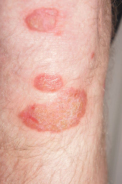
Image Source: Wikimedia Commons (2013). Psoriasis am Unterschenkel, Licensed under a creative commons licence. Available for free at: https://commons.wikimedia.org/wiki/File:Psoriasis_am_Unterschenkel.JPG (Accessed: 30 June 2021).
Patterns of presentation
The Moll and Wright diagnostic criterion is the simplest and most often used classification system to describe the 5 main patterns of presentations in psoriatic arthritis:
- Symmetric polyarthritis (30-40%, most common type)- symmetric polyarthritis most commonly occurs in women and can mimic the pattern seen in rheumatoid arthritis. Wrists, hands, feet, and ankles are usually affected. Factors which can help in differentiating psoriatic arthritis from rheumatoid arthritis are distal interphalangeal joint involvement rather than metacarpophalangeal joints, absence of skin nodules and absence of rheumatoid factor or anti-CCP in blood results.
- Asymmetric oligoarthritis (20-30%)- most commonly affected areas are hands and feet, initially with enthesitis causing dactylitis (“sausage fingers”). Usually, less than 5 joints are involved.
- Distal interphalangeal joint (DIP) arthritis (10%)- sole DIP joint involvement usually occurs in men. The nail and surrounding tissues can also be involved.
- Arthritis mutilans (5%)- arthritis mutilans is a rare type of severe and deformative DIP disease, which gives the appearance of “telescoping fingers” due to resorption of the terminal phalanx. The classic “pencil in cup” appearance on radiographs can be seen. This is the most severe and debilitating form of psoriatic arthritis.
- Psoriatic spondylitis with sacroiliac and spinal involvement- spondylitis most commonly occurs in men and presents with morning stiffness and limited range of back movement. Patients may be asymptomatic, and there may not be any obvious radiological findings. This can mimic ankylosing spondylitis; however, tends to affect vertebrae asymmetrically rather than symmetrically.

Image Source: Wikimedia Commons (2010). Psoriatic arthritis2010, Licensed under a creative commons licence. Available for free at: https://commons.wikimedia.org/wiki/File:Psoriatic_arthritis2010.JPG (Accessed: 30 June 2021).
Complete Guide to Clinical Genetics ST3 Application and Recruitment (2027)
Written by Medibuddy. Last updated: October 2026. Clinical Genetics is

Complete Guide to Clinical Radiology ST3 Application and Recruitment (2027)
Recruitment paused: NHS England is not running Clinical Radiology ST3+ recru

Complete Guide to Paediatric Surgery ST3 Application and Recruitment in United Kingdom (2026)
Written by Medibuddy. Last updated: October 2026. If you’re planning


Take your subscriptions with you
Our mobile app allows you to access your interview and exam question banks wherever you are.

