


 Close
Close
 7th February 2024
Cardiac Electrophysiology
There are four chambers of the heart. The function of the heart is reliant on the coordinated contraction of heart chambers. This coordination is via the cardiac conduction pathway.
Cardiac conduction starts with pacemaker cells. Cardiac pacemaker cells are specialised cells. They undergo spontaneous depolarisation (automaticity) regularly (rhythmicity). They are found in the sinoatrial (SA) nodes, atrioventricular (AV) nodes, bundle of His, and ventricles. They have different rates of spontaneous depolarisation, with the SA node having the fastest (70 times per minute) and ventricular pacemaker cells having the slowest (40 times per minute).
Generation of pacemaker action potential
The electrophysiology of action potential generation of cardiac pacemaker cells follows a cycle:
Following an action potential, the pacemaker cell returns to a resting membrane potential of -60mV
- Phase 4: Increasing membrane permeability to sodium ions causes slow leakage of sodium ions via sodium channels into the cell, causing membrane potential to become less negative. This results in spontaneous depolarisation of the pacemaker cells.
- This is referred to as the funny current, If. The funny current is found in cardiac areas supporting spontaneous depolarisation, including the sinoatrial node, atrioventricular node and Purkinje fibres.
- Phase 0: When the membrane potential reaches a threshold potential of -40mV, voltage-gated T-type calcium channels open, and rapid Ca2+ influx into the cell occurs, causing depolarisation to the membrane potential of +20mV (Phase 0)
- Phase 3: Following depolarisation, the voltage-gated T-type calcium channels close. Voltage-gated potassium channels open, causing efflux of K+ from the cell, returning membrane potential to resting potential of -60mV (Phase 3).
 Image Source: Wikimedia Commons (2010). Pacemaker potential annotated, Licensed under a creative commons licence. Available for free at: https://upload.wikimedia.org/wikipedia/commons/8/81/Pacemaker_potential_annotated.gif (Accessed: 4 January 2024).
Conduction of electrical signals of cardiac myocytes
- Phase 0: Action potentials from pacemaker cells or nearby myocytes induce transient opening of Na+ channels, resulting in an influx of Na+ into myocytes. This produces rapid depolarisation to the membrane potential of +20mV
- Phase 1: Immediately following phase 0, Na+ channels close in response to depolarisation, voltage-gated K+ channel transiently opens, and electrochemical gradient causes transient efflux of K+ ions.
- Phase 2: Membrane potential is stable at this phase. Inward and outward currents are balanced. K+ channels remain open. L-type Ca2+ channels open. This results in the influx of Ca2+ ions, balanced by the efflux of K+ ions. The influx of Ca2+ ions initiates the release of intracellular Ca2+ stores in excitation-contraction coupling (see later).
- Phase 3: Repolarisation begins due to the closing of L-type Ca2+ channels. K+ channels remain open, resulting in the outward flow of K+.
- Phase 4: Membrane potential is maintained at resting membrane potential at -85mV. This phase also allows an interval between action potentials to return Ca2+ into intracellular stores.
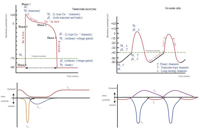
Image Source: Wikimedia Commons (2019). Heart rhythm ionic flow, Licensed under a creative commons licence. Available for free at: https://upload.wikimedia.org/wikipedia/commons/b/bc/Heart_rhythm_ionic_flow.svg (Accessed: 4 January 2024).
References
Oberman, R., Shumway, K. R., & Bhardwaj, A. (2023). Physiology, Cardiac. In StatPearls. StatPearls Publishing. http://www.ncbi.nlm.nih.gov/books/NBK526089/
Complete Guide to Clinical Genetics ST3 Application and Recruitment (2027)
Written by Medibuddy. Last updated: October 2026. Clinical Genetics is

Complete Guide to Clinical Radiology ST3 Application and Recruitment (2027)
Recruitment paused: NHS England is not running Clinical Radiology ST3+ recru

Complete Guide to Paediatric Surgery ST3 Application and Recruitment in United Kingdom (2026)
Written by Medibuddy. Last updated: October 2026. If you’re planning


Take your subscriptions with you
Our mobile app allows you to access your interview and exam question banks wherever you are.

