


 Close
Close
 7th February 2024
Conduction of electrical signals of cardiac myocytes
- Phase 0: Action potentials from pacemaker cells or nearby myocytes induce transient opening of Na+ channels, resulting in an influx of Na+ into myocytes. This produces rapid depolarisation to the membrane potential of +20mV
- Phase 1: Immediately following phase 0, Na+ channels close in response to depolarisation, voltage-gated K+ channel transiently opens, and electrochemical gradient causes transient efflux of K+ ions.
- Phase 2: Membrane potential is stable at this phase. Inward and outward currents are balanced. K+ channels remain open. L-type Ca2+ channels open. This results in the influx of Ca2+ ions, balanced by the efflux of K+ ions. The influx of Ca2+ ions initiates the release of intracellular Ca2+ stores in excitation-contraction coupling (see later).
- Phase 3: Repolarisation begins due to the closing of L-type Ca2+ channels. K+ channels remain open, resulting in the outward flow of K+.
- Phase 4: Membrane potential is maintained at resting membrane potential at -85mV. This phase also allows an interval between action potentials to return Ca2+ into intracellular stores.
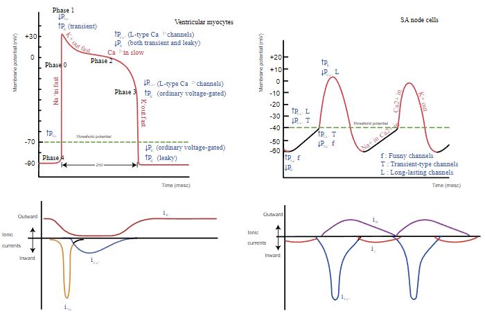
Image Source: Wikimedia Commons (2019). Heart rhythm ionic flow, Licensed under a creative commons licence. Available for free at: https://upload.wikimedia.org/wikipedia/commons/b/bc/Heart_rhythm_ionic_flow.svg (Accessed: 4 January 2024).
Complete Guide to Clinical Genetics ST3 Application and Recruitment (2027)
Written by Medibuddy. Last updated: October 2026. Clinical Genetics is
Clinical Genetics ST3

Complete Guide to Clinical Radiology ST3 Application and Recruitment (2027)
Recruitment paused: NHS England is not running Clinical Radiology ST3+ recru
Clinical Radiology ST3

Complete Guide to Paediatric Surgery ST3 Application and Recruitment in United Kingdom (2026)
Written by Medibuddy. Last updated: October 2026. If you’re planning
Paediatric Surgery ST3


Take your subscriptions with you
Our mobile app allows you to access your interview and exam question banks wherever you are.

