


 Close
Close
 16th February 2024
Cardiac and Respiratory System
Thoracic cavity
Thoracic cage
The bony thoracic wall, aka thoracic cage, comprises the thoracic vertebrae, ribs, costal cartilage and sternum. There are a total of 12 ribs. The first seven ribs are true ribs, connected to the vertebral column on the back and directly with the sternum anteriorly. The next three are false ribs, as their front attachment with the sternum is indirectly via combined costal cartilage.
The last two are the floating ribs, which do not have anterior attachment to the sternum. Inferior and just internal to each rib is the costal groove. The costal groove contains the intercostal vein, artery and nerve from superior to inferior.

Image Source: Wikimedia Commons (2014). Endothoracic fascia, Licensed under a creative commons licence. Available for free at: https://upload.wikimedia.org/wikipedia/commons/e/e2/Endothoracic_fascia.svg (Accessed: 9 January 2024).
Pleural lining
The pleura is a thin double membrane which lines the lungs on each side. The outer membrane is called the parietal pleural; the inner is known as the visceral pleura. The parietal pleural is closely attached to the thoracic wall and the visceral pleura to the lungs. A tiny physiological space contains fluid between the two layers, which helps maintain the negative pressure for the lungs to expand.
Somatic nerves innervate the visceral layer; hence, it is sensitive to touch, pain, temperature and pressure. The visceral layer is only sensitive to stretch due to its visceral nerve innervation.
Lungs
The respiratory system is divided into the conducting zone and the respiratory zone. The conducting zone starts from the nose and ends at the bronchioles. The trachea is part of the conducting zone and starts in the neck from the larynx and ends at the carina, at the level of the sternal angle and the T4/T5 vertebral level, by dividing into the right and left main bronchus. The right main bronchus is shorter, wider and more vertical than the left. Each of these main bronchi enters its respective lungs at the hilum and further divides into secondary lobar, then tertiary segmental bronchioles, which supply bronchopulmonary segments. Each bronchopulmonary segment is functionally and structurally independent.
 Image Source: Wikimedia Commons (2013). 2312 Gross Anatomy of the Lungs, Licensed under a creative commons licence. Available for free at: https://upload.wikimedia.org/wikipedia/commons/7/7e/2312_Gross_Anatomy_of_the_Lungs.jpg (Accessed: 9 January 2024).
The respiratory zone includes the respiratory bronchioles and alveoli and moves the respiratory gases, that is, oxygen and carbon dioxide, in and out of the blood. Oxygenation occurs in the respiratory zone, from where the oxygenated blood is taken by the alveolar capillaries in the connective tissue surrounding the bronchopulmonary segment and drains to the segmental and main pulmonary veins.
The right lung is divided into three lobes: the upper, middle and lower lobes. The horizontal and oblique fissures delineate these. The left lung has only two lobes, upper and lower, divided by a single oblique fissure. The parietal pericardium lies directly in contact with the left lower lobe.
The pulmonary plexus supplies the lung tissue, providing parasympathetic, sympathetic and visceral afferent innervation. Two pulmonary veins from each lung bring the oxygenated blood to the left atrium. Deoxygenated blood is supplied to the lungs by the pulmonary arteries, accounting for the majority of blood supplied to the lungs. Oxygen supplies for the tissues are delivered via the bronchial arteries.
Lymphatic drainage
The thoracic duct brings the lymph from the lower half and the left side of the upper half of the body. It starts from the cisterna chyli at the level of L1; it is initially to the right of the midline in the thorax but then crosses the midline to the left at T4-6 and drains at the origin of the left brachiocephalic vein. The lymph drainage from the right upper half of the body is via the right lymphatic duct, which drains at the origin of the right brachiocephalic vein.
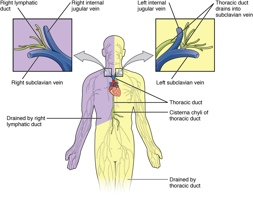 Image Source: Wikimedia Commons (2013). 2203 Lymphatic Trunks and Ducts System, Licensed under a creative commons licence. Available for free at :https://upload.wikimedia.org/wikipedia/commons/4/43/2203_Lymphatic_Trunks_and_Ducts_System.jpg (Accessed: 9 January 2024).
References
Wineski, L. E., & Snell, R. S. (2019). Snell’s clinical anatomy by regions.
Chaurasia, B. D. (2019). Human anatomy: Regional and applied, dissection and clinical. New Delhi, India: CBS Publishers & Distributors.
Moore, K. L., Dalley, A. F., & Agur, A. M. R. (2014). Moore’s Clinically Orientated Anatomy (7th ed.). China: Lippincott.
Mediastinum
Summary
The mediastinum is bounded anteriorly by the sternum, posteriorly by the spine and laterally by the parietal pleura. This is then divided into superior and inferior mediastinum at the level of sternal angle anteriorly and inferior border of the fourth thoracic vertebra.
The inferior mediastinum is subdivided into anterior, middle and posterior mediastinum. The anterior mediastinum is bordered by the sternum anteriorly and the pericardial sac posteriorly. The posterior mediastinum is bordered by the pericardial sac anteriorly and the thoracic vertebrae posteriorly.
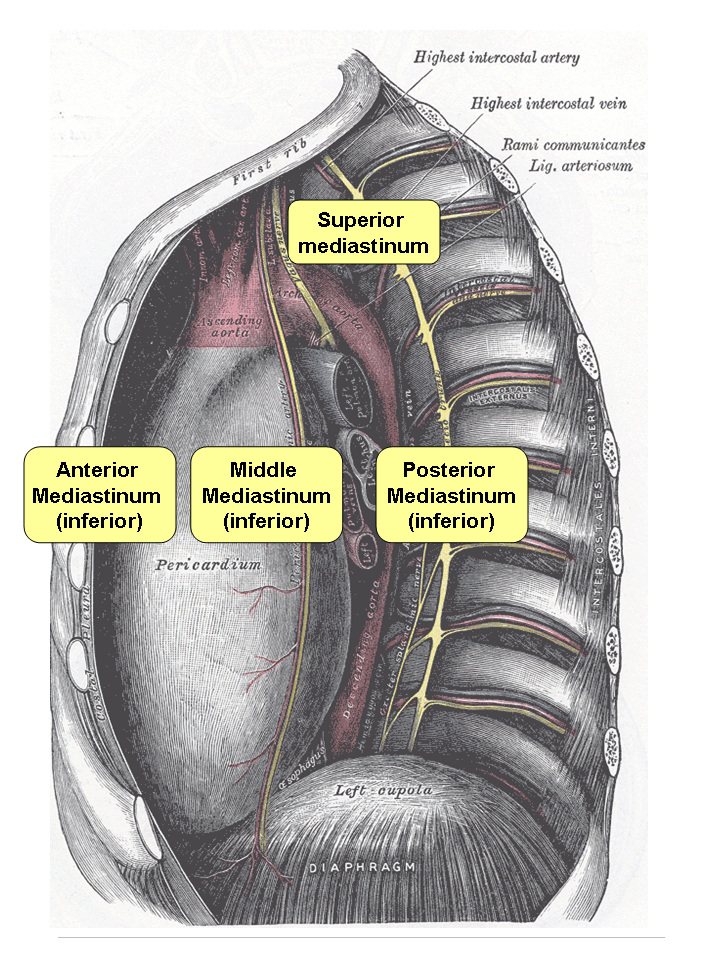 Image Source: Wikimedia Commons (2006). Mediastinum, Licensed under a creative commons licence. Available for free at: https://upload.wikimedia.org/wikipedia/commons/a/ab/Mediastinum.png (Accessed: 9 January 2024).
Superior mediastinum
- Arch of the aorta and associated branches
- Right brachiocephalic artery
- Left common carotid artery
- Left subclavian artery
- Major veins
- Superior vena cava
- Right and left brachiocephalic vein
- Left superior intercostal veins
- Nerves
- Left laryngeal nerve
- Phrenic nerve
- Vagus nerve
- Sympathetic trunk
- Other major organs
- Thymus gland ( in most adults)
- Thoracic duct
- Trachea
- Oesophagus
- Lymph node group
Anterior mediastinum
- Thymus gland ( extension into the anterior mediastinum is possible)
- Lymph node groups
Middle mediastinum
- Heart and origin of the great vessels
- Nerves
- Cardiac plexus
- Phrenic nerves ( descends from the superior mediastinum)
- Tracheobronchial lymph nodes
- Trachea and the main bronchi.
Posterior mediastinum
- Thoracic aorta and its associated branches
- Bronchial artery
- Intercoastal arteries
- Oesophageal arteries
- Oesophagus
- Thoracic duct
- Veins
- Azygous vein
- Hemizygous vein
- Accessory hemiazygous vein
- Thoracic sympathetic trunk and oesophageal vagal plexus
References
Wineski, L. E., & Snell, R. S. (2019). Snell’s clinical anatomy by regions.
Chaurasia, B. D. (2019). Human anatomy: Regional and applied, dissection and clinical. New Delhi, India: CBS Publishers & Distributors.
Moore, K. L., Dalley, A. F., & Agur, A. M. R. (2014). Moore’s Clinically Orientated Anatomy (7th ed.). China: Lippincott.
Cardiac system
Pericardium
There are two layers of the pericardium: the outer tough fibrous (parietal) pericardium and the inner membranous serous pericardium, with a trace of pericardial fluid between the layers. This acts as a physical barrier between the heart and the other organs. It also prevents overfilling and fixes the heart within the mediastinum.
Cardiac anatomy
The heart consists of four chambers: two atria and two ventricles. The right atrium receives deoxygenated blood from the body via the superior and inferior vena cava and pumps into the right ventricle through the tricuspid valve. The right ventricle then pumps this blood through the pulmonary trunk into the pulmonary circulation for oxygenation, and the oxygenated blood is returned via the pulmonary veins into the left atrium.
Blood flows from the left atrium through the mitral valve into the left ventricle. Then, this oxygenated blood is supplied to the body, starting from the aorta to arteries, arterioles and the capillaries. The atria contract, and the AV valves (mitral and tricuspid) open in diastole. The ventricles contract, and outlet valves (Pulmonic and aortic) open in systole.
The heart’s conduction system begins at the sinoatrial node within the right atrial wall. The pacemaker cells spontaneously generate an electrical impulse, and these travel across the atrial walls. The impulses arrive at the atrioventricular (AV) node within the atrioventricular wall septum close to the coronary sinus. The impulses will travel from the AV node into the atrioventricular bundle (Bundle of His). The AV bundle will divide into the left and right bundle branches to supply the left and right ventricles. These bundles terminate as the Purkinje fibres as the electrical impulses are transmitted across the ventricular walls.

Image Source: Wikimedia Commons (2015). Cardiac conduction system, Licensed under a creative commons licence. Available for free at: https://upload.wikimedia.org/wikipedia/commons/3/3d/Cardiac_conduction_system.jpg (Accessed: 9 January 2024).
Cardiac blood supply
The blood supply to the heart is by the left and right coronary arteries. The left coronary artery comes off the left coronary cusp, while the right coronary artery comes off the right coronary cusp above the aortic valve. The left coronary artery starts as the left main stem and then divides into the left anterior descending (LAD) and left circumflex artery. The LAD lies within the anterior interventricular groove and travels towards the apex of the heart. The left marginal artery travels along the coronary sulcus towards the posterior heart surface.
The right coronary artery travels along the coronary sulcus, with the right marginal artery branching off the artery. The right coronary artery continues as the posterior interventricular artery within the posterior interventricular groove.
The distribution of the arteries’ blood supply in a typical right-dominant system is as follows:
- Right coronary artery – Right atrium, SA node, AV node
- Right marginal artery – Lateral right ventricular wall.
- Right interventricular artery – Inferior aspect of the heart
- LAD – Anterior interventricular septum and anterior left ventricular wall
- Left circumflex artery – Lateral left ventricular wall
Venous return from the heart is through the coronary sinus into the right atrium. The main tributaries for the coronary sinus include the great cardiac, small, middle, and posterior cardiac veins.
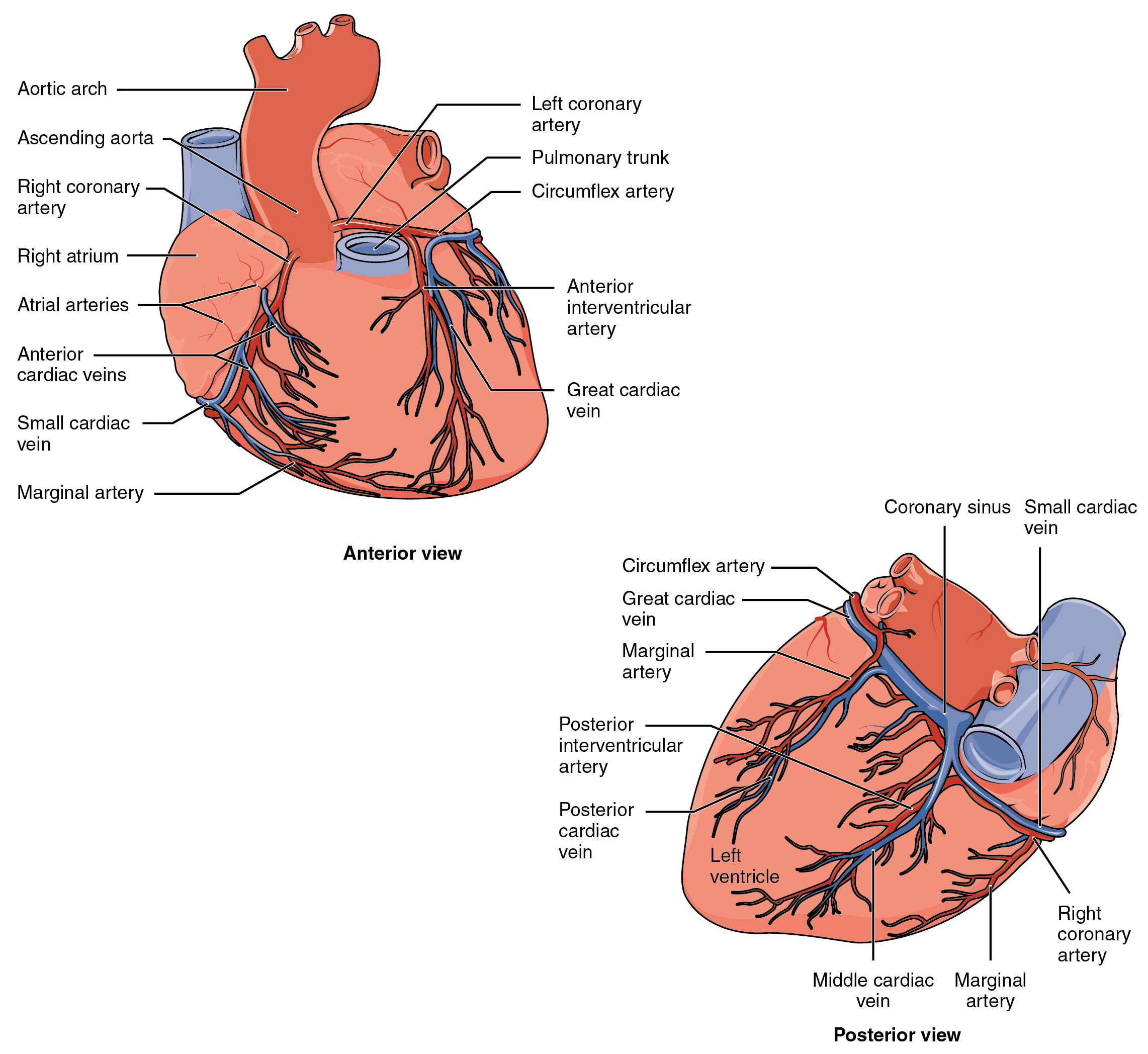 Image Source: Wikimedia Commons (2013). 2014ab Coronary Blood Vessels, Licensed under a creative commons licence. Available for free at: https://upload.wikimedia.org/wikipedia/commons/7/7c/2014ab_Coronary_Blood_Vessels.jpg (Accessed: 9 January 2024).
References
Wineski, L. E., & Snell, R. S. (2019). Snell’s clinical anatomy by regions.
Chaurasia, B. D. (2019). Human anatomy: Regional and applied, dissection and clinical. New Delhi, India: CBS Publishers & Distributors.
Moore, K. L., Dalley, A. F., & Agur, A. M. R. (2014). Moore’s Clinically Orientated Anatomy (7th ed.). China: Lippincott.
Complete Guide to Clinical Genetics ST3 Application and Recruitment (2027)
Written by Medibuddy. Last updated: October 2026. Clinical Genetics is

Complete Guide to Clinical Radiology ST3 Application and Recruitment (2027)
Recruitment paused: NHS England is not running Clinical Radiology ST3+ recru

Complete Guide to Paediatric Surgery ST3 Application and Recruitment in United Kingdom (2026)
Written by Medibuddy. Last updated: October 2026. If you’re planning


Take your subscriptions with you
Our mobile app allows you to access your interview and exam question banks wherever you are.

