


 Close
Close
 19th February 2024
Autonomic nervous system
Sympathetic nervous system
Main structure
The preganglionic neurons of the sympathetic nervous system arise within the thoracolumbar section of the spinal cord. These can be found within the lateral horns of the spinal cord within spinal levels T1-L2. The sympathetic fibres will exit the spinal cord with the spinal nerves. However, they will quickly diverge from the spinal nerve as the white rami communicans and synapses onto the sympathetic chain.
The sympathetic chain consists of 3 separate structures: 2 paravertebral ganglion chains that run alongside the spinal cord and a central prevertebral ganglion located anterior to the aorta. The paravertebral chains extend along the entire length of the spinal cord. The prevertebral ganglion is closely associated with the origin of the major blood vessels within the abdominal aorta.
The preganglionic sympathetic neurons will follow one of the following paths once it has entered the sympathetic chain:
- Ascend and synapse with a ganglion at a higher vertebral level
- It is usual for sympathetic fibres to leave the T1-T5 levels as they supply the cervical ganglion
- Three main cervical ganglion exist – Superior, middle and inferior
- Inferior ganglion can be merged with the first thoracic ganglion to form the stellate ganglion.
- Descend and synapse with a ganglion at a lower vertebral level
- Usual for fibres from T5-L2 as it supplies the lumbosacral ganglions
- Synapse within the same vertebral level
- Fibres will leave the chain as a grey ramus communicans and merge with the spinal nerve’s anterior rami to arrive at its target organ.
- Travel without synapsing towards its target organ
- This usually occurs with the T5 -L2 preganglionic fibres as they travel towards the prevertebral ganglion to contribute towards the splanchnic nerves.
Once the sympathetic fibre synapses within the sympathetic chain, the postganglionic nerve fibres will leave and join the course of the peripheral nerves to reach their end-organ targets. These include the sweat glands, smooth muscle walls, erector pili muscles, respiratory lining, cardiac tissues, GI tract, urinary tracts, ciliary muscles within the eye and the adrenal glands.
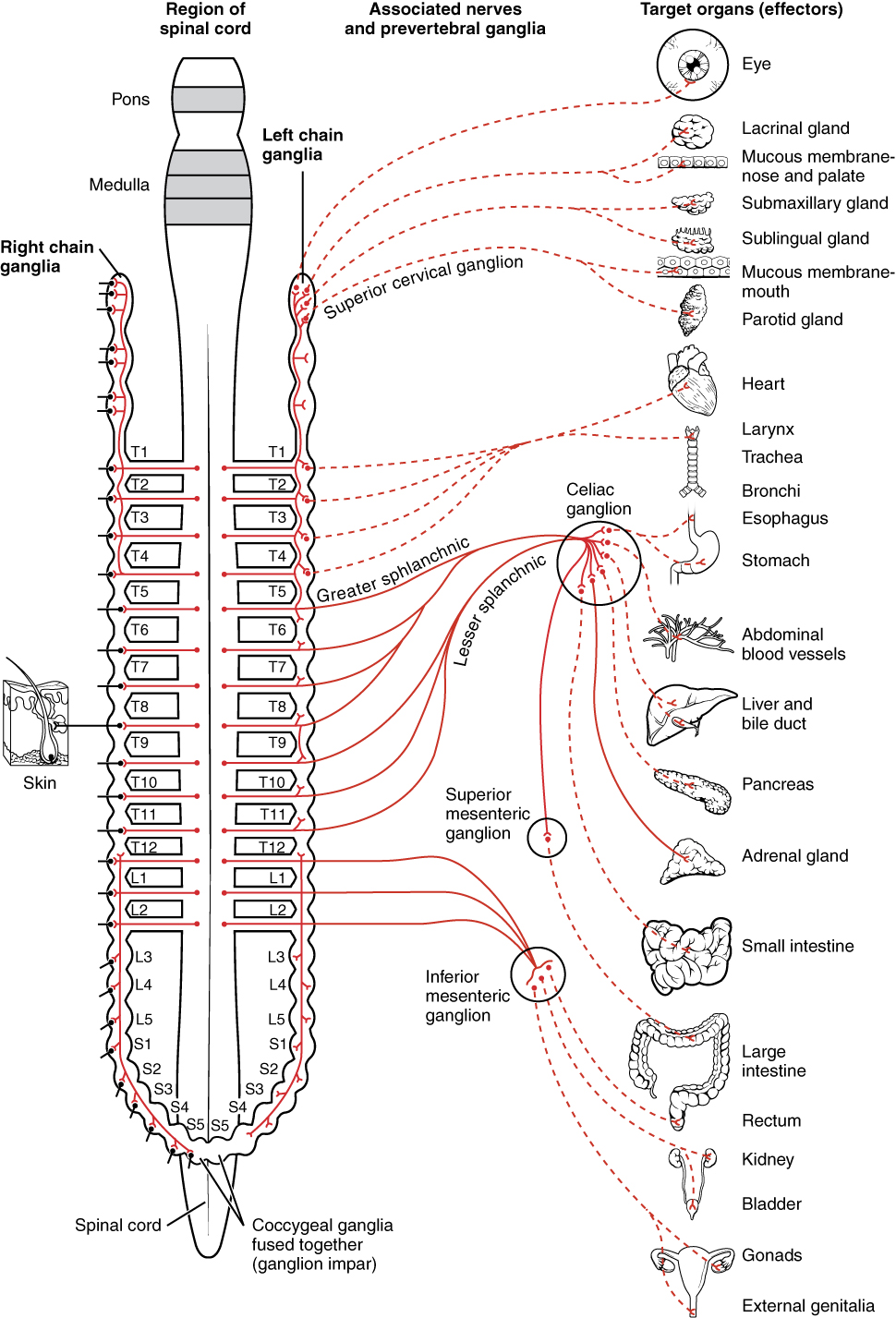 Image Source: Wikimedia Commons (2013). 1501 Connections of the Sympathetic Nervous System, Licensed under a creative commons licence. Available for free at: https://upload.wikimedia.org/wikipedia/commons/f/fd/1501_Connections_of_the_Sympathetic_Nervous_System.jpg (Accessed: 9 January 2024).
Parasympathetic nervous system
Main structure
The preganglionic neurons of the parasympathetic nervous system are concentrated within several cranial nerve nuclei within the medulla oblongata and the sacral spinal cord. In contrast to the sympathetic nervous system, the preganglionic neurons have long courses and will travel close to their effector organ before they synapse with their postganglionic neuron.
The cranial parasympathetic nervous system originates from CN III, VII, IX, and X. CN III supplies the parasympathetic fibres destined for the ciliary ganglion. Fibres from the Edinger-Westphal nucleus will travel towards the ciliary ganglion before it synapses and supplies the ciliary muscle and pupils with parasympathetic fibres via the short ciliary nerves.
CN VII carries parasympathetic fibres from the superior salivary nucleus and synapses with the pterygopalatine and submandibular ganglion. It will then supply the lacrimal gland and nasal mucosa via the greater petrosal nerve, while the submandibular nerve will supply the submandibular gland.
CN IX carries fibres from the inferior salivary nucleus with its synapse within the otic ganglion. The auricular nerve will then carry the parasympathetic fibres towards the parotid gland.
CN X supplies most of the parasympathetic innervation within the body, with the abdominal and thoracic cavities receiving its parasympathetic innervation from the vagus nerve. The origin of the fibres is from the nucleus ambiguous within the medulla. The presynaptic fibres travel down into the thorax and abdomen to supply the lungs, heart and abdominal organs via various parasympathetic ganglions close to the aforementioned organs. The abdominal organs are supplied up to the level of the splenic flexure of the large intestine.
The sacral portion of the parasympathetic nervous system arises from the S2-S4 levels. These provide the parasympathetic innervation to the descending colon, sigmoid colon, rectum and pelvic organs.
 Image Source: Wikimedia Commons (2014). Blausen 0703 Parasympathetic Innervation, Licensed under a creative commons licence. Available for free at: https://upload.wikimedia.org/wikipedia/commons/f/f0/Blausen_0703_Parasympathetic_Innervation.png (Accessed: 9 January 2024).
References
Moore, K. L., Dalley, A. F., & Agur, A. M. R. (2014). Introduction to Clinically Orientated Anatomy. In Moore’s Clinically Orientated Anatomy (7th ed.). China: Lippincott.
Hall, J. E. (2011). The Nervous System: C. Motor and Integrative Neurophysiology. In Guyton and Hall Textbook of Medical Physiology (12th ed.). USA: Saunders Elsevier.
Waxenbaum JA, Reddy V, Varacallo M. Anatomy, Autonomic Nervous System. [Updated 2021 Jul 29]. In: StatPearls [Internet]. Treasure Island (FL): StatPearls Publishing; 2022 Jan-. Available from: https://www.ncbi.nlm.nih.gov/books/NBK539845/
Tindle J, Tadi P. Neuroanatomy, Parasympathetic Nervous System. [Updated 2021 Nov 5]. In: StatPearls [Internet]. Treasure Island (FL): StatPearls Publishing; 2022 Jan-. Available from: https://www.ncbi.nlm.nih.gov/books/NBK553141/
Patel, T. R. (2015). Chapter 36 – Anatomy of the Sympathetic Nervous System. In R. S. Tubbs, E. Rizk, M. M. Shoja, M. Loukas, N. Barbaro, & R. J. Spinner (Eds.), Nerves and Nerve Injuries (pp. 495–506). San Diego: Academic Press.
Complete Guide to Clinical Genetics ST3 Application and Recruitment (2027)
Written by Medibuddy. Last updated: October 2026. Clinical Genetics is

Complete Guide to Clinical Radiology ST3 Application and Recruitment (2027)
Recruitment paused: NHS England is not running Clinical Radiology ST3+ recru

Complete Guide to Paediatric Surgery ST3 Application and Recruitment in United Kingdom (2026)
Written by Medibuddy. Last updated: October 2026. If you’re planning


Take your subscriptions with you
Our mobile app allows you to access your interview and exam question banks wherever you are.

