


 Close
Close
 20th February 2024
Karyotyping
Karyotyping involves whole chromosomal analysis using microscopy. Cells are cultured within a medium, and colchicine arrests cell division during metaphase. The chromosomes are made visible using various stains and arranged based on their appearance to form the karyotype.
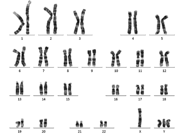
Image Source: Wikimedia Commons (2017). Peripheral blood karyotype result belonging to the subject compatible with 48-XYYY, Licensed under a creative commons licence. Available for free at: https://commons.wikimedia.org/wiki/File:Peripheral-blood-karyotype-result-belonging-to-the-subject-compatible-with-48-XYYY.png (Accessed: 4 January 2024).
Karyotypes have a role in identifying large genetic abnormalities, including aneuploidy, reciprocal and Robertsonian translocation, and large microdeletions or duplications. Its use has largely been superseded by microarray analysis but still has a role in identifying balanced translocations.
Complete Guide to Clinical Genetics ST3 Application and Recruitment (2027)
Written by Medibuddy. Last updated: October 2026. Clinical Genetics is

Complete Guide to Clinical Radiology ST3 Application and Recruitment (2027)
Recruitment paused: NHS England is not running Clinical Radiology ST3+ recru

Complete Guide to Paediatric Surgery ST3 Application and Recruitment in United Kingdom (2026)
Written by Medibuddy. Last updated: October 2026. If you’re planning


Take your subscriptions with you
Our mobile app allows you to access your interview and exam question banks wherever you are.

