


 Close
Close
 20th February 2024
Vascular and Muscular Anatomy
Arterial anatomy
Aorta
The arterial supply of the body begins at the aortic root. Coronary circulation is covered in the “Cardiac and Respiratory Anatomy” section. The aortic root continues as the ascending aorta, which becomes the arch of the aorta at the level of the second sternocostal joint. Three main branches arise from the aortic arch before it becomes the descending aorta: brachiocephalic trunk, left common carotid artery and left subclavian artery.
The descending aorta can be divided into the thoracic and abdominal aorta. At the level of L4, the abdominal aorta terminates as it bifurcates into the common iliac arteries.
 Image Source: Wikimedia Commons (2017). Aorta branches, Licensed under a creative commons licence. Available for free at: https://upload.wikimedia.org/wikipedia/commons/f/f1/Aorta_branches.jpg (Accessed: 9 January 2024).
Thoracic, abdominal and pelvic cavity
The thoracic aorta supplies several branches, including (from superior to inferior):
- Bronchial artery – Supplies the bronchus, peribronchial tissue and visceral pleura
- Mediastinal artery
- Oesophageal artery – Supplies the oesophagus
- Pericardial artery – Supplies the dorsal pericardium
- Superior phrenic artery – Supplies the superior diaphragm
- Intercostal artery and subcostal artery – Supplies the intercostal space.
The abdominal aorta begins at the T12 vertebral level, and multiple branches arise from the abdominal aorta, including (from superior to inferior):
- Inferior phrenic artery
- Coeliac artery
- Superior mesenteric artery
- Middle suprarenal artery – Supplies the adrenal glands
- Renal arteries
- Gonadal arteries
- Inferior mesenteric artery
- Lumbar and sacral arteries
The coeliac axis, superior mesenteric artery and inferior mesenteric artery supply the GI tract and the arteries supply the foregut, midgut and hindgut, respectively. The coeliac artery has several branches:
- Left gastric artery
- Splenic artery – Further branches include the left gastroepiploic artery, short gastric arteries and the pancreatic branches
- Common hepatic artery – Further branches to the proper hepatic artery and gastroduodenal artery
- The proper hepatic artery supplies the right gastric, right hepatic and left hepatic artery
- The gastroduodenal artery supplies the right gastroepiploic and superior pancreaticoduodenal artery.
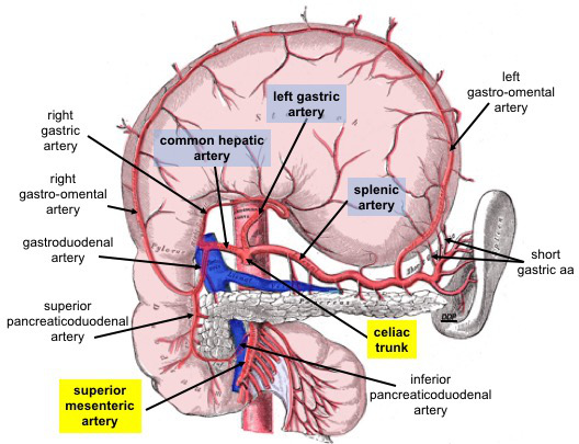
Image Source: Wikimedia Commons (2019). Celiac Trunk and Branches, Licensed under a creative commons licence. Available for free at: https://upload.wikimedia.org/wikipedia/commons/3/36/Celiac_Trunk_and_Branches.jpg (Accessed: 9 January 2024).
The superior mesenteric artery supplies blood from the level of the ampulla of Vater within the duodenum to the proximal 2/3rd of the transverse colon. The inferior mesenteric artery supplies the remainder 1/3rd of the transverse colon up to the anal canal at the level of the pectinate line.
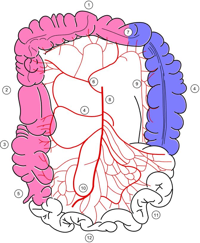
Image Source: Wikimedia Commons (2008). Colonic blood supply, Licensed under a creative commons licence. Available for free at: https://upload.wikimedia.org/wikipedia/commons/9/9f/Colonic_blood_supply.svg (Accessed: 9 January 2024).
Legend: 4 – Right colic artery; 6 – Middle colic artery; 8 – Superior mesenteric artery; 9 – Marginal artery; 10 – Ileocolic artery.

Image Source: Wikimedia Commons (2005). Inferior mesenteric, Licensed under a creative commons licence. Available for free at: https://upload.wikimedia.org/wikipedia/commons/6/64/Inferior_mesenteric_a.gif (Accessed: 9 January 2024).
It is important to note the watershed area created within the splenic flexure, as this is the point of anastomosis between the superior and inferior mesenteric arteries.
The pelvic blood supply mainly arises from the internal iliac artery. The internal iliac artery will provide branches to the bladder, rectum, perineum, and muscles surrounding the gluteal and adductor muscles. It also supplies arterial blood to the vagina and uterus.
Head and neck
The arterial supply to the head and neck arises from the common carotid artery. This arises directly from the arch of the aorta on the left, while the right common carotid artery arises from the brachiocephalic artery. The common carotid artery will bifurcate into the internal and external carotid artery.
The internal carotid artery mainly supplies the intracranial structures, with the ophthalmic artery supplying the structures within the orbit.
The external carotid artery supplies the remainder of the neck and head. The main branches of this include the facial artery, maxillary artery and superficial temporal artery. The external carotid artery branches will provide arterial blood to the structures of the face, tongue, external ear, pharynx, maxilla, mandible, nasal structures and scalp.
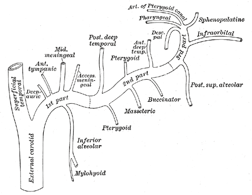
Image Source: Wikimedia Commons (2012). Gray511, Licensed under a creative commons licence. Available for free at: https://upload.wikimedia.org/wikipedia/commons/a/aa/Gray511.png (Accessed: 9 January 2024).
Upper limb
The arterial supply to the upper limb arises from the subclavian artery bilaterally. It travels laterally through the axillae and becomes the axillary artery after it crosses the lateral border of the first rib. The axillary artery provides the anterior and posterior circumflex artery, which wraps around the surgical neck of the humerus, and it can be damaged during fractures of the surgical neck of the humerus. The axillary artery becomes the brachial artery after it crosses the teres major.
The brachial artery travels with the radial nerve within the radial groove of the humerus and provides the bulk of the arterial supply to the structures of the upper arm. As the artery crosses the bicipital aponeurosis, it bifurcates into the radial and ulnar arteries. The radial artery supplies the structures within the posterolateral forearm, while the ulnar artery supplies the anteromedial forearm. Within the hand, the radial and ulnar arteries form two anastomoses, giving rise to the deep and superficial palmar arteries.

Image Source: Wikimedia Commons (2013). Thoracic Upper Limb Arteries Chart, Licensed under a creative commons licence. Available for free at: https://upload.wikimedia.org/wikipedia/commons/c/c0/2128_Thoracic_Upper_Limb_Arteries_Chart.jpg (Accessed: 9 January 2024).
Lower limb
The femoral artery is the lower limb’s main artery, which acts as a direct continuation of the external iliac artery as it crosses the inguinal ligament. Several branches arise from the femoral artery, which supplies the femur and muscles around the thigh. It becomes the popliteal artery as it enters the posterior compartment of the leg close to the knee.
The popliteal artery later divides into the anterior tibial and tibioperoneal trunk. The tibioperoneal artery will further divide into the posterior tibial and fibular arteries. The anterior tibial artery continues along the anterior aspect of the leg and becomes the dorsalis pedis artery. The posterior tibial artery enters the foot via the tarsal tunnel and crosses posterior to the medial malleolus. This contributes to the circulation of the foot via the medial and lateral plantar arteries.
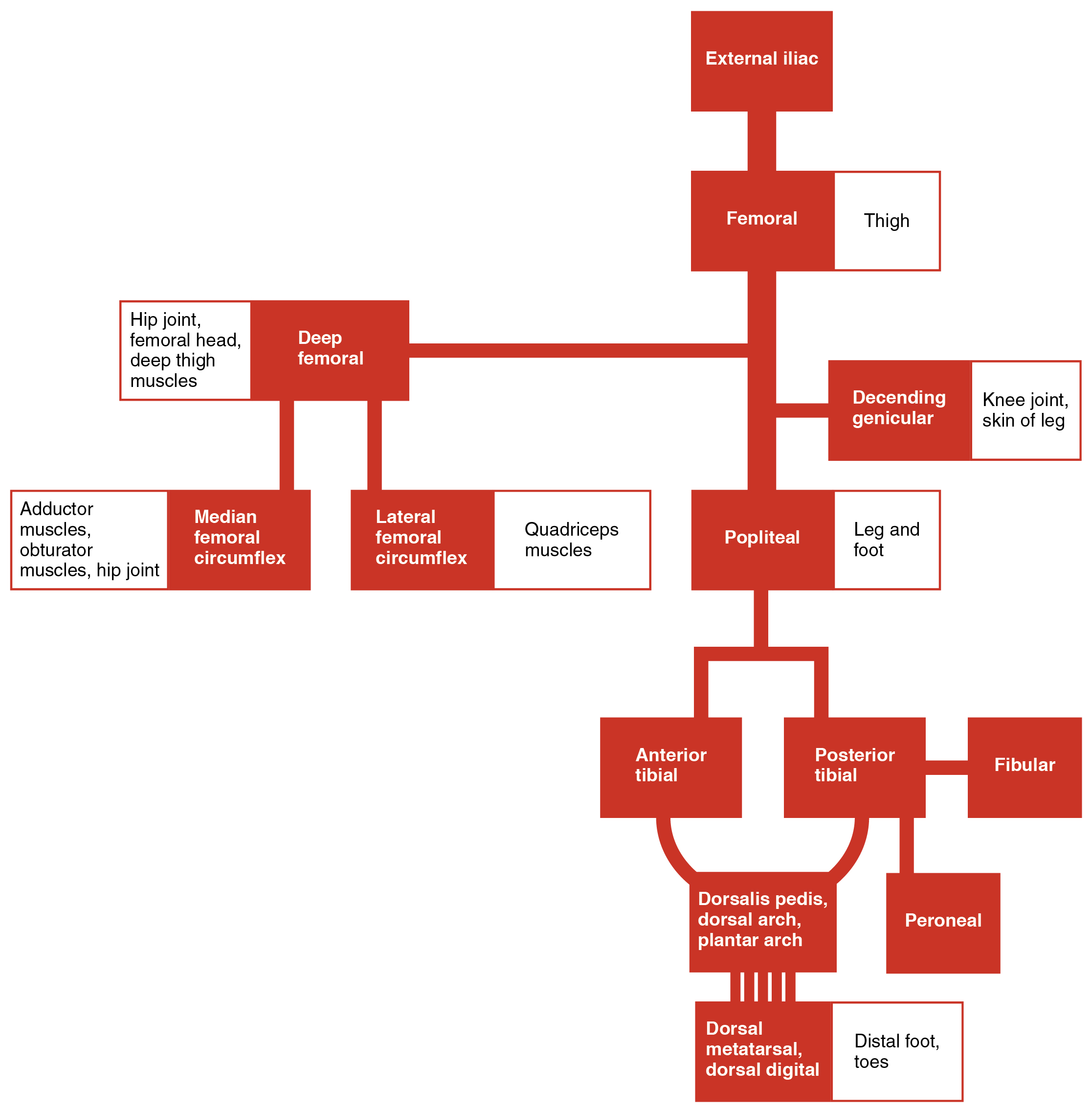 Image Source: Wikimedia Commons (2017). 2130 Lower Limb Arteries Chart, Licensed under a creative commons licence. Available for free at: https://upload.wikimedia.org/wikipedia/commons/0/0d/2130_Lower_Limb_Arteries_Chart.jpg (Accessed: 9 January 2024).
References
- Moore, K. L., Dalley, A. F., & Agur, A. M. R. (2014). Moore’s Clinically Orientated Anatomy (7th ed.). China: Lippincott.
- Kumar & M. Clark (2012). (Eds.), Kumar & Clark’s Clinical Medicine (12 ed.). Spain: Elsevier.
- Wineski, L. E., & Snell, R. S. (2019). Snell’s clinical anatomy by regions.
Venous anatomy
Upper limb
The venous drainage of the upper limbs can be divided into the superficial and deep systems. The basilic and cephalic veins will drain blood from the dorsal aspect of the hand and pass within the arm’s medial and anterolateral aspects. They are connected within the elbow by the median cubital vein. Both veins drain into the axillary vein.
The deep venous system is situated beside their accompanying arteries and is formed by the forearm’s radial and ulnar veins. These drain into the brachial vein and eventually into the axillary vein. Both axillary veins will drain into the subclavian and, eventually, the brachiocephalic vein.
Lower limb
The venous drainage of the foot starts with the dorsal venous arch dorsally, with the medial and lateral plantar veins located on the plantar aspect. The dorsal venous arch will travel deep into the leg to form the anterior tibial vein. The medial and lateral plantar veins will combine to form the posterior tibial and the fibular veins. All three veins will unite to form the popliteal vein.
As it travels up the leg through the adductor canal, this becomes the femoral vein. The thigh muscles will drain via the deep vein of the thigh, and it will empty into the femoral vein. As the femoral vein travels past the inguinal canal, it becomes the external iliac vein.
Superficial veins are also found in the lower limb and are made of great saphenous and small saphenous veins. The great saphenous vein originates from the dorsal venous arch of the foot and travels superiorly past the medial malleolus and medial condyle of the femur. This empties into the femoral vein inferior to the inguinal ligament. The small saphenous vein travels past the lateral malleolus along the two heads of the gastrocnemius muscle to empty into the popliteal vein via the popliteal fossa.

Image Source: Wikimedia Commons (2013). 2137 Lower Limb Veins Chart, Licensed under a creative commons licence. Available for free at: https://upload.wikimedia.org/wikipedia/commons/8/86/2137_Lower_Limb_Veins_Chart.jpg (Accessed: 9 January 2024).
The superior and inferior gluteal veins will drain gluteal structures and will drain into the internal iliac vein.
Head and neck
The Nervous System section covers the venous return from the cranium, and these vessels will drain into the internal jugular vein. The external face drains into the external jugular vein, and its tributaries are the posterior auricular and retromandibular veins.
The external jugular veins will empty into the subclavian vein. The internal jugular veins combine with the subclavian veins to form the brachiocephalic veins. The brachiocephalic veins will unite at the right side of the superior mediastinum to form the SVC, and this drains directly into the right atrium.
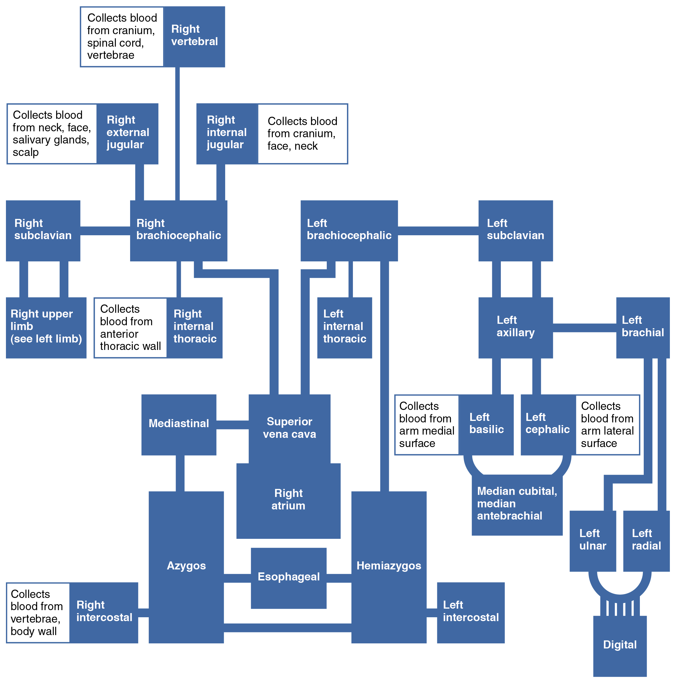 Image Source: Wikimedia Commons (2013). 2135 Veins Draining into Superior Vena Cava Chart, Licensed under a creative commons licence. Available for free at: https://upload.wikimedia.org/wikipedia/commons/d/df/2135_Veins_Draining_into_Superior_Vena_Cava_Chart.jpg (Accessed: 9 January 2024).
Thoracic, abdominal and pelvic cavity
Within the thoracic cavity, the main veins that pass through are the inferior vena cava and the azygos venous system. The azygous venous system consists of the azygous vein, hemiazygos vein and accessory hemiazygos vein. The azygos vein can arise from the lumbar azygos vein or directly from the inferior vena cava at the renal vein level. It courses within the posterior mediastinum and arches over the right pulmonary hilum before draining into the SVC. Its major tributaries are the hemiazygos and accessory hemiazygos vein. The hemiazygos vein drains from the lumbar ascending and subcostal veins from the 9-11 ribs. This will drain back to the azygos vein at the level of T8. The accessory hemiazygos vein will drain blood from the 5th-8th ribs and the left bronchial vein. It empties into the azygos vein at T7.
The pelvic venous drainage occurs through the internal iliac vein. It receives the venous return from the majority of the pelvic organs. It will merge with the external iliac vein at the level of the pubic symphysis to form the common iliac vein.
The systemic venous drainage from the abdomen occurs through the IVC. The IVC is formed from the union of the common iliac veins at the level of L5. This travels superiorly and will drain into the right atrium. It receives blood from the common iliac, lumbar, renal, right gonadal, right suprarenal, and hepatic veins.
 Image Source: Wikimedia Commons (2013). 2132 Thoracic Abdominal Veins, Licensed under a creative commons licence. Available for free at: https://upload.wikimedia.org/wikipedia/commons/2/28/2132_Thoracic_Abdominal_Veins.jpg (Accessed: 9 January 2024).
Venous drainage of the GI tract, spleen, pancreas and gallbladder is carried out via the portal venous system. The union of the splenic vein and superior mesenteric vein forms the portal vein. The portal vein divides into the right and left portal vein as it enters the liver, delivering nutrient-rich venous blood into the liver.
 Image Source: Wikimedia Commons (2013). 2138 Hepatic Portal Vein System, Licensed under a creative commons licence. Available for free at: https://upload.wikimedia.org/wikipedia/commons/c/cf/2138_Hepatic_Portal_Vein_System.jpg (Accessed: 9 January 2024).
References
- Moore, K. L., Dalley, A. F., & Agur, A. M. R. (2014). Moore’s Clinically Orientated Anatomy (7th ed.). China: Lippincott.
- Kumar & M. Clark (2012). (Eds.), Kumar & Clark’s Clinical Medicine (12 ed.). Spain: Elsevier.
- Wineski, L. E., & Snell, R. S. (2019). Snell’s clinical anatomy by regions.
Relevant upper and lower limb anatomy
While an in-depth knowledge of muscular anatomy is optional in the MRCP syllabus, some major muscle groups in the upper and lower limbs are worth noting.
Upper limb
The pectoral region of the upper limb consists of the pectoralis major, pectoralis minor, subclavius and the serratus anterior. The serratus anterior dysfunction contributes to scapular winging, which occurs with damage to the long thoracic nerve.
The extrinsic shoulder muscles include the trapezius, latissimus dorsi, rhomboid major, rhomboid minor and the levator scapulae. The trapezius is useful to test the function of CN XI. These muscles help with the scapula’s movement and allow movement between the upper limb and back.
 Image Source: Wikimedia Commons (2016). 118 Muscles that Position the Pectoral Girdle, Licensed under a creative commons licence. Available for free at: https://upload.wikimedia.org/wikipedia/commons/9/91/1118_Muscles_that_Position_the_Pectoral_Girdle.jpg (Accessed: 9 January 2024).
The intrinsic muscles of the shoulder include the deltoids, teres major and the rotator cuff muscles. These muscles are helpful in the movement of the shoulder joint. Of note, abduction of the shoulder is performed predominantly by the supraspinatus (up to the initial 15°) and the middle deltoids (beyond the initial 15°). The subscapularis achieves internal rotation of the shoulder, while external rotation is achieved by the teres minor. Rotator cuff tendonitis, especially the supraspinatus muscle, is relatively common with repetitive motion of the shoulder joint.
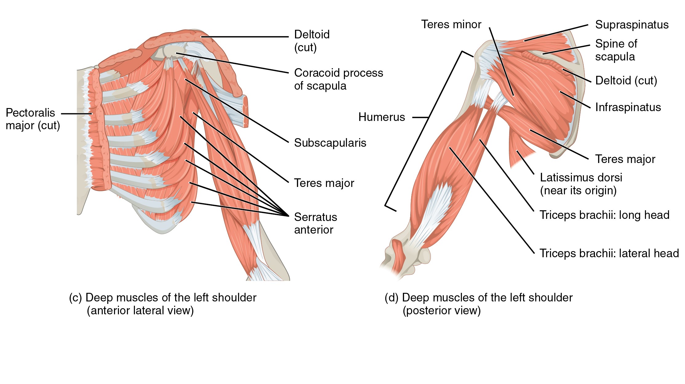
Image Source: Wikimedia Commons (2016). 1119 Muscles that Move the Humerus, Licensed under a creative commons licence. Available for free at: https://upload.wikimedia.org/wikipedia/commons/7/7e/1119_Muscles_that_Move_the_Humerus.jpg (Accessed: 9 January 2024).
Within the arm, the anterior compartment contains the biceps brachii, coracobrachialis and the brachialis. The posterior compartment contains the triceps brachii.
The forearm contains multiple muscles responsible for the movement and manipulation of the hand and digits. These can be broadly divided into the flexor muscles that are found within the anterior compartment and the extensor muscles within the posterior compartment. The muscles within the anterior compartment share a common tendon attaching to the medial epicondyle of the humerus and are predominantly innervated by the median nerve. These muscles help with pronation of the forearm along with flexion of the wrist and digits.
The muscles of the posterior compartment are generally extensors and will act to help extend the wrist and fingers. They are predominantly innervated by the radial nerve and mostly share a common tendon that attaches to the lateral epicondyle of the humerus. The exception is the brachioradialis, which acts as a flexor for the elbow despite its position within the posterior compartment.
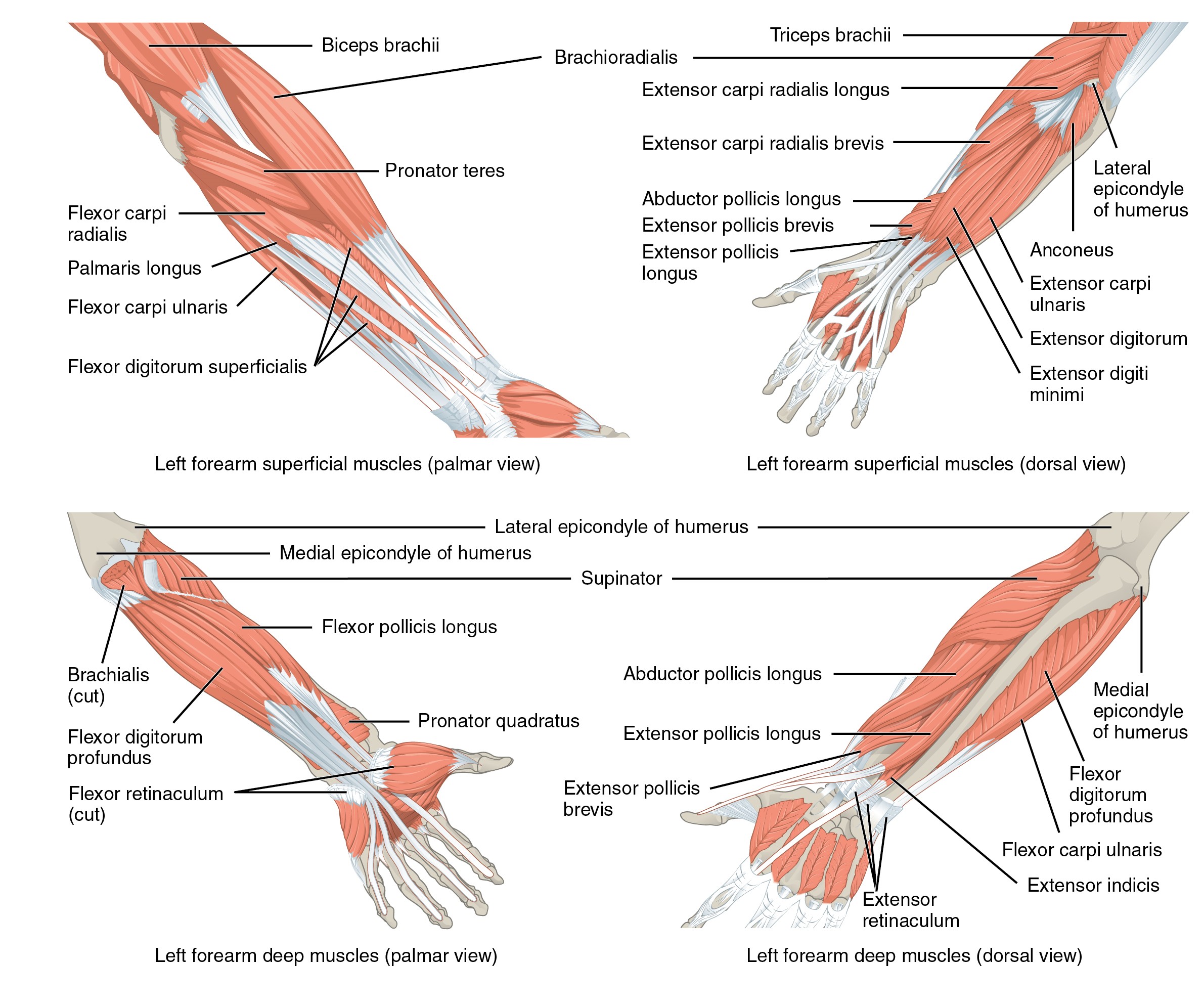
Image Source: Wikimedia Commons (2015). 1120 Muscles that Move the Forearm, Licensed under a creative commons licence. Available for free at: https://upload.wikimedia.org/wikipedia/commons/7/73/1120_Muscles_that_Move_the_Forearm.jpg (Accessed: 9 January 2024).
Within the hand, intrinsic muscles allow fine movements of the hand. The thenar eminence contains the opponens pollicis, abductor pollicis brevis and flexor pollicis brevis. These aid with opposition, abduction and flexion of the thumb, respectively. The median nerve supplies these. The ulnar nerve innervates the hypothenar eminence, which helps with the little finger’s opposition, abduction and flexion. Lastly, the lumbricals and interossei muscles are also important muscles within the hand. The lumbricals are innervated by both the median and ulnar nerve and allow flexion at the MCP joint while allowing extension of the IP joint. The dorsal interossei muscles aid in the abduction of the fingers and are innervated by the ulnar nerve. The palmar interossei muscles aid in the adduction of the fingers.
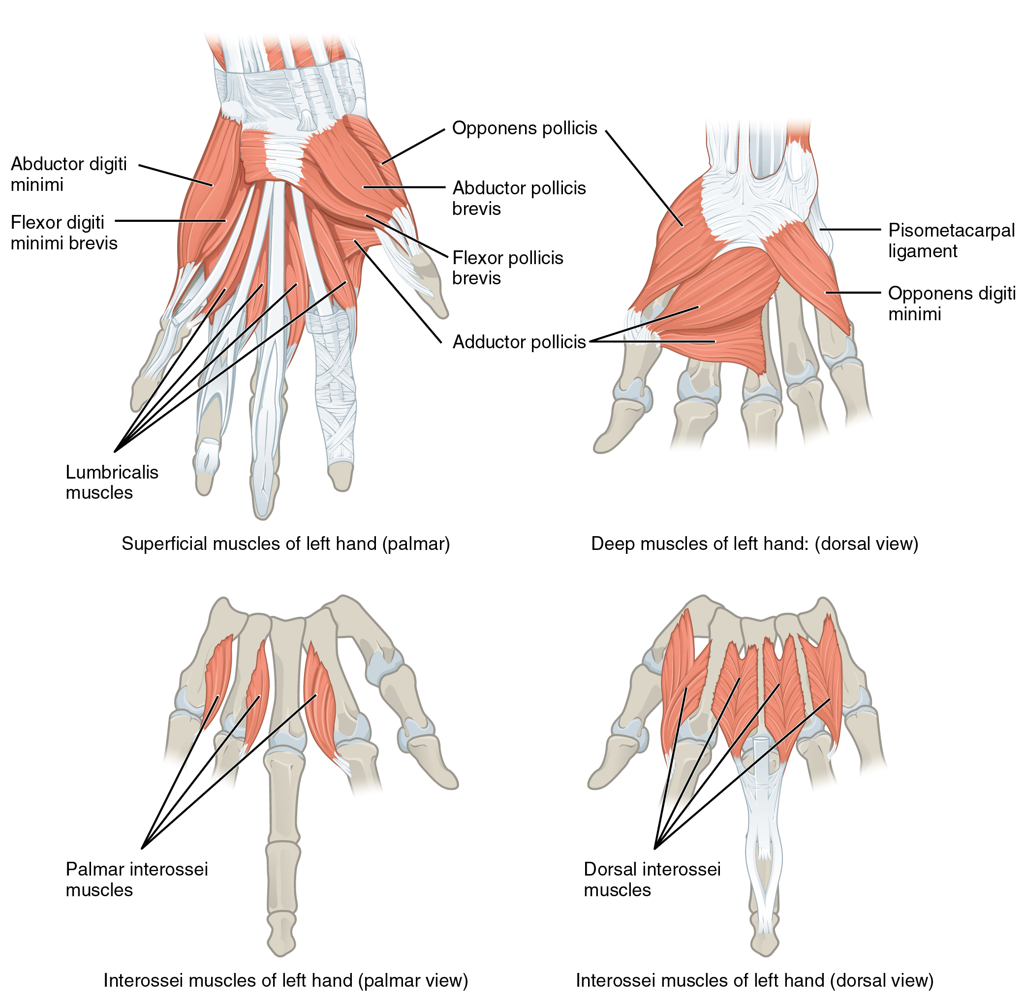
Image Source: Wikimedia Commons (2016). 1121 Intrinsic Muscles of the Hand, Licensed under a creative commons licence. Available for free at: https://upload.wikimedia.org/wikipedia/commons/0/0e/1121_Intrinsic_Muscles_of_the_Hand.jpg (Accessed: 9 January 2024).
Lower limb
The gluteal region contains the superficial and deep muscles. The superficial muscles consist of the gluteal muscles and the tensor fascia lata. The gluteal muscles aid in the extension and abduction of the femur. The tensor fascia lata acts to flex, abduct and internally rotate the hip. The iliotibial band is a continuation of the fascia lata and airs to extend, abduct and laterally rotate the hip. Deep muscles within the gluteal region are located underneath the gluteus minimus and generally act to rotate the hip laterally and provide stability to the hip joint.
The thigh can be divided into three anatomical compartments. The femoral nerve innervates the anterior compartment and grossly acts to extend the leg at the level of the knee joint. The medial compartment acts as the hip adductor and is innervated by the obturator nerve. The posterior compartment acts as the knee flexors and hip extensors. The sciatic nerve innervates these muscles.
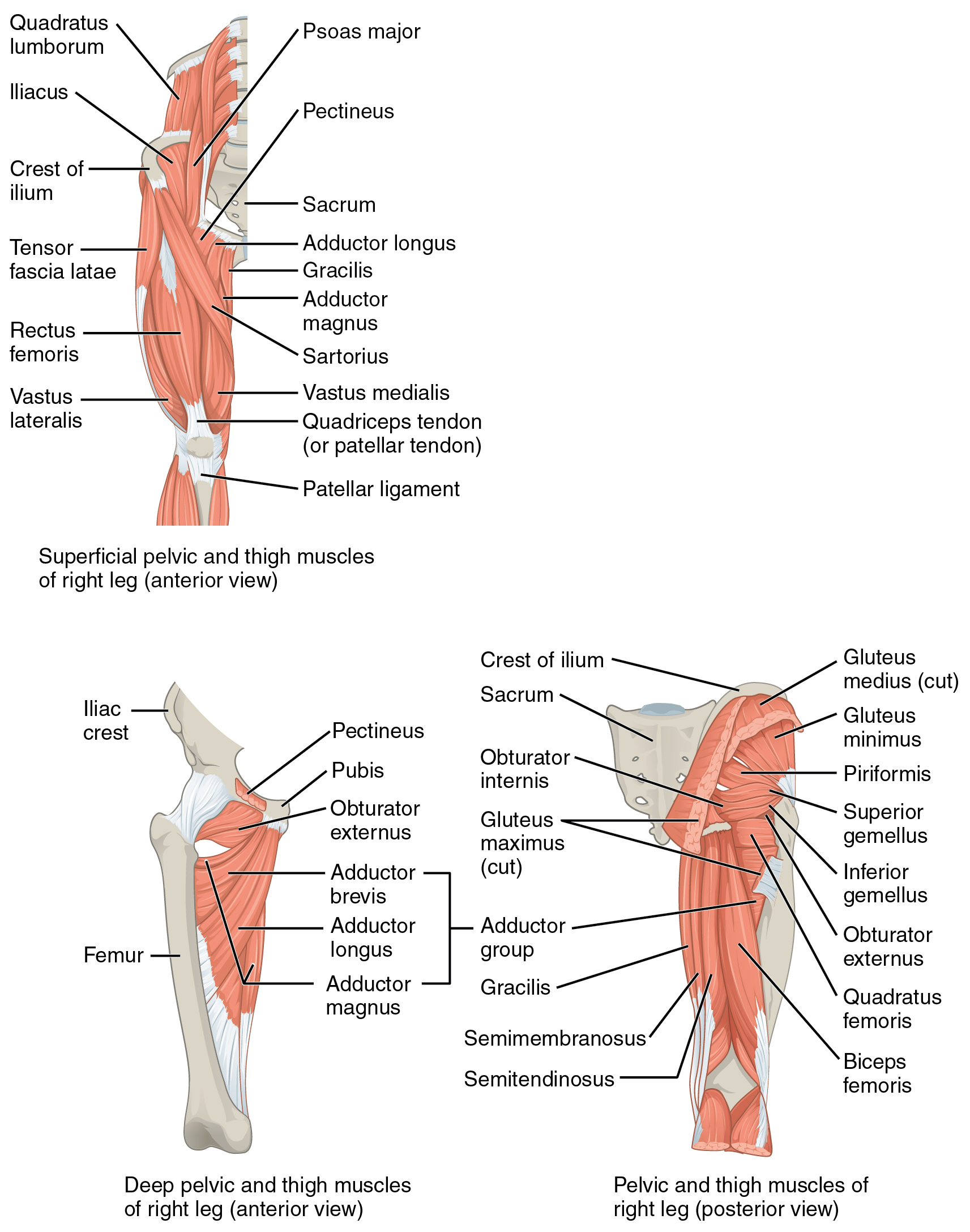 Image Source: Wikimedia Commons (2016). 122 Gluteal Muscles that Move the Femur, Licensed under a creative commons licence. Available for free at: https://upload.wikimedia.org/wikipedia/commons/8/8f/1122_Gluteal_Muscles_that_Move_the_Femur.jpg (Accessed: 9 January 2024).
The leg is also divided into three compartments. The anterior compartment of the leg helps to dorsiflex and invert the foot and is typically supplied by the deep fibular nerve. The lateral compartment of the leg helps evert the foot and is innervated by the superficial fibular nerve. Lastly, the posterior compartment aids in both plantarflexion and inversion of the foot. The foot’s musculature predominantly controls the fine movement of the great toe and digits. The medial and lateral plantar nerves innervate these.

Image Source: Wikimedia Commons (2018). 1123 Muscles of the Leg that Move the Foot and Toes, Licensed under a creative commons licence. Available for free at: https://upload.wikimedia.org/wikipedia/commons/4/47/1123_Muscles_of_the_Leg_that_Move_the_Foot_and_Toes.jpg (Accessed: 9 January 2024).
References
- Moore, K. L., Dalley, A. F., & Agur, A. M. R. (2014). Moore’s Clinically Orientated Anatomy (7th ed.). China: Lippincott.
- Kumar & M. Clark (2012). (Eds.), Kumar & Clark’s Clinical Medicine (12 ed.). Spain: Elsevier.
- Wineski, L. E., & Snell, R. S. (2019). Snell’s clinical anatomy by regions.
Complete Guide to Clinical Genetics ST3 Application and Recruitment (2027)
Written by Medibuddy. Last updated: October 2026. Clinical Genetics is

Complete Guide to Clinical Radiology ST3 Application and Recruitment (2027)
Recruitment paused: NHS England is not running Clinical Radiology ST3+ recru

Complete Guide to Paediatric Surgery ST3 Application and Recruitment in United Kingdom (2026)
Written by Medibuddy. Last updated: October 2026. If you’re planning


Take your subscriptions with you
Our mobile app allows you to access your interview and exam question banks wherever you are.

