


 Close
Close
 20th February 2024
Relevant upper and lower limb anatomy
While an in-depth knowledge of muscular anatomy is optional in the MRCP syllabus, some major muscle groups in the upper and lower limbs are worth noting.
Upper limb
The pectoral region of the upper limb consists of the pectoralis major, pectoralis minor, subclavius and the serratus anterior. The serratus anterior dysfunction contributes to scapular winging, which occurs with damage to the long thoracic nerve.
The extrinsic shoulder muscles include the trapezius, latissimus dorsi, rhomboid major, rhomboid minor and the levator scapulae. The trapezius is useful to test the function of CN XI. These muscles help with the scapula’s movement and allow movement between the upper limb and back.
 Image Source: Wikimedia Commons (2016). 118 Muscles that Position the Pectoral Girdle, Licensed under a creative commons licence. Available for free at: https://upload.wikimedia.org/wikipedia/commons/9/91/1118_Muscles_that_Position_the_Pectoral_Girdle.jpg (Accessed: 9 January 2024).
The intrinsic muscles of the shoulder include the deltoids, teres major and the rotator cuff muscles. These muscles are helpful in the movement of the shoulder joint. Of note, abduction of the shoulder is performed predominantly by the supraspinatus (up to the initial 15°) and the middle deltoids (beyond the initial 15°). The subscapularis achieves internal rotation of the shoulder, while external rotation is achieved by the teres minor. Rotator cuff tendonitis, especially the supraspinatus muscle, is relatively common with repetitive motion of the shoulder joint.
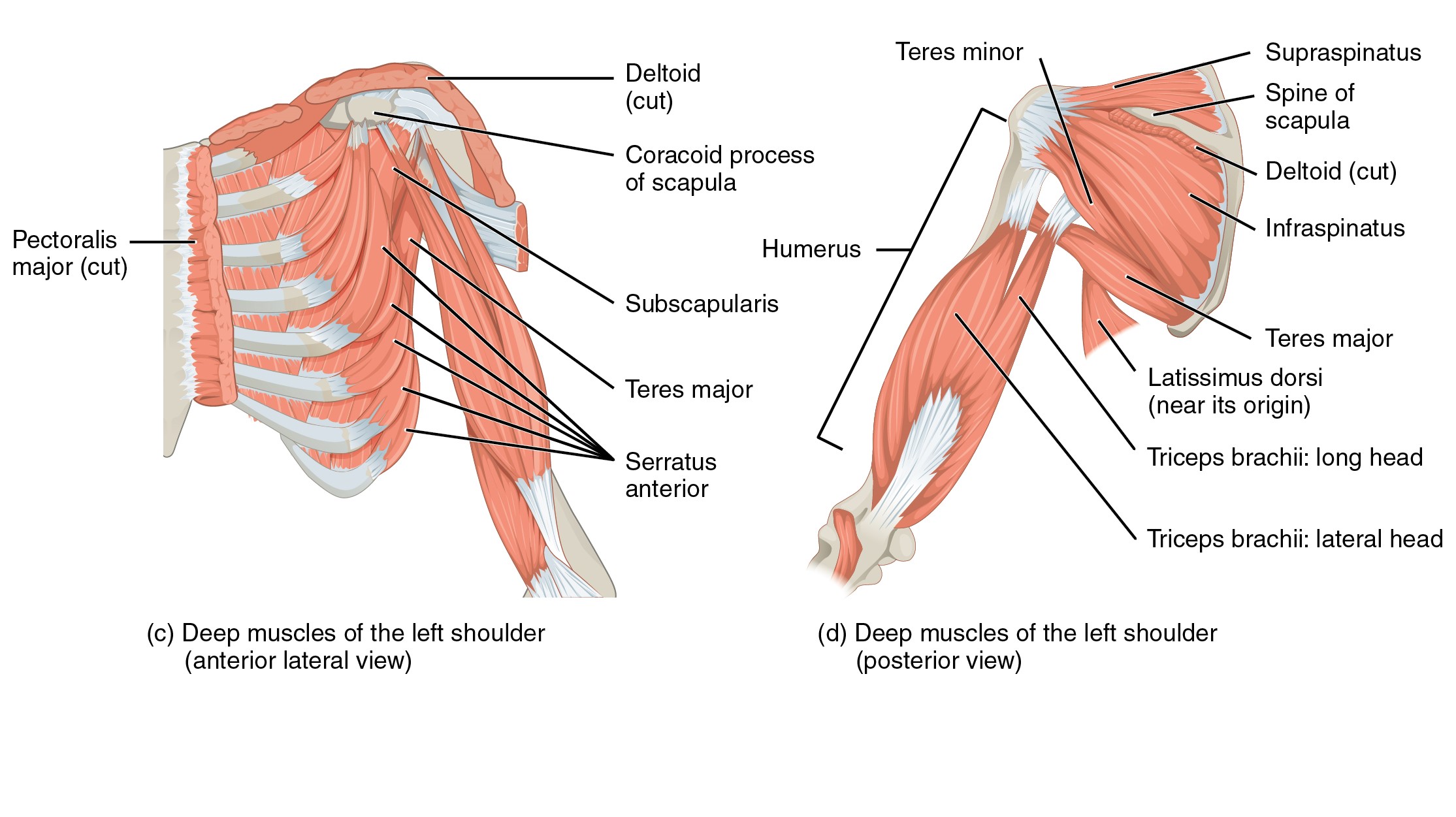 Image Source: Wikimedia Commons (2016). 1119 Muscles that Move the Humerus, Licensed under a creative commons licence. Available for free at: https://upload.wikimedia.org/wikipedia/commons/7/7e/1119_Muscles_that_Move_the_Humerus.jpg (Accessed: 9 January 2024).
Within the arm, the anterior compartment contains the biceps brachii, coracobrachialis and the brachialis. The posterior compartment contains the triceps brachii.
The forearm contains multiple muscles responsible for the movement and manipulation of the hand and digits. These can be broadly divided into the flexor muscles that are found within the anterior compartment and the extensor muscles within the posterior compartment. The muscles within the anterior compartment share a common tendon attaching to the medial epicondyle of the humerus and are predominantly innervated by the median nerve. These muscles help with pronation of the forearm along with flexion of the wrist and digits.
The muscles of the posterior compartment are generally extensors and will act to help extend the wrist and fingers. They are predominantly innervated by the radial nerve and mostly share a common tendon that attaches to the lateral epicondyle of the humerus. The exception is the brachioradialis, which acts as a flexor for the elbow despite its position within the posterior compartment.
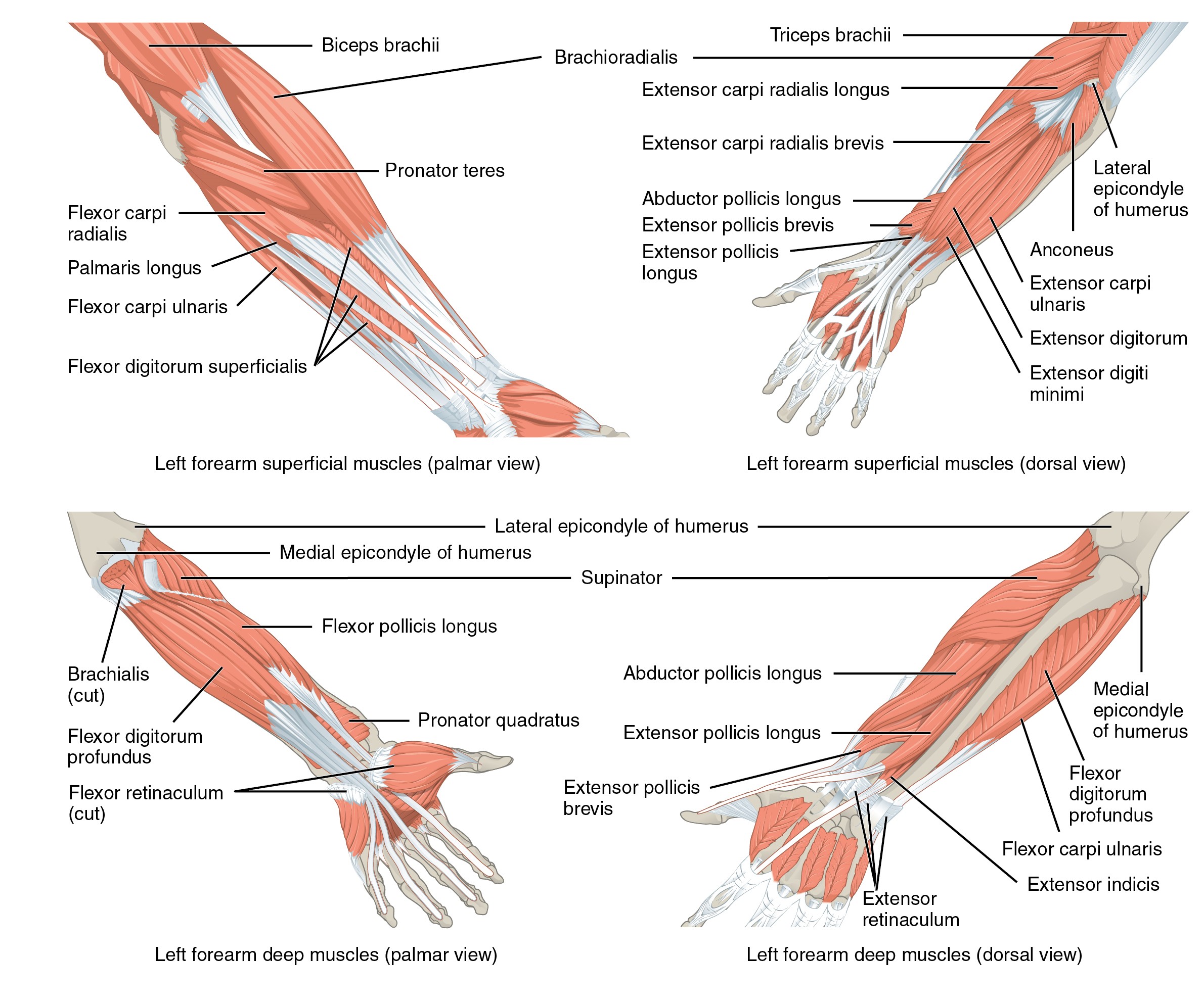
Image Source: Wikimedia Commons (2015). 1120 Muscles that Move the Forearm, Licensed under a creative commons licence. Available for free at: https://upload.wikimedia.org/wikipedia/commons/7/73/1120_Muscles_that_Move_the_Forearm.jpg (Accessed: 9 January 2024).
Within the hand, intrinsic muscles allow fine movements of the hand. The thenar eminence contains the opponens pollicis, abductor pollicis brevis and flexor pollicis brevis. These aid with opposition, abduction and flexion of the thumb, respectively. The median nerve supplies these. The ulnar nerve innervates the hypothenar eminence, which helps with the little finger’s opposition, abduction and flexion. Lastly, the lumbricals and interossei muscles are also important muscles within the hand. The lumbricals are innervated by both the median and ulnar nerve and allow flexion at the MCP joint while allowing extension of the IP joint. The dorsal interossei muscles aid in the abduction of the fingers and are innervated by the ulnar nerve. The palmar interossei muscles aid in the adduction of the fingers.
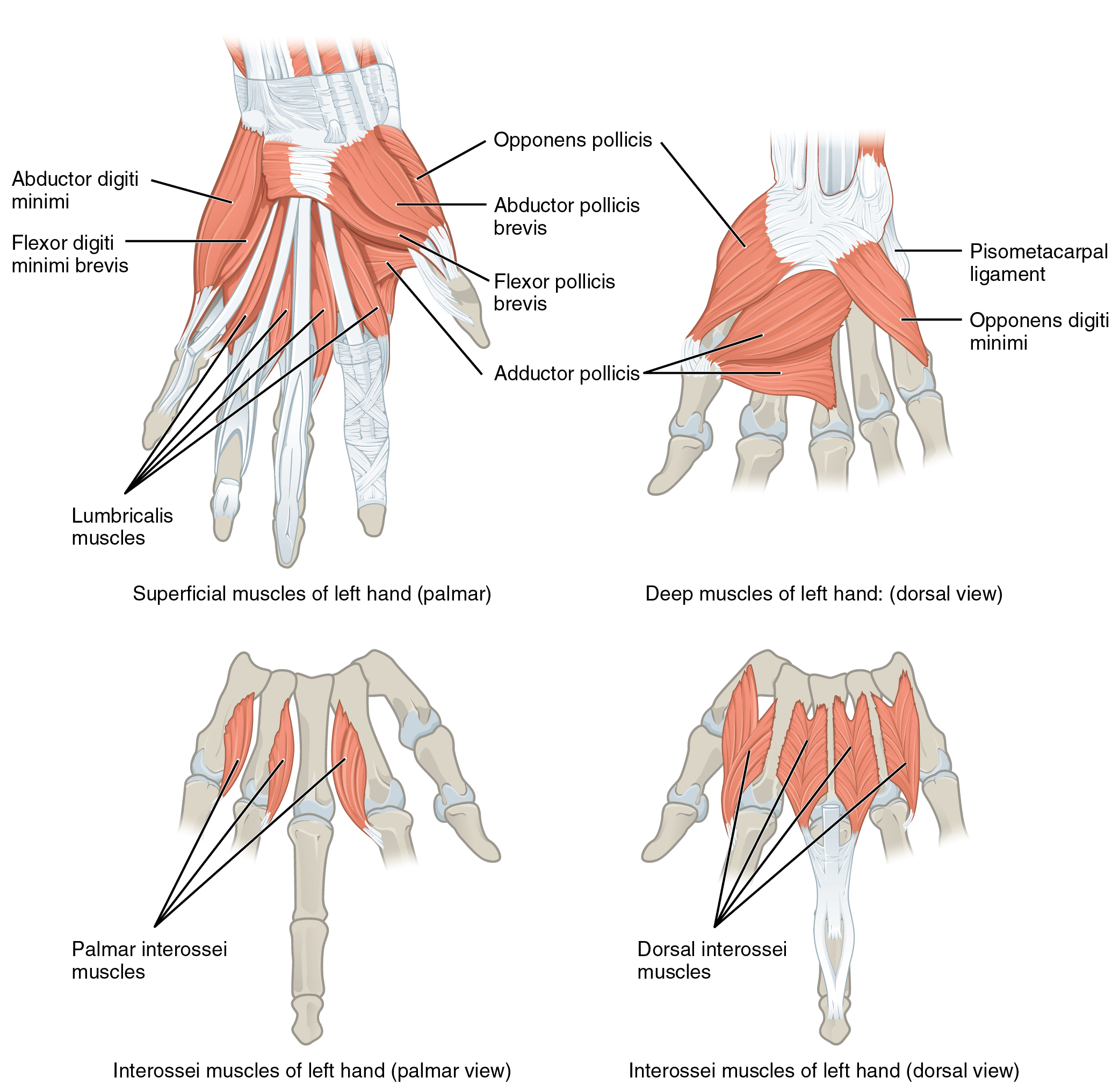 Image Source: Wikimedia Commons (2016). 1121 Intrinsic Muscles of the Hand, Licensed under a creative commons licence. Available for free at: https://upload.wikimedia.org/wikipedia/commons/0/0e/1121_Intrinsic_Muscles_of_the_Hand.jpg (Accessed: 9 January 2024).
Lower limb
The gluteal region contains the superficial and deep muscles. The superficial muscles consist of the gluteal muscles and the tensor fascia lata. The gluteal muscles aid in the extension and abduction of the femur. The tensor fascia lata acts to flex, abduct and internally rotate the hip. The iliotibial band is a continuation of the fascia lata and airs to extend, abduct and laterally rotate the hip. Deep muscles within the gluteal region are located underneath the gluteus minimus and generally act to rotate the hip laterally and provide stability to the hip joint.
The thigh can be divided into three anatomical compartments. The femoral nerve innervates the anterior compartment and grossly acts to extend the leg at the level of the knee joint. The medial compartment acts as the hip adductor and is innervated by the obturator nerve. The posterior compartment acts as the knee flexors and hip extensors. The sciatic nerve innervates these muscles.
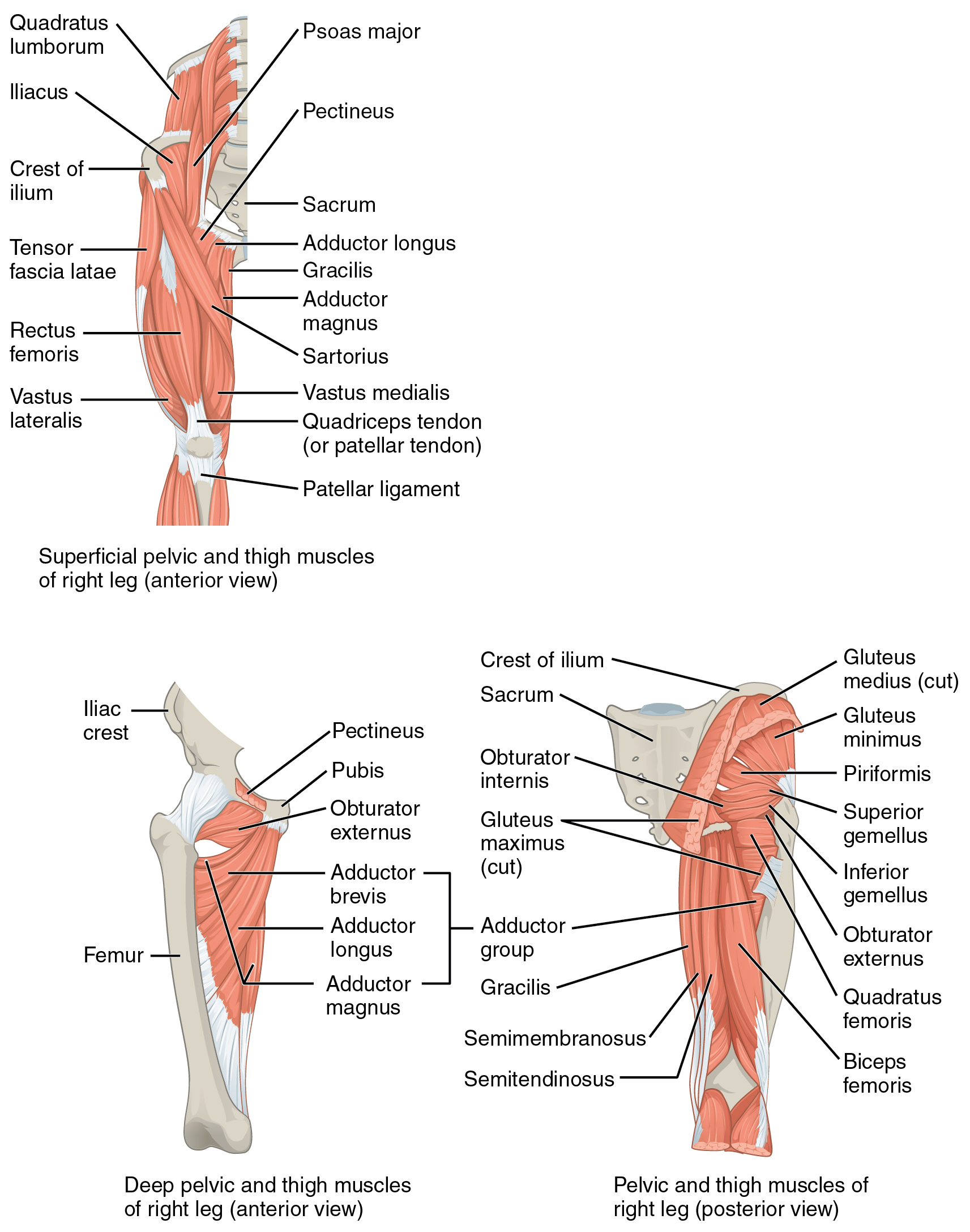 Image Source: Wikimedia Commons (2016). 122 Gluteal Muscles that Move the Femur, Licensed under a creative commons licence. Available for free at: https://upload.wikimedia.org/wikipedia/commons/8/8f/1122_Gluteal_Muscles_that_Move_the_Femur.jpg (Accessed: 9 January 2024).
The leg is also divided into three compartments. The anterior compartment of the leg helps to dorsiflex and invert the foot and is typically supplied by the deep fibular nerve. The lateral compartment of the leg helps evert the foot and is innervated by the superficial fibular nerve. Lastly, the posterior compartment aids in both plantarflexion and inversion of the foot. The foot’s musculature predominantly controls the fine movement of the great toe and digits. The medial and lateral plantar nerves innervate these.
 Image Source: Wikimedia Commons (2018). 1123 Muscles of the Leg that Move the Foot and Toes, Licensed under a creative commons licence. Available for free at: https://upload.wikimedia.org/wikipedia/commons/4/47/1123_Muscles_of_the_Leg_that_Move_the_Foot_and_Toes.jpg (Accessed: 9 January 2024).
References
- Moore, K. L., Dalley, A. F., & Agur, A. M. R. (2014). Moore’s Clinically Orientated Anatomy (7th ed.). China: Lippincott.
- Kumar & M. Clark (2012). (Eds.), Kumar & Clark’s Clinical Medicine (12 ed.). Spain: Elsevier.
- Wineski, L. E., & Snell, R. S. (2019). Snell’s clinical anatomy by regions.
Complete Guide to Clinical Genetics ST3 Application and Recruitment (2027)
Written by Medibuddy. Last updated: October 2026. Clinical Genetics is

Complete Guide to Clinical Radiology ST3 Application and Recruitment (2027)
Recruitment paused: NHS England is not running Clinical Radiology ST3+ recru

Complete Guide to Paediatric Surgery ST3 Application and Recruitment in United Kingdom (2026)
Written by Medibuddy. Last updated: October 2026. If you’re planning


Take your subscriptions with you
Our mobile app allows you to access your interview and exam question banks wherever you are.

