


 Close
Close
 21st February 2024
Methods of Genetic Inheritance
Basics of inheritance and the pedigree
Inheritance
Our fundamental understanding of inheritance stems from experiments performed by Gregor Mendel on peas. Several essential concepts arose from these experiments:
- Dominant and recessive genes – Dominant gene variants (alleles) tend to impact the phenotype when present (Homozygous dominant or heterozygote); Recessive alleles affect the phenotype when the genotype is homozygous recessive.
- Segregation – Each gamete only obtains one allele of a gene during meiosis.
- Independent assortment – Genes from different loci are transmitted independently from each other.
These principles of inheritance can be visualised using a Punnett square. When drawing a Punnett square, capital letters are used to denote the dominant allele, while lowercase letters are used to represent the recessive allele.
 Image Source: Wikimedia Commons (2013). Punnett hetero homoblue, Licensed under a creative commons licence. Available for free at: https://commons.wikimedia.org/wiki/File:Punnett_hetero_homoblue.svg (Accessed: 5 January 2024).
Pedigree
A thorough history, including family history, will provide vital information about the inheritance pattern and valuable clues to the underlying genetic condition. Geneticists will use a pedigree to visualise the information gathered from the family history. The pedigree will highlight the relationship between the index patient and all other family members. While candidates will not be expected to draw a pedigree during the MRCP exam, it is a helpful tool to visualise the family history as it can reveal valuable clues to the pattern of inheritance of the conditions.
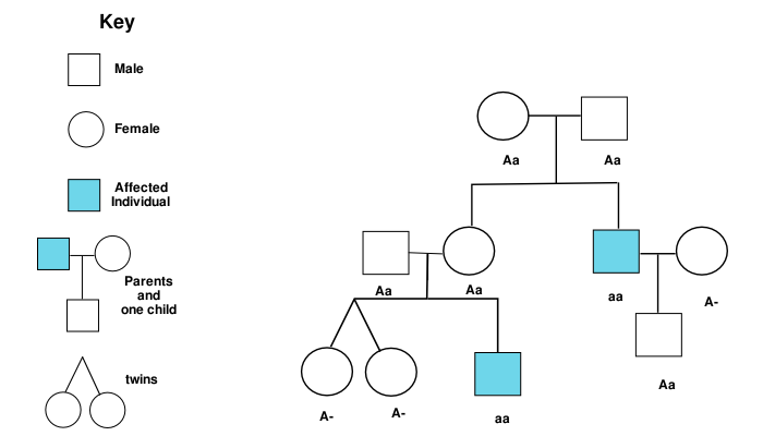
Image Source: Wikimedia Commons (2006). Pedigree chart B, Licensed under a creative commons licence. Available for free at: https://commons.wikimedia.org/wiki/File:PedigreechartB.png (Accessed: 5 January 2024).
References
- Read, A., & Donnai, D. (2021). Chapter 1: What can we learn from family history? In New Clinical Genetics: A guide to genomic medicine (4th ed.). United Kingdom: Scoin Publishing Limited.
- Turnpenny, P. D., & Ellard, S. (2017). Patterns of inheritance. In Emery’s Elements of Medical Genetics. China: Elsevier.
Mendelian inheritance
Complications in inheritance
Mendelian inheritance refers to the inheritance of genetic disorders predominantly through the inheritance of a single gene. This can be through the autosomal or sexual chromosomes, leading to the four main classes of inheritance: autosomal dominant, autosomal recessive, X-linked dominant and X-linked recessive.
Genetic inheritance can be complicated by several factors, making the pattern of inheritance less clear:
- Pleiotropy – A single gene mutation results in phenotypes affecting multiple organ systems. This can result in one gene mutation presenting as seemingly unrelated symptoms.
- Penetrance – The degree to which a genotype is expressed towards an individual’s phenotype, resulting in some carriers not displaying the mutation’s phenotype.
- Variable expressivity – The degree to which a phenotype is expressed for a given genotype, resulting in different severity of symptoms between patients with a similar genotype.
Autosomal dominant
Autosomal dominant traits can present themselves in individuals with the heterozygous genotype. A vertical pedigree will be observed, with most generations having at least one affected individual. Each affected individual will have one parent affected by the same condition. Male-to-male transmission is possible with equal incidence between males and females within the family tree. Each child will have a 1 in 2 chance of inheriting the mutation if one partner is unaffected by the condition.
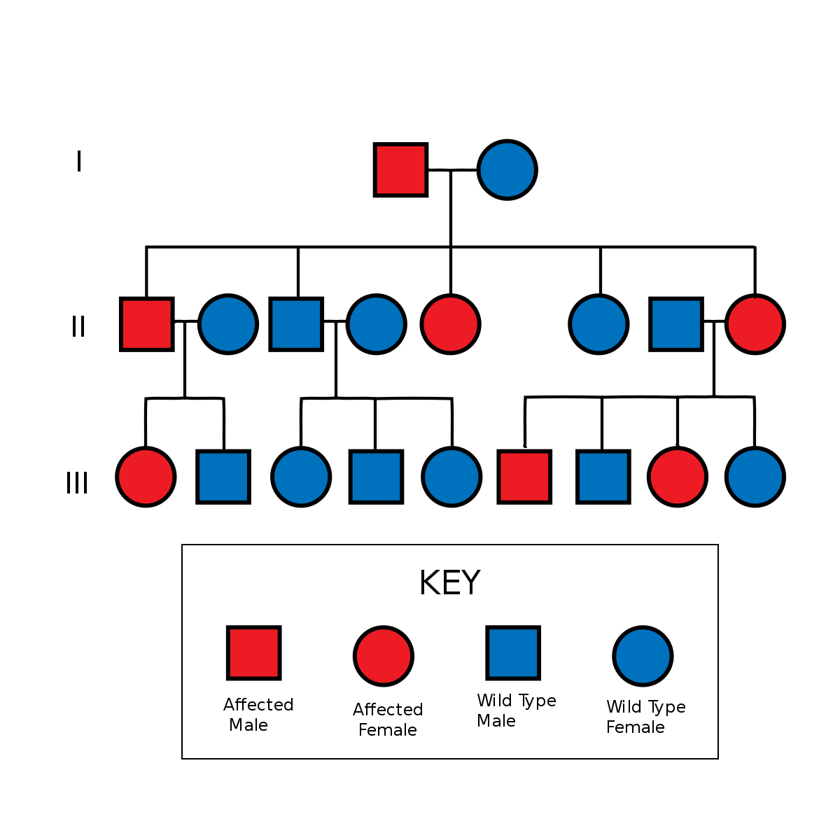 Image Source: Wikimedia Commons (2006). Autosomal Dominant Pedigree Chart, Licensed under a creative commons licence. Available for free at: https://commons.wikimedia.org/wiki/File:Autosomal_Dominant_Pedigree_Chart.svg (Accessed: 5 January 2024).
A homozygous autosomal dominant genotype is possible, and this typically presents with a more severe phenotype than the heterozygous genotype.
Autosomal recessive
Recessive traits only present themselves in the homozygous genotype. Heterozygote genotypes will typically not display the phenotype of the condition and will serve as carriers for the condition. In a typical pedigree, the parents of the affected patient will not show the phenotype of the condition. In addition, there will be equal incidence between male and female patients. The condition will typically present within a single-family unit without affecting other family units within the family tree. Heterozygote parents will have a 1 in 4 chance of having a child affected by the condition. A recessive condition can be amplified within a consanguineous relationship due to the higher chances of the recessive alleles pairing up.
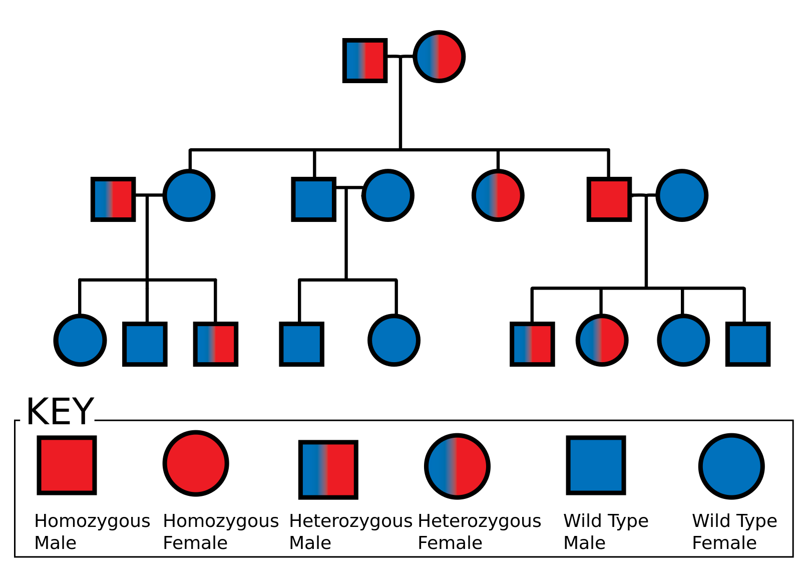
Image Source: Wikimedia Commons (2006). Autosomal Recessive Pedigree Chart, Licensed under a creative commons licence. Available for free at: https://commons.wikimedia.org/wiki/File:Autosomal_Recessive_Pedigree_Chart.svg (Accessed: 5 January 2024).
X-linked dominant
The sex chromosomes ( X and Y chromosomes) are the primary chromosomes that determine the sex of the developing embryo. Males only possess one copy of the X chromosome, while female somatic cells undergo the process of X-inactivation, creating mosaicism for the X-chromosome activity in females.
The X-linked dominant condition presents similarly to an autosomal dominant pedigree due to the heterozygote genotype being sufficient to produce the phenotype. Due to the impact of mosaicism, females will typically present with a milder phenotype. Males usually have a more severe phenotype with only one X chromosome copy, and some conditions may result in a lethal phenotype.
Affected males will not pass on their genotype to their male children, while all female children will be affected. Affected females will have a 50% chance of passing the condition to both male and female children.

Image Source: Wikimedia Commons (2019). Drawing – X-Linked Dominant (1), Licensed under a creative commons licence. Available for free at: https://commons.wikimedia.org/wiki/File:Wiki_Drawing_-_X- Linked_Dominant_(1).svg (Accessed: 5 January 2024).
X-linked recessive
X-linked recessive conditions are more commonly seen in males, with females often acting as carriers for the gene mutations. Males cannot pass on the mutation to their male children, but females will always act as carriers. A pedigree can show affected males in the generation above the index case. Female carriers will have a 50% chance of bearing a male affected by the condition or a female acting as a carrier for the condition.
Females can occasionally display the diseases’ phenotype through skewed X-inactivation ( X-inactivation favouring the expression of the mutant allele) or if they are homozygous for the mutant allele. Variable expressivity secondary to mosaicism, variable penetrance, and de novo mutations within X-linked mutations can result in an atypical pedigree, preventing the visualisation of a clear inheritance pattern.
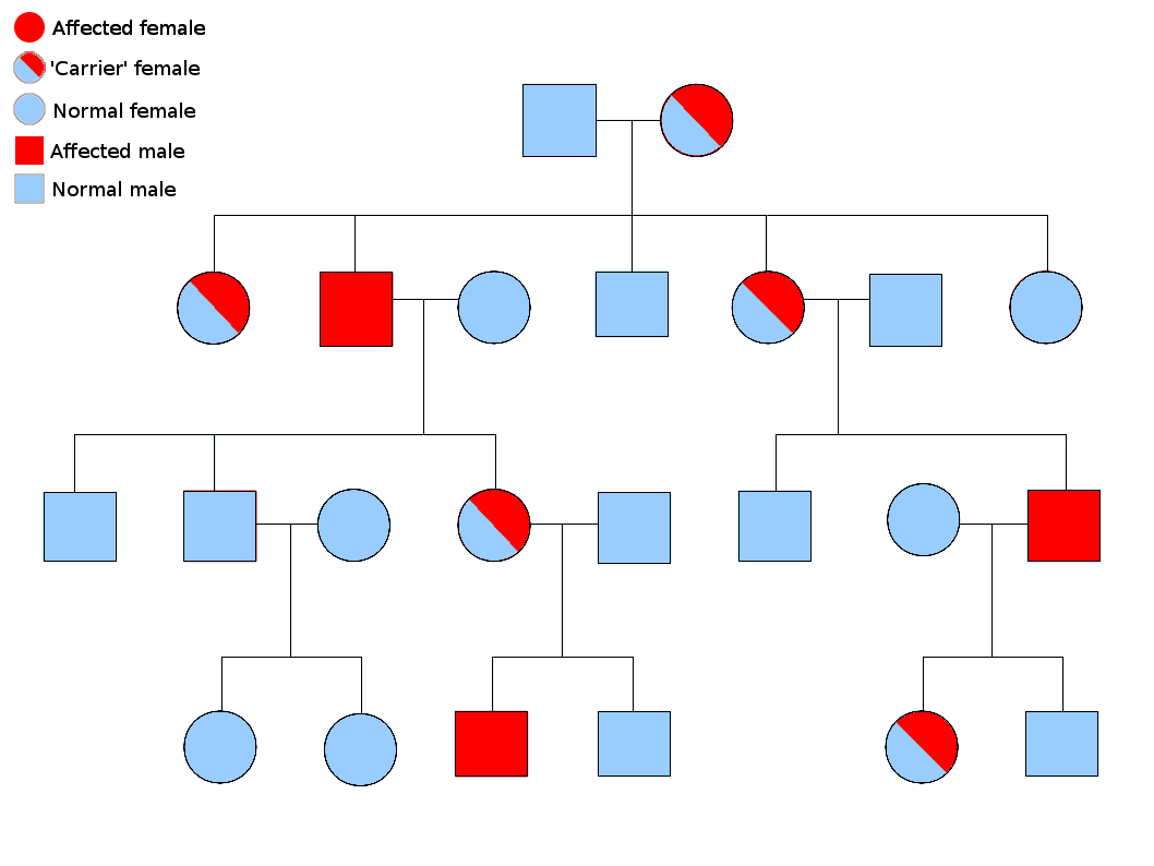 Image Source: Wikimedia Commons (2013). Sex linked inheritance, Licensed under a creative commons licence. Available for free at: https://commons.wikimedia.org/wiki/File:Sex_linked_inheritance.png (Accessed: 5 January 2024).
Mitochondrial
Mitochondrial disorders are unique due to mitochondrial DNA mutations acting as the driver for the phenotype. Mitochondrial DNA displays high mutation rates, and mutant mitochondria populations can vary between cells and tissues, resulting in significant variability in phenotypic expression between affected individuals.
Mitochondrial conditions typically show an exclusive maternal inheritance because embryonic mitochondria are derived from the ovum. Male and female children are equally affected. An affected male will not pass the condition to any children.
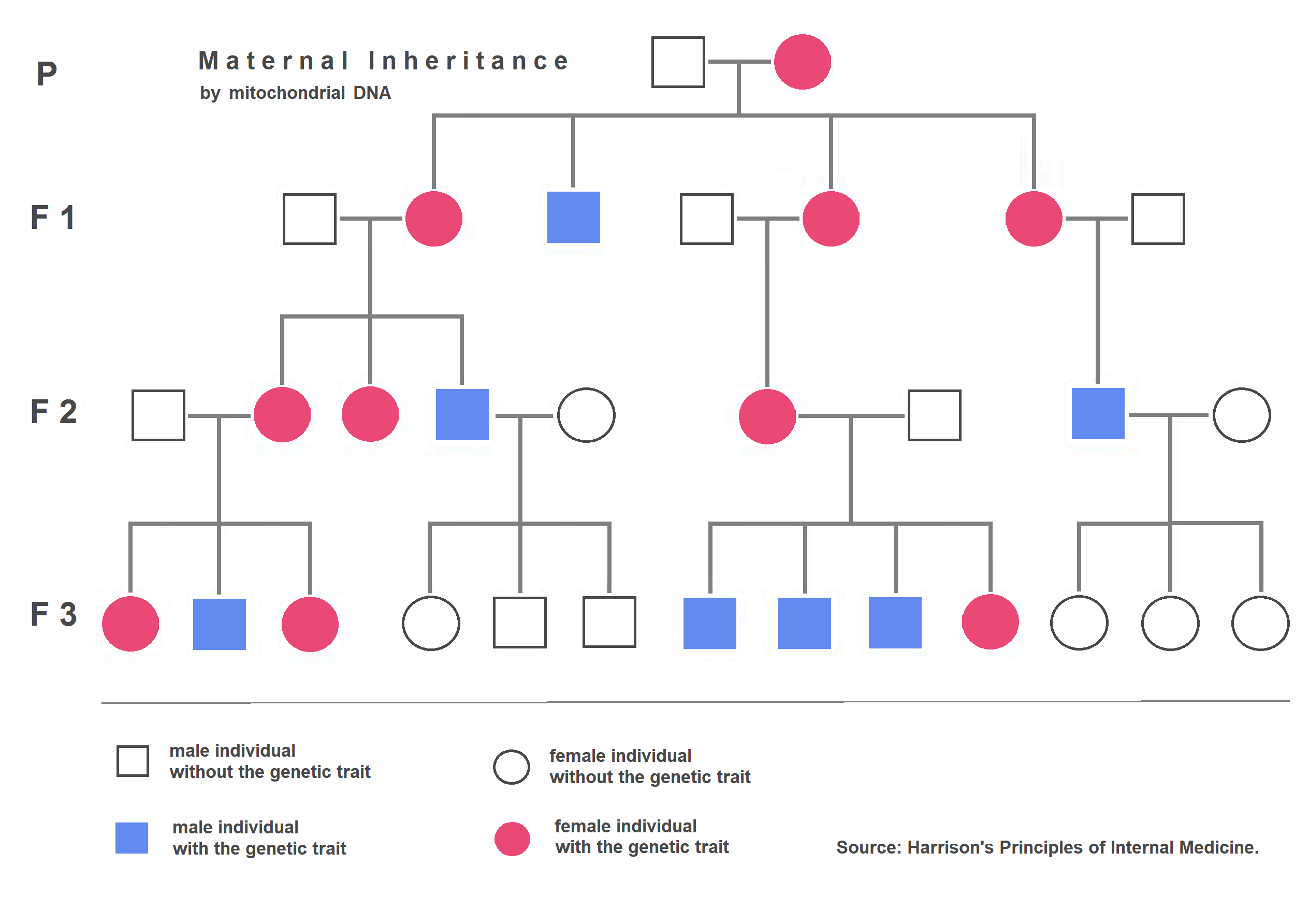 Image Source: Wikimedia Commons (2019). Maternal Inheritance – mitochondrial DNA, Licensed under a creative commons licence. Available for free at: https://upload.wikimedia.org/wikipedia/commons/7/76/Maternal_Inheritance_-_mitochondrial_DNA.png (Accessed: 5 January 2024).
References
- Turnpenny, P. D., & Ellard, S. (2017). Patterns of inheritance. In Emery’s Elements of Medical Genetics. China: Elsevier.
- Read, A., & Donnai, D. (2021). Chapter 1: What can we learn from family history? In New Clinical Genetics: A guide to genomic medicine (4th ed.). United Kingdom: Scoin Publishing Limited.
- Mensink, K. A., & Highsmith, W. E. (2009). Chapter 5 – Basic Concepts in Human Molecular Genetics. In W. B. Coleman & G. J. Tsongalis (Eds.), Molecular Pathology (pp. 89–107). San Diego: Academic Press.
- Jorde, L. B., Carey, J. C., & Bamshad, M. J. (2016). Chapter 4: Autosomal Dominant and Recessive Inheritance. In Medical Genetics (5th ed.). United States: Elsevier.
Non-mendelian inheritance
Epigenetics
Gene expression is a tightly controlled process, and many factors affect gene transcription. Epigenetics refers to the various factors that alter the expression of a gene. This process allows selective gene expression between different cell types, allowing the differentiation of cells.
X-inactivation
This process is predominantly seen in females, whereby the presence of 2 X chromosomes would result in lethal aneuploidy. Female cells compensate for this by inactivating one copy of the X-chromosome within its cells, ensuring only one X chromosome is actively involved in gene transcription. The inactivated X-chromosome can be seen as a Barr body. Therefore, females will naturally display mosaicism for the X chromosome as half of their somatic cells with an active maternal X chromosome, and the other half contains an active paternal X chromosome. The female’s children will not inherit the pattern of X-inactivation as both X chromosomes are active within gonadal tissues.
The impact of X-inactivation depends on the gene product:
Milder phenotype displayed – For example, heterozygous carriers of haemophilia will display deranged clotting time, but no clinical significance is seen.
The patchy distribution of normal and abnormal tissue throughout the body – For example, patchy skin pigmentation in incontinentia pigmenti in heterozygous females.
Genetic imprinting
Genetic imprinting is the process of silencing an allele from a particular parent, leaving only one active copy of the gene within all somatic cells. Imprinting occurs due to DNA methylation and histone modification to condense the affected allele, preventing the transcription of genes within that allele.
Classically, this is exemplified by Prader-Willi syndrome and Angelman syndrome. Both syndromes are caused by a deletion within the long arm of chromosome 15, and the phenotype displayed depends on the parent from which the mutation is inherited. Paternal inheritance of the deletion results in Prader-Willi syndrome, while maternal inheritance results in Angelman syndrome. This is due to genetic imprinting preventing any gene product from being produced.
Prader-Willi syndrome is typically characterised by short stature, obesity and learning difficulties. Severe learning difficulties and persistent happy affect with ataxia characterise Angelman syndrome.
Other conditions that are caused by genetic imprinting include Beckwith-Wiedemann syndrome and Russell-Silver syndrome; both conditions are caused by abnormal methylation of imprinted genes within the maternal and paternal chromosome 11, respectively, resulting in increased or decreased expression of insulin-like growth factor 2 (IGF2) respectively.
Trinucleotide repeats and anticipation
Anticipation is a phenomenon seen with some autosomal dominant conditions whereby the condition affects the younger generation at an earlier age with increased severity. This often occurs in diseases caused by trinucleotide repeats, such as Huntington’s disease, myotonic dystrophy, spinocerebellar ataxia and Fragile X syndrome.
The inherently unstable nature of the trinucleotide repeat allows expansion to occur during meiosis, which has been linked to increased severity of the condition between generations.
Multifactorial inheritance
Multifactorial inheritance is a catch-all term whereby a condition appears to be influenced by multiple genetic loci. This is commonly associated with many common conditions with multiple predisposing factors like cancer, cardiac conditions, asthma, and mental illness.
The liability threshold theory has been proposed to explain multifactorial inheritance. Genetic and environmental factors must be combined before the phenotype is expressed.
Large Scale population studies can be used to study and estimate the risk of recurrence within a family, but environmental factors prevent the generalisation of such results. Studies between monozygotic and dizygotic twins have been utilised to study various traits and allow the heritability of various conditions to be estimated.
References
- Jorde, L. B., Carey, J. C., & Bamshad, M. J. (2016). Chapter 12: Multifactorial Inheritance and Common Diseases. In Medical Genetics (5th ed.). United States: Elsevier.
- Mensink, K. A., & Highsmith, W. E. (2009). Chapter 5 – Basic Concepts in Human Molecular Genetics. In W. B. Coleman & G. J. Tsongalis (Eds.), Molecular Pathology (pp. 89–107). San Diego: Academic Press.
- Turnpenny, P. D., & Ellard, S. (2017). Patterns of inheritance. In Emery’s Elements of Medical Genetics. China: Elsevier.
Complete Guide to Clinical Genetics ST3 Application and Recruitment (2027)
Written by Medibuddy. Last updated: October 2026. Clinical Genetics is

Complete Guide to Clinical Radiology ST3 Application and Recruitment (2027)
Recruitment paused: NHS England is not running Clinical Radiology ST3+ recru

Complete Guide to Paediatric Surgery ST3 Application and Recruitment in United Kingdom (2026)
Written by Medibuddy. Last updated: October 2026. If you’re planning


Take your subscriptions with you
Our mobile app allows you to access your interview and exam question banks wherever you are.

