


 Close
Close
 21st February 2024
Basic Anatomy of the Eye
The Orbit
The orbit is a complex structure that protects the eye. It is pyramidal in shape, with the base sitting anteriorly and the apex of the pyramid situated posteriorly. A detailed overview of the structure of the orbit is detailed below.
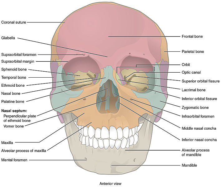
Image Source: Wikimedia Commons (2013). 704_Skull-01, Licensed under a creative commons licence. Available for free at: https://commons.wikimedia.org/wiki/File:704_Skull-01.jpg (Accessed: 28 September 2023).
Bone Anatomy
The orbit is primarily composed of seven bones, which include the frontal bone, zygomatic bone, maxilla, palatine bone, ethmoid bone, lacrimal bone, and sphenoid bone. These seven bones come together to create a protective socket for the eye. Their intricate arrangement provides structural support, safeguards the eye from external forces, and offers pathways for various nerves and blood vessels crucial for vision and eye function.
Frontal Bone
The frontal bone forms the upper part of the orbit (the roof) along with the lesser wing of the sphenoid bone. It is a ‘shell-shaped’ bone which consists of squamous, orbital and nasal parts. It extends from the forehead down to the eyebrow region and contributes to the roof of the eye socket. The frontal sinus, an air-filled cavity within this bone, lies just above the orbit. The frontal bone separates the orbit from the anterior cranial fossa.
Clinical relevance: frontal sinus fractures can occur following vehicle accidents or falls, with the characteristic clinical sign being the frontal depression in the anterior table of the skull.
Zygomatic Bone
The zygomatic bone forms the lateral and inferior (‘the floor’) border of the orbit. It plays a vital role in maintaining the overall shape of the face and protecting the eye from lateral impacts.
Clinical relevance: Zygomatic arch fracture is associated with trauma to the side of the face. This type of fracture can displace the nearby infraorbital nerve, leading to ipsilateral paralysis of the cheek, nose and lip.
Maxilla
The maxilla contributes to the orbital floor, providing structural support beneath the eye. The maxilla also houses the maxillary sinus, which is located below the orbit.
Palatine Bone
The palatine bone is a small, L-shaped bone located near the back of the orbit. It forms a portion of the orbital floor and is situated posteriorly to the maxilla.
Ethmoid Bone
The ethmoid bone forms the medial wall of the orbit. It is a complex bone with numerous thin, bony plates that help create the ethmoidal air cells. These air cells are important for reducing the weight of the skull and enhancing voice resonance. The ethmoid bone contains the cribriform plate, through which olfactory nerve fibres pass.
Clinical relevance: the ethmoid bone can be fractured in facial trauma – most commonly following a dashboard injury or a fall from a height. Fracture to the cribriform plate can cause an anastomosis between the nasal cavity and the central nervous system, which can present as watery discharge from a nostril (rhinorrhoea). Spontaneous CSF rhinorrhoea often requires surgical input.
Lacrimal Bone
The lacrimal bone is a small, fragile bone that contributes to the medial wall of the orbit. It contains the lacrimal sac, which is involved in tear drainage, and has a small groove for the nasolacrimal duct.
Sphenoid Bone
The sphenoid bone is a critical component of the orbit. Structurally, it is butterfly-shaped and consists of a body, a greater wing, a lesser wing and pterygoid processes. It forms the posterior part of the orbit and contributes to the main skull foramina, including the superior orbital fissure, the foramen rotundum and the foramen ovale. The important bony landmarks of the sphenoid bone include the sella turcica and the chiasmatic groove, a sulcus formed by the optic chiasm. The sphenoid bone also houses the sphenoidal sinus.
Clinical relevance: the sphenoid bone is closely associated with the pituitary gland; the pituitary can be accessed endoscopically through the sphenoid bone and sinus. Transsphenoidal surgery is usually the treatment choice for pituitary adenoma rather than an extensive craniotomy.
Borders of the Orbit
- Roof: Frontal and Sphenoid (lesser wing)
- Floor: Maxilla, Palatine, Zygomatic
- Medial: Ethmoid, Lacrimal, Maxilla, Sphenoid
- Lateral: Zygomatic and Sphenoid (greater wing)

Image Source: Wikimedia Commons (2013). 713 Bones Forming Orbit, Licensed under a creative commons licence. Available for free at: https://commons.wikimedia.org/wiki/File:713_Bones_Forming_Orbit.jpg (Accessed: 28 September 2023).
Orbital Fissures and Foramina
Optic Canal
The optic canal, within the lesser wing of the sphenoid bone, serves as a crucial passage for the optic nerve (cranial nerve II) and the ophthalmic artery. Any compression or damage to the optic canal can lead to vision impairment.
Superior Orbital Fissure
Positioned between the greater and lesser wings of the sphenoid bone, the superior orbital fissure accommodates several critical structures, including cranial nerves III (oculomotor), IV (trochlear), and VI (abducens), along with the ophthalmic division of the trigeminal nerve (cranial nerve V1). These nerves are responsible for eye movement, pupil constriction, and sensory innervation to various parts of the face and eye. A lesion or compression within the superior orbital fissure can result in debilitating eye movement disorders or sensory deficits.
Inferior Orbital Fissure
The inferior orbital fissure is located at the intersection of the sphenoid, maxilla, and zygomatic bones. It provides a pathway for branches of the maxillary nerve (cranial nerve V2) and blood vessels. Disorders affecting this fissure can lead to sensory disturbances in the face and orbit, particularly the lower eyelid and cheek.
Ethmoidal Foramina
The ethmoidal foramina, found in the ethmoid bone, permit the passage of anterior and posterior ethmoidal vessels and nerves. These structures play a role in the sensory innervation and vascular supply to the nasal cavity and ethmoidal air cells. Blockages or damage to these foramina can disrupt normal nasal physiology and potentially affect the eye due to their proximity.
Supraorbital and Infraorbital Foramen
The supraorbital foramen is the small opening at the superior orbital margin in the frontal bone just below the superciliary arches that transmits the supra-orbital nerve, artery and vein. The infraorbital foramen is within the maxillary bone. It is the anterior opening of the infraorbital canal, which is the anterior continuation of the infraorbital groove, which runs through the floor of the orbit.
References
- The Bony Orbit – Borders – Contents – Fractures – TeachMeAnatomy [Internet]. Available from: https://teachmeanatomy.info/head/organs/eye/bony-orbit/
- Kaplan HJ. Anatomy and function of the eye. Immune Response and the Eye. 2007; 92:4-10.
- Gaillard F. Orbit | Radiology Reference Article | Radiopaedia.org [Internet]. Radiopaedia. Available from: https://radiopaedia.org/articles/orbit
- Snell RS, Lemp MA. Clinical anatomy of the eye. Malden, Mass. Blackwell Science; 1998.
Periorbital Structures
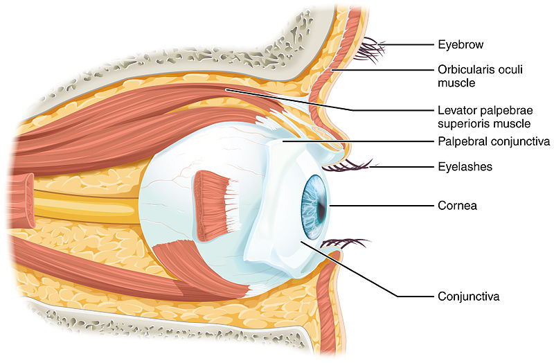
Image Source: Wikimedia Commons (2013). 1411 Eye in The Orbit, Licensed under a creative commons licence. Available for free at: https://commons.wikimedia.org/wiki/File:1411_Eye_in_The_Orbit.jpg (Accessed:28 September 2023).
The Eyelid
The eyelids are thin and movable layers that envelop the front part of the eyeball. They serve as protection against excessive light and injury while ensuring adequate lubrication by spreading tears across the eye’s surface.
The eyelids are divided into upper and lower sections, which come together at the medial canthus and lateral canthus of the eye. The palpebral aperture refers to the opening between these upper and lower sections. The important structures of the eyelid are as follows.
Skin and Subcutaneous Tissue
The most superficial layer of the eyelid consists of the skin and subcutaneous tissue. The skin in this region is one of the thinnest on the human body. Within the subcutaneous layer, there is loose connective tissue but no subcutaneous fat. Consequently, the eyelids can easily swell due to oedema or blood accumulation. The eyelashes, along with the modified sweat glands (ciliary glands of Moll) and sebaceous glands (glands of Zeiss), are found here.
Orbicularis Oculi
The Orbicularis Oculi muscle encircles the eye and has 3 parts: orbital, which forcefully closes the eyelid; palpebral, which gently closes the eyelids; and lacrimal, which drains tears. It is responsible for blinking and maintaining closure to protect the eye. It originates from the medial orbital margin and inserts into the skin around the orbit, as well as the superior and inferior tarsal plates. It is innervated by the temporal and zygomatic branches of the facial nerve.
Tarsal Plates
The tarsal plate is a dense strip of connective tissue within each eyelid. It provides structural support, ensuring they maintain their shape and position over the eye. The superior tarsus (upper eyelid) and inferior tarsus (lower eyelid) are located deep to the palpebral region of the Orbicularis Oculi. The Meibomian glands found within the tarsal plate secrete oil to the eye’s surface to slow the evaporation of the tear film and prevent the eyelids from sticking together when shut. These glands can become blocked, leading to blepharitis and stye and chalazion formation.
Levator Palpebrae Superioris (LPS)
The LPS, supplied by the superior branch of the oculomotor nerve, works to open the eyelid. It originates from the lesser wing of the sphenoid and inserts into the upper eyelid and superior tarsal plate. Muller’s muscle assists the LPS in opening the eyelid and is supplied by the sympathetic fibres from the superior cervical ganglion.
Innervation and Vasculature of the Eyelid
The eyelid is supplied by the branches of the trigeminal nerve, with V1 supplying the upper eyelid and V2 supplying the lower eyelid. The ophthalmic, facial and superficial temporal artery supply the eyelid, and it is drained by a rich network of veins split medially and laterally. Medially, it drains via the medial palpebral vein into the angular and ophthalmic veins. Laterally, blood drains from the lateral palpebral vein to the superficial temporal vein.
Extraocular Muscles
The extraocular muscles are a group of six muscles responsible for the precise movement and control of the eyeball within the orbit. They are attached to the wall of the orbit and work in conjunction to allow coordinated eye movements, maintaining binocular vision.
The recti muscles (superior, inferior, lateral and medial) all originate from the common tendinous ring, a ring of fibrous tissue found at the back of the orbit which surrounds the optic canal. The oblique muscles (superior and inferior) take an angled approach into the eyeball and attach to the posterior surface of the sclera.
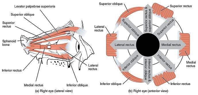
Image Source: Wikimedia Commons (2015). 1107 The Extrinsic Eye Muscles right eye lat, Licensed under a creative commons licence. Available for free at: https://commons.wikimedia.org/wiki/File:1107_The_Extrinsic_Eye_Muscles_right_eye_lat.png (Accessed: 28 September 2023).
Superior Rectus
Originates from the superior part of the common tendinous ring and attaches to the superior and anterior sclera. Its main function is to elevate, and it also contributes to adduction and medial rotation. Its supply is the oculomotor nerve.
Inferior Rectus
Originates from the inferior part of the common tendinous ring and attaches to the inferior and anterior sclera. Its main function is to depress the eyeball, but it also contributes to adduction and lateral rotation. It is supplied by the oculomotor nerve.
Medial Rectus
Originates from the medial part of the common tendinous ring and attaches to the medial and anterior sclera. Its main function is to adduct the eyeball. It is supplied by the oculomotor nerve.
Lateral Rectus
Originates from the lateral part of the common tendinous ring and attaches to the lateral and anterior sclera. Its main function is to abduct the eyeball. It is supplied by the abducens nerve.
Superior Oblique
Originates from the sphenoid bone; its tendon passes through a trochlea and attaches just behind the superior rectus. Its main function is to depress, abduct and medially rotate the eyeball.
Inferior Oblique
Originates from the inferomedial aspect of the orbital floor, passes above the inferior rectus and attaches posteriorly to the lateral rectus.
The Lacrimal Apparatus
The lacrimal apparatus is the system responsible for the removal of tears from the eye’s orbit.
Following lacrimal fluid secretion from the lacrimal gland, tears flow over the eye’s surface and collect in the lacrimal lake, situated at the medial canthus. From this point, they travel through a network of canals into the lacrimal sac. The lacrimal sac, which serves as the widened part of the nasolacrimal duct, is found within a groove formed by the lacrimal bone and the frontal process of the maxilla. Tears then descend through the nasolacrimal duct and ultimately discharge into the inferior meatus of the nasal cavity.
The lacrimal gland is positioned within the superior and lateral aspects of the orbit, above the outer edge of the eye. The gland receives its blood supply primarily from branches of the ophthalmic artery. Parasympathetic nerve fibres, carried by the facial nerve, stimulate tear production, while sympathetic fibres regulate blood vessel constriction within the gland. The primary purpose of the gland is to secrete lacrimal fluid, which protects, lubricates and supplies the cornea with nutrients.
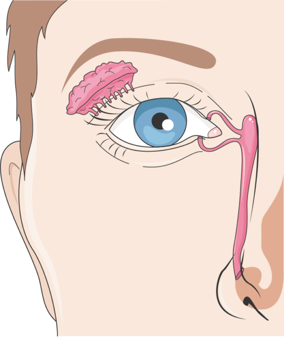
Image Source: Tear-duct, Licensed under a creative commons licence. Available for free at: https://smart.servier.com/smart_image/tear-duct/ (Accessed: 28 September 2023).
Clinical relevance: Disorders of the lacrimal gland can have significant implications for ocular health. Dry Eye Syndrome can occur from reduced tear production from the lacrimal gland, causing discomfort and potential corneal damage. Dacryoadenitis is inflammation of the lacrimal gland, often due to infection or autoimmune diseases, which can result in pain and swelling. Tumours can develop within the lacrimal gland, which may require surgical intervention. In certain cases, lacrimal gland surgery is necessary to treat various conditions or alleviate excessive tearing (epiphora).
The Conjunctiva
The conjunctiva is a thin, transparent membrane that lines the inner surface of the eyelids and covers the anterior part of the eyeball’s sclera. Its function primarily is to protect and lubricate the eye. The conjunctiva secretes mucin, which provides a stable tear film to keep the eye moist.
The conjunctiva consists of three layers: the palpebral conjunctiva lines the inner surface and is the deepest layer of the eyelid; the bulbar conjunctiva covers the sclera; and the fornix is the loose, folded portion where the palpebral and bulbar conjunctiva meet.
Clinical relevance: Conjunctiva can commonly become inflamed due to several causes, including bacterial, viral, and allergic conjunctivitis. It usually presents with a pink eye with redness, itching and discharge. Pinguecula and pterygium are growths on the conjunctiva that usually develop following chronic exposure to ultraviolet light.
References
- The Eye – TeachMeAnatomy [Internet]. teachmeanatomy.info. Available from: https://teachmeanatomy.info/head/organs/eye/
- Snell RS, Lemp MA. Clinical anatomy of the eye. Malden, Mass. Blackwell Science; 1998.
- Jones J. Extraocular muscles | Radiology Reference Article | Radiopaedia.org [Internet]. Radiopaedia. [cited 2023 Oct 5]. Available from: https://radiopaedia.org/articles/extraocular-muscles-1
Internal Structures of The Eye
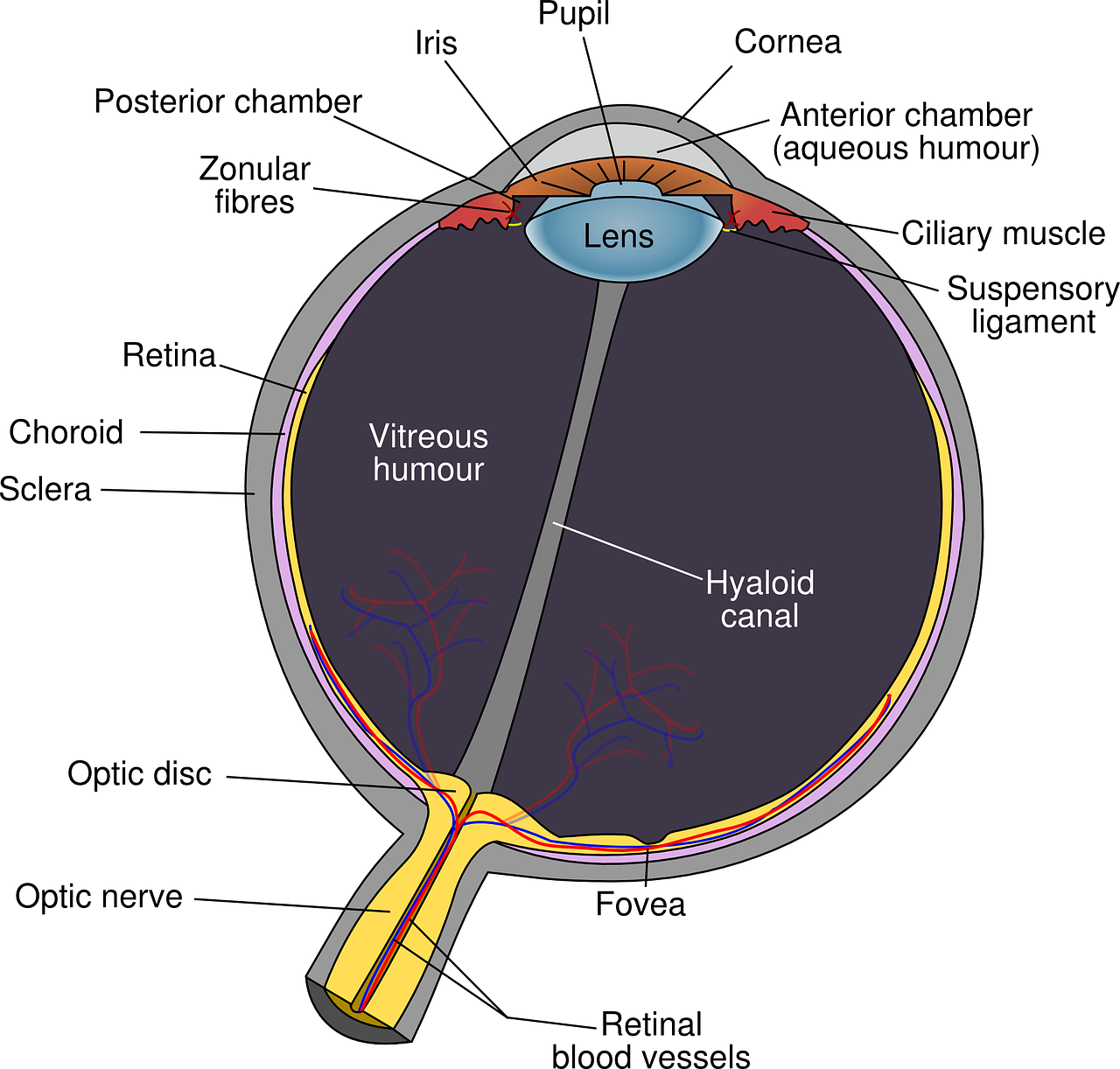 Image Source: Eye diagram eyeball -body pupil 39998, Licensed under a creative commons licence. Available for free at: https://pixabay.com/vectors/eye-diagram-eyeball-body-pupil-39998/(Accessed: 28 September 2023).
Layers Of the Eyeball – Fibrous Layer
The Sclera
The fibrous layer is the outermost layer and consists of the sclera and cornea, which are continuous with one another. The sclera is the white part of the eyeball, and it is primarily composed of collagen fibres. It is important for the protection of the eyeball, to maintain the shape of the eye and to allow attachment of the extraocular muscles to facilitate eye movement.
The sclera has three main layers: the episclera, scleral stroma and lamina fusca. The episclera is a thin connective tissue that covers the scleral surface and contains blood vessels. It provides vasculature for the conjunctiva. Scleral stroma forms the bulk of the sclera. It is composed of collagen fibres, fibroblasts, and proteoglycans. Its unique arrangement of collagen fibres contributes to the sclera’s strength while maintaining transparency.
The Cornea
The cornea is a transparent, dome-shaped structure at the front of the eye. It is responsible for approximately two-thirds of the eye’s refractive power to allow light to focus on the retina. To maintain clarity and transparency, the cornea lacks blood vessels and receives oxygen and nutrients from tear fluid and aqueous humour.
The cornea has five distinct layers:
- Corneal Epithelium – The outermost layer acts as a protective barrier and provides a smooth surface for the tear film.
- Bowman’s Layer – Located beneath the epithelium, it consists of collagen fibres and offers structural support.
- Corneal Stroma – makes up most of the corneal thickness; it consists of a network of collagen fibres arranged for transparency.
- Descemet’s Membrane – Found beneath the stroma, it provides a basement membrane for the corneal endothelium.
- Corneal Endothelium – The innermost layer helps maintain corneal transparency by regulating fluid balance
Layers of the Eyeball – Vascular Layer
The vascular layer (uveal tract) is the middle layer of the eye that includes the choroid, ciliary body, and iris. It plays several essential roles in supporting and nourishing the eye.
The Choroid
Positioned between the sclera and retina, the choroid is highly vascularised and rich in blood vessels. Its functions include providing nutrients and oxygen to the retina, absorbing excess light via melanocytes and thermoregulation.
Ciliary Body
The ciliary body, positioned just behind the iris, is a crucial structure for adjusting the eye’s focus. The ciliary muscle contracts and relaxes regularly, which alters the shape of the lens, allowing the eye to focus on objects at different distances. The muscle has smooth muscle fibres which attach to the lens of the eye by the ciliary processes. The ciliary body produces aqueous humour, which nourishes the cornea and lens while maintaining intraocular pressure.
Iris
The iris is the coloured part of the eye, and it regulates the amount of light entering the eye through its central opening, the pupil. Its muscles control the pupil’s size in response to changes in light intensity. The iris is a thin, circular diaphragm suspended between the cornea and the lens. It divides the space between these structures into two chambers: the anterior chamber (in front of the iris) and the posterior chamber (behind the iris).
The iris has two types of smooth muscle fibres: dilator and sphincter pupillae muscles. Dilator pupillae, when contracted, enlarges the pupil, which allows more light to enter. Sphincter pupillae, when contracted, constrict the pupil, which prevents more light from entering.
Layers of the Eyeball – Inner Layer (Retina)
The innermost layer of the eye is the retina, responsible for detecting light. The retina has two distinct layers: The pigmented (outer) layer, consisting of a single layer of cells, is closely connected to the choroid. Its primary function is to support the choroid in absorbing light, preventing light from scattering within the eyeball. This layer extends around the entire inner surface of the eye. The neural (inner) layer is positioned at the back and sides of the eye. This layer contains photoreceptors, specialised cells responsible for detecting light.
At the centre of the retina lies the macula, distinguished by its yellowish colour and abundant pigmentation. Within the macula is the fovea centralis, a specialised depression that houses a high density of light-detecting cells. This region is responsible for achieving precise and detailed vision. Conversely, where the optic nerve enters the retina is termed the optic disc; it notably lacks any light-sensitive cells.
Lens
The lens is a transparent, biconvex structure positioned directly behind the iris, suspended by the ciliary body. The lens serves a vital role in the accommodation reflex and refraction.
The lens is primarily composed of specialised lens fibres. These fibres lack cell nuclei and organelles, contributing to the lens’s transparency and optical clarity. It has an outer capsule that encapsulates the lens fibres, and its outermost portion is known as the lens capsule. The lens fibres are arranged concentrically like the layers of an onion.
The ability of the lens to change its shape is called accommodation. This process is controlled by the ciliary muscle and the zonular fibres. When focusing on near objects, the ciliary muscle contracts, causing the lens to thicken and increase its refractive power. Conversely, for distant objects, the ciliary muscle relaxes, causing the lens to flatten.
Vitreous Body
The vitreous body is a gel-like, transparent substance that occupies the rear part of the eyeball, located posterior to the lens. It serves to support the lens anteriorly and the retina posteriorly. It also contributes to the magnifying power of the eye.
Vitreous humour consists mainly of water (approximately 99%) along with a network of fine collagen fibres, hyaluronic acid, and other macromolecules. It has a gel-like consistency, allowing it to maintain the shape of the eye and support delicate structures such as the retina. Structurally, the vitreous can be divided into two regions: the vitreous cortex, which is adjacent to the retina, and the vitreous core, located deeper within the eye. The core is more liquid in nature than the cortex.
Anterior Chamber of The Eye
The anterior chamber is a space located between the cornea and the iris. The anterior chamber’s size varies from person to person but is typically around 3 mm in depth. It plays a crucial role in maintaining the eye’s shape and intraocular pressure. The aqueous humour is a clear, watery fluid that circulates through the anterior chamber. It nourishes the cornea and lens, provides nutrients to the avascular tissues, and helps maintain the eye’s shape.
Production and drainage of Aqueous Humour Image Source: Aqueous humor pathway, Licensed under a creative commons licence. Available for free at: https://openclipart.org/detail/276637/aqueous-humor-pathway (Accessed: 28 September 2023).
The aqueous humour undergoes a continuous cycle of production and drainage within the anterior chamber. Aqueous humour is produced by the ciliary processes of the ciliary body located behind the iris. The ciliary body secretes aqueous humour into the posterior chamber, and it then flows into the anterior chamber through the pupil.
Aqueous humour drains from the anterior chamber through the trabecular meshwork, a specialised drainage system located in the angle between the cornea and the iris. From there, it enters Schlemm’s canal and eventually reaches the bloodstream.
The aqueous humour delivers nutrients and oxygen to the avascular structures of the cornea and lens. The balance between aqueous humour production and drainage helps maintain a stable intraocular pressure within the eye. Abnormal pressure can lead to glaucoma.
Posterior Chamber of the Eye
The posterior chamber is situated at the back of the eye, extending from the posterior surface of the iris to the front surface of the vitreous body. It comprises the iris, the ciliary body, the lens and the vitreous humour. The posterior chamber contributes to accommodation and refraction, as well as regulation of the intra-ocular pressure.
References
- The Eye – TeachMeAnatomy [Internet]. teachmeanatomy.info. Available from: https://teachmeanatomy.info/head/organs/eye/
- Snell RS, Lemp MA. Clinical anatomy of the eye. Malden, Mass. Blackwell Science; 1998.
- Knipe H, Yang V. Retina [Internet]. Radiopaedia.org; 2018 [cited 2023 Oct 5]. Available from: https://radiopaedia.org/articles/retina?lang=gb
Complete Guide to Clinical Genetics ST3 Application and Recruitment (2027)
Written by Medibuddy. Last updated: October 2026. Clinical Genetics is

Complete Guide to Clinical Radiology ST3 Application and Recruitment (2027)
Recruitment paused: NHS England is not running Clinical Radiology ST3+ recru

Complete Guide to Paediatric Surgery ST3 Application and Recruitment in United Kingdom (2026)
Written by Medibuddy. Last updated: October 2026. If you’re planning


Take your subscriptions with you
Our mobile app allows you to access your interview and exam question banks wherever you are.

