


 Close
Close
 21st February 2024
Internal Structures of The Eye
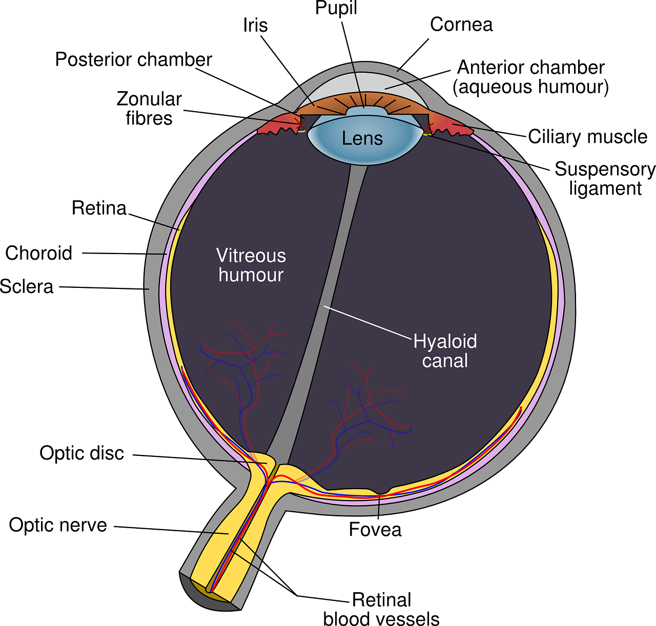
Image Source: Eye diagram eyeball -body pupil 39998, Licensed under a creative commons licence. Available for free at: https://pixabay.com/vectors/eye-diagram-eyeball-body-pupil-39998/(Accessed: 28 September 2023).
Layers Of the Eyeball – Fibrous Layer
The Sclera
The fibrous layer is the outermost layer and consists of the sclera and cornea, which are continuous with one another. The sclera is the white part of the eyeball, and it is primarily composed of collagen fibres. It is important for the protection of the eyeball, to maintain the shape of the eye and to allow attachment of the extraocular muscles to facilitate eye movement.
The sclera has three main layers: the episclera, scleral stroma and lamina fusca. The episclera is a thin connective tissue that covers the scleral surface and contains blood vessels. It provides vasculature for the conjunctiva. Scleral stroma forms the bulk of the sclera. It is composed of collagen fibres, fibroblasts, and proteoglycans. Its unique arrangement of collagen fibres contributes to the sclera’s strength while maintaining transparency.
The Cornea
The cornea is a transparent, dome-shaped structure at the front of the eye. It is responsible for approximately two-thirds of the eye’s refractive power to allow light to focus on the retina. To maintain clarity and transparency, the cornea lacks blood vessels and receives oxygen and nutrients from tear fluid and aqueous humour.
The cornea has five distinct layers:
- Corneal Epithelium – The outermost layer acts as a protective barrier and provides a smooth surface for the tear film.
- Bowman’s Layer – Located beneath the epithelium, it consists of collagen fibres and offers structural support.
- Corneal Stroma – makes up most of the corneal thickness; it consists of a network of collagen fibres arranged for transparency.
- Descemet’s Membrane – Found beneath the stroma, it provides a basement membrane for the corneal endothelium.
- Corneal Endothelium – The innermost layer helps maintain corneal transparency by regulating fluid balance
Layers of the Eyeball – Vascular Layer
The vascular layer (uveal tract) is the middle layer of the eye that includes the choroid, ciliary body, and iris. It plays several essential roles in supporting and nourishing the eye.
The Choroid
Positioned between the sclera and retina, the choroid is highly vascularised and rich in blood vessels. Its functions include providing nutrients and oxygen to the retina, absorbing excess light via melanocytes and thermoregulation.
Ciliary Body
The ciliary body, positioned just behind the iris, is a crucial structure for adjusting the eye’s focus. The ciliary muscle contracts and relaxes regularly, which alters the shape of the lens, allowing the eye to focus on objects at different distances. The muscle has smooth muscle fibres which attach to the lens of the eye by the ciliary processes. The ciliary body produces aqueous humour, which nourishes the cornea and lens while maintaining intraocular pressure.
Iris
The iris is the coloured part of the eye, and it regulates the amount of light entering the eye through its central opening, the pupil. Its muscles control the pupil’s size in response to changes in light intensity. The iris is a thin, circular diaphragm suspended between the cornea and the lens. It divides the space between these structures into two chambers: the anterior chamber (in front of the iris) and the posterior chamber (behind the iris).
The iris has two types of smooth muscle fibres: dilator and sphincter pupillae muscles. Dilator pupillae, when contracted, enlarges the pupil, which allows more light to enter. Sphincter pupillae, when contracted, constrict the pupil, which prevents more light from entering.
Layers of the Eyeball – Inner Layer (Retina)
The innermost layer of the eye is the retina, responsible for detecting light. The retina has two distinct layers: The pigmented (outer) layer, consisting of a single layer of cells, is closely connected to the choroid. Its primary function is to support the choroid in absorbing light, preventing light from scattering within the eyeball. This layer extends around the entire inner surface of the eye. The neural (inner) layer is positioned at the back and sides of the eye. This layer contains photoreceptors, specialised cells responsible for detecting light.
At the centre of the retina lies the macula, distinguished by its yellowish colour and abundant pigmentation. Within the macula is the fovea centralis, a specialised depression that houses a high density of light-detecting cells. This region is responsible for achieving precise and detailed vision. Conversely, where the optic nerve enters the retina is termed the optic disc; it notably lacks any light-sensitive cells.
Lens
The lens is a transparent, biconvex structure positioned directly behind the iris, suspended by the ciliary body. The lens serves a vital role in the accommodation reflex and refraction.
The lens is primarily composed of specialised lens fibres. These fibres lack cell nuclei and organelles, contributing to the lens’s transparency and optical clarity. It has an outer capsule that encapsulates the lens fibres, and its outermost portion is known as the lens capsule. The lens fibres are arranged concentrically like the layers of an onion.
The ability of the lens to change its shape is called accommodation. This process is controlled by the ciliary muscle and the zonular fibres. When focusing on near objects, the ciliary muscle contracts, causing the lens to thicken and increase its refractive power. Conversely, for distant objects, the ciliary muscle relaxes, causing the lens to flatten.
Vitreous Body
The vitreous body is a gel-like, transparent substance that occupies the rear part of the eyeball, located posterior to the lens. It serves to support the lens anteriorly and the retina posteriorly. It also contributes to the magnifying power of the eye.
Vitreous humour consists mainly of water (approximately 99%) along with a network of fine collagen fibres, hyaluronic acid, and other macromolecules. It has a gel-like consistency, allowing it to maintain the shape of the eye and support delicate structures such as the retina. Structurally, the vitreous can be divided into two regions: the vitreous cortex, which is adjacent to the retina, and the vitreous core, located deeper within the eye. The core is more liquid in nature than the cortex.
Anterior Chamber of The Eye
The anterior chamber is a space located between the cornea and the iris. The anterior chamber’s size varies from person to person but is typically around 3 mm in depth. It plays a crucial role in maintaining the eye’s shape and intraocular pressure. The aqueous humour is a clear, watery fluid that circulates through the anterior chamber. It nourishes the cornea and lens, provides nutrients to the avascular tissues, and helps maintain the eye’s shape.
Production and drainage of Aqueous Humour

Image Source: Aqueous humor pathway, Licensed under a creative commons licence. Available for free at: https://openclipart.org/detail/276637/aqueous-humor-pathway (Accessed: 28 September 2023).
The aqueous humour undergoes a continuous cycle of production and drainage within the anterior chamber. Aqueous humour is produced by the ciliary processes of the ciliary body located behind the iris. The ciliary body secretes aqueous humour into the posterior chamber, and it then flows into the anterior chamber through the pupil.
Aqueous humour drains from the anterior chamber through the trabecular meshwork, a specialised drainage system located in the angle between the cornea and the iris. From there, it enters Schlemm’s canal and eventually reaches the bloodstream.
The aqueous humour delivers nutrients and oxygen to the avascular structures of the cornea and lens. The balance between aqueous humour production and drainage helps maintain a stable intraocular pressure within the eye. Abnormal pressure can lead to glaucoma.
Posterior Chamber of the Eye
The posterior chamber is situated at the back of the eye, extending from the posterior surface of the iris to the front surface of the vitreous body. It comprises the iris, the ciliary body, the lens and the vitreous humour. The posterior chamber contributes to accommodation and refraction, as well as regulation of the intra-ocular pressure.
References
- The Eye – TeachMeAnatomy [Internet]. teachmeanatomy.info. Available from: https://teachmeanatomy.info/head/organs/eye/
- Snell RS, Lemp MA. Clinical anatomy of the eye. Malden, Mass. Blackwell Science; 1998.
- Knipe H, Yang V. Retina [Internet]. Radiopaedia.org; 2018 [cited 2023 Oct 5]. Available from: https://radiopaedia.org/articles/retina?lang=gb
Complete Guide to Clinical Genetics ST3 Application and Recruitment (2027)
Written by Medibuddy. Last updated: October 2026. Clinical Genetics is

Complete Guide to Clinical Radiology ST3 Application and Recruitment (2027)
Recruitment paused: NHS England is not running Clinical Radiology ST3+ recru

Complete Guide to Paediatric Surgery ST3 Application and Recruitment in United Kingdom (2026)
Written by Medibuddy. Last updated: October 2026. If you’re planning


Take your subscriptions with you
Our mobile app allows you to access your interview and exam question banks wherever you are.

