


 Close
Close
 8th March 2024
Adult Congenital Heart Disease
Acyanotic Heart Disease
Introduction
Congenital heart disease is the commonest form of congenital anomaly, affecting about 8 in every 1000 live births in the United Kingdom. Diagnostic and management outcomes have significantly improved, which has led to an increasing population of adults with the condition. Adult congenital heart disease (ACHD) can be categorised into acyanotic; cyanotic heart disease and coarctation of the aorta.
Acyanotic heart diseases are congenital cardiac malformations that lead to a left-to-right shunt. Cyanosis is not typically a feature as patients retain a normal level of oxyhaemoglobin saturation in the systemic circulation. However, uncorrected left-to-right shunts may eventually lead to Eisenmenger syndrome. The main types of acyanotic heart defects include:
- Atrial Septal Defect
- Ventricular Septal Defect
- Patent Ductus Arteriosus
Atrial Septal Defect
Atrial septal defect (ASD) is the commonest form of acyanotic heart disease in which a pathological communication is formed that allows shunting of blood between the left and right atrium. It constitutes approximately 5-10% of congenital heart diseases and co-exists with other congenital defects in 30-50% of cases.
A patent foramen ovale (PFO) is a different form of interatrial shunt caused by failure of the foramen ovale to close completely after birth. PFOs are present in 25% of the general population and the majority of adults remain asymptomatic. PFOs may result in paradoxical embolisation of thrombus from the venous circulation into the systemic circulation and the risk increases in the presence of an atrial septal aneurysm.
ASD can be divided into the following subtypes: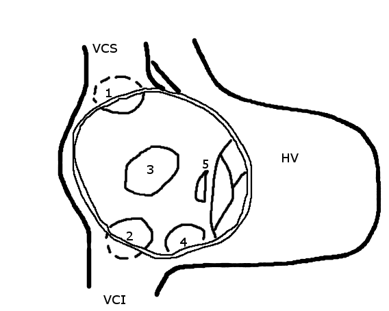
Image Source: Wikimedia Commons (2005). Atrial septal defect, Licensed under a creative commons licence. Available for free at: https://en.wikipedia.org/wiki/Atrial_septal_defect svg (Accessed: 26 January 2024).
Ostium secundum
These constitute 80% of ASDs and are located within, or surrounding, the fossa ovalis. They form as a result of poor development of the septum primum of varying degrees during embryologic development and have associations with other forms of congenital defects, including mitral valve prolapse, pulmonary stenosis and partial anomalous pulmonary venous drainage.
Ostium primum
They account for 15-20% of ASDs and are part of the spectrum of atrioventricular septal defects (AVSD). Primum ASD is caused by failure of the septum primum to fuse with the endocardial cushions at the interatrial septal base and are frequently associated with a cleft in the anterior leaflet of the mitral valve, leading to varying degrees of mitral regurgitation. Partial AVSD includes primum ASD, cleft mitral valve, and ventricular septal defect (VSD). Complete AVSD is characterised by a common AV valve with primum ASD and inlet VSD, often seen in Down’s syndrome.
Sinus venosus defect
Sinus venosus defects constitute 5-10% of ASDs and involve a portion of the atrial wall derived from the sinus venosus but there is no true communication between the right and left atria. Transoesophageal echocardiography (TOE) is generally required for assessment in adults, especially when there is unexplained right chamber dilatation. They can be further subdivided into superior and inferior defects. The commonest of the two is a superior sinus venosus, located below the orifice of the superior vena cava (SVC). The right upper lobe and middle pulmonary veins connect to the junction of the SVC and right atrium or on the SVC itself. An inferior sinus venosus results from the breakdown between the right atrial wall and the inferior right-sided pulmonary veins.
Unroofed coronary sinus
Caused by the absence of the common wall between the coronary sinus and left atrium, this defect accounts for less than 1% of ASDs and is often associated with complete AV septal defect; absence of coronary sinus and a persistent left sided SVC.
Pathophysiology
ASDs cause a left-to-right shunt, which results in volume overload of the RA and RV. The severity depends on the size of the ASD and filling pressures of the ventricles. Factors which heighten the degree of shunting include elevated LV filling pressures in hypertensive cardiomyopathy or increased LA pressure seen in mitral stenosis. In contrast, factors that reduce the degree of left-to-right shunt include pulmonary hypertension or increased RA pressure in tricuspid stenosis.
Clinical presentation
Patients commonly remain asymptomatic until adulthood, where 30% begin to develop breathlessness on exertion by the third decade and more than 75% of patients by the fifth decade. Late complications include supraventricular tachyarrhythmias, severe pulmonary hypertension and right-sided heart failure. Paradoxical embolism causing systemic events may be the first clue to an ASD.
Auscultation will reveal fixed splitting of S2 and a systolic flow murmur at the left second intercostal space. A loud P2 component of the second heart sound indicates pulmonary hypertension. If cyanosis develops, this generally suggests reversal of the shunt (Eisenmenger syndrome).
Investigations
On an ECG, both primum and secundum ASDs display first degree AV block and RSR’ pattern in V1 but can be differentiated by right axis deviation in secundum ASD and left axis deviation in primum ASD.
A chest x-ray may show cardiomegaly and prominent vascular markings.
The first line diagnostic modality is transthoracic echocardiography (TTE), where intravenous contrast studies can be performed to elicit the shunt and colour-flow Doppler can identify intra-arterial flow. The degree of shunting can be assessed using the shunt fraction (Qp/Qs), which is the ratio of pulmonary flow (Qp) to systemic flow (Qs). A left-to-right shunt is considered significant when the ratio of pulmonary to systemic blood flow (Qp/Qs) is >1.5 or when the RV dilates. There may also be functional tricuspid regurgitation, RA and RV enlargement, as well as abnormal septal motion. TOE is optional for further anatomical definition.
Cardiac magnetic resonance (CMR) imaging provides high quality assessment of RV size and function and clear views of the pulmonary veins. It is useful for quantification of pulmonary to systemic flow ratio (Qp:Qs) where either a ratio > 1.5 or evidence of RV dilation confirms a significant shunt.
Cardiac catheterisation is not routinely required for diagnostic purposes except to assess for pulmonary pressures or coronary artery disease. Additional investigations involve oximetry sampling that can demonstrate an oxygen step-up from the vena cava to the right atrium.
Management
Medical management is not typically required as most patients are asymptomatic. Congestive cardiac failure is managed with diuretics and atrial tachyarrhythmias with rate, rhythm, and anticoagulation therapy.
Treatment options include percutaneous and surgical closure and indications for intervention include:
- Dilatation of right chambers without pulmonary artery hypertension (PAH), regardless of symptoms
- Paradoxical embolism
- PAH with marked left-to-right shunt (Qp:Qs>1.5)
A percutaneous approach is the first choice for secundum ASD with adequate margins from AV valves, coronary sinus, origin of vena cava and right pulmonary veins. Surgical repair has a low mortality and a good long-term outcome when performed early during childhood and in the absence of pulmonary hypertension.
ASD closure is contraindicated in irreversible pulmonary hypertension (systolic pulmonary pressure > 2/3 systolic BP) and in Eisenmenger physiology.
Pregnancy
Women with uncomplicated ASD can have uncomplicated pregnancies despite the physiological increase in stroke volume and cardiac output. Venous thromboembolism prophylaxis is important as there exists a risk of paradoxical embolisation.
Antibiotic prophylaxis
The risk of infective endocarditis is higher in those with an ASD than in the general population but the European Society of Cardiology (ESC) guidelines recommend antibiotic prophylaxis in the following:
- Cyanotic congenital heart disease
- Previous infective endocarditis
- Repaired defect with prosthetic material for the first 6 months
- Residual defect after intervention
Ventricular Septal Defect
A ventricular septal defect (VSD) is one of the most common congenital cardiac defects (second to bicuspid aortic valve) with a prevalence of about 20-25% of all congenital heart diseases. Males and females are affected equally. Acquired VSDs, e.g., from myocardial infarction, are not discussed here.
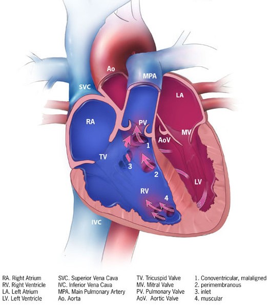
Image Source: Wikimedia Commons (2013). Ventricular septal defect, Licensed under a creative commons licence. Available for free at: https://en.wikipedia.org/wiki/Ventricular_septal_defect (Accessed: 26 January 2024).
There are four subtypes of VSD the commonest of which is membranous VSD, accounting for 80% of defects. They occur within the membranous septum with potential involvement of the tricuspid and aortic valves.
Muscular VSDs are completely surrounded by muscle. They make up to 15-20% of VSDs.
5% of VSDs are outlet/infundibular defects located below the aortic and pulmonary valves. They may be associated with aortic regurgitation.
Inlet VSD, also known as AV canal/AVSD type, typically occur in Down’s syndrome and is found below the AV valves.
Clinical presentation
Most patients with a small VSD will remain asymptomatic and the natural history will depend on the location and size of the defect as these impact the degree of left-to-right shunting.
On auscultation of the heart, a small VSD will produce a holosystolic murmur at the left parasternal border and a palpable systolic thrill. A moderate sized VSD may elicit a mitral diastolic rumble and S3 gallop due to LA and LV dilatation with increased flow across the mitral valve. Large VSD can progress to pulmonary hypertension and Eisenmenger syndrome. Signs of pulmonary hypertension, such as a left parasternal heave and loud P2, may be present. Other findings in large VSD include S4 heart sound, tricuspid regurgitation and Graham Steele murmur.
Complications of VSD include:
- Left heart failure
- Pulmonary hypertension (Cor pulmonale due to pressure and volume overload)
- Tricuspid regurgitation
- Arrhythmias
- Infective endocarditis
- Aortic regurgitation due to leaflet prolapse
- Subaortic stenosis
- Double-chambered right ventricle where a muscle band develops as a result of VSD jet leading to infundibular pulmonary stenosis.
Investigations
The ECG in VSD can show LVH and biphasic QRS complexes in the precordial leads. Echocardiography is useful for colour-flow Doppler, which shows a high-velocity systolic jet across the VSD. If echocardiography provides insufficient data, then CMR is a useful modality. Cardiac catheterisation allows measurement of Qp:Qs, pulmonary pressure and pulmonary vascular resistance.
Management
Asymptomatic patients with small defects do not require intervention apart from routine cardiology follow-up. The following indications warrant intervention with surgical or percutaneous closure:
- Haemodynamically significant VSD in patients with evidence of LV dysfunction
- Prolapse of aortic valve causing moderate aortic regurgitation
- In the presence of severe pulmonary hypertension, closure should only be considered in the case of left-to-right shunt (Qp:Qs ratio >1.5)
- Double chamber RV
- Infective endocarditis
Surgical closure can be performed with low operative mortality and good long-term outcomes. Percutaneous closure can be considered in selected cases, such as muscular and membranous VSDs that are located away from the tricuspid and aortic valves.
Patent Ductus Arteriosus
The patent ductus arteriosus (PDA) is a remnant of the normal embryonic vessel called the ductus arteriosus, which connects the left pulmonary artery to the aorta. If the PDA fails to close after birth, the direction of blood flow leads to a left-to-right shunt. The reported incidence is around 1 in 2000 live births with a 2:1 female predominance. Important risk factors include congenital rubella; premature birth, genetic predisposition and birth at high altitude.

Image Source: Wikimedia Commons (2012). Patent ductus arteriosus, Licensed under a creative commons licence. Available for free at: https://en.wikipedia.org/wiki/Patent_ductus_arteriosus (Accessed: 26 January 2024).
Clinical presentation
Signs and symptoms depend on the size and the length of the PDA (Table 1):
| Silent | Incidental and asymptomatic |
| Small | Qp:Qs <1.5
Asymptomatic with a flow murmur |
| Moderate | Qp:Qs between 1.5 to 2.2
May present with left or right heart failure Continuous machinery murmur at the first left intercostal space +/- thrill |
| Large | Qp:Qs > 2.2
A large PDA initially causes left ventricular overload If uncorrected, severe pulmonary hypertension develops (Eisenmenger physiology) The classic machinery murmur diminishes as pulmonary pressure increases Patients develop cyanosis and clubbing |
Table 1: Clinical presentation of different sized PDA
The complications of PDA include congestive cardiac failure, progressive pulmonary HTN and Eisenmenger syndrome; infective endocarditis, arrhythmias and ductus aneurysms.
Investigations
Echocardiography is important to quantify the degree of LA and LV enlargement; pulmonary artery pressure and size, right heart changes and diastolic flow reversal in the descending aorta. Further evaluation of PDA anatomy can be obtained using CMR. Cardiac catheterisation may be performed to measure pulmonary pressures, pulmonary vascular resistance and flow ratio (Qp:Qs). Oximetry sampling can demonstrate an oxygen step-up at the level of the pulmonary artery.
Management
This depends on the age of the patient, whether they are an infant, child or adult and the size and degree of left-to-right shunting. In preterm infants, pharmacological therapy using cyclo-oxygenase inhibitors such as Indomethacin is used. For adults, treatment is with transcatheter device closure rather than surgical closure, which is reserved for very large PDA, aneurysms or endarteritis.
Indications for PDA closure include small, moderate and large PDAs without Eisenmenger’s physiology and all patients with LV failure and no pulmonary hypertension, regardless of symptoms. Closure is not recommended in severe pulmonary hypertension and Eisenmenger’s physiology with right-to-left shunt due to the risk of life threatening pulmonary oedema.
Cardiac Haemodynamic Assessment
The heart has a dual circulation with deoxygenated blood received via the SVC/IVC into the right atrium, down the AV valve into the RV, and to the lungs via the pulmonary arteries. Once oxygenated, the blood travels to the left atrium via pulmonary veins. This process prevents mixing of oxygen depleted blood from oxygenated blood. The differences in oxygen saturations are shown in image.

Image Source: Wikimedia Commons (2016). Diagram of the human heart_(cropped, Licensed under a creative commons licence. Available for free at: https://commons.wikimedia.org/wiki/File:Diagram_of_the_human_heart_(cropped).svg (Accessed: 26 January 2024).
Cardiac catheterisation with oxygen studies is useful to identify and localise intracardiac shunts by detecting differences in oxygen saturation values. The normal values for oxygen saturation and pressures at different cardiac sites are presented in Table 2. A left to right shunt is considered if there is a significant increase (>75%) in blood oxygen saturation higher than normal values. Left heart saturations < 94% are suggestive of a right to left shunt.
| Site | O2 saturation | End systolic mmHg | End diastolic mmHg | Mean
mmHg |
|---|---|---|---|---|
| Inferior vena cava | < 75% | – | – | – |
| Superior vena cava | < 75% | – | – | – |
| Right atrium | < 75% | – | – | 1-5 |
| Right ventricle | < 75% | 15-30 | 1-7 | – |
| Pulmonary artery | < 75% | 15-30 | 4-12 | 9-19 |
| Pulmonary capillary wedge pressure | – | – | – | 4-12 |
| Left atrium | 98% | – | – | 2-12 |
| Left ventricle | 98% | 90-140 | 5-12 | – |
| Aorta | 98% | 90-140 | 60-90 | 70-105 |
Table 2: Normal oxygen saturations and pressures at different cardiac sites. Cited from Braunwald’s Heart Disease: A Textbook of Cardiovascular Medicine (Bonow et al, 2022)
Intracardiac shunts can be quantified using the ratio of pulmonary flow (Qp) and total systemic (Qs) blood flow (Table 2).
| Qp/Qs <1.5 | Qp/Qs: 1.5 – 2 | Qp/Qs > 2 |
|---|---|---|
| Small left-to-right shunt | Moderate left-to-right shunt | Large left-to-right shunt |
Table 3. Quantification of intra cardiac shunts using pulmonary blood flow (Qp) and total systemic blood flow (Qs)
References
- Baumgartner H, De Backer J, Babu-Narayan SV, et al. 2020 ESC Guidelines for the management of adult congenital heart disease. Eur Heart J. 2021;42(6):563-645. doi:10.1093/eurheartj/ehaa554
- Hoffman JI, Kaplan S. The incidence of congenital heart disease. J Am Coll Cardiol. 2002;39(12):1890-1900. doi:10.1016/s0735-1097(02)01886-7
- Teshome MK, Najib K, Nwagbara CC, Akinseye OA, Ibebuogu UN. Patent Foramen Ovale: A Comprehensive Review. Curr Probl Cardiol. 2020;45(2):100392. doi:10.1016/j.cpcardiol.2018.08.004
- Geva T, Martins JD, Wald RM. Atrial septal defects. Lancet. 2014;383(9932):1921-1932. doi:10.1016/S0140-6736(13)62145-5
- Bonow Robert O et al. Braunwald’s Heart Disease: A Textbook of Cardiovascular Medicine. Vol. 1. 12th ed. Elsevier 2022.
- Griffin BP. Manual of Cardiovascular Medicine: Wolters Kluwer Health 2018.
Cyanotic Heart Disease
Introduction
Cyanotic heart disease is the other subdivision of congenital heart disease. They result due to mixed de/oxygenated blood in the systemic circulation or when deoxygenated blood bypasses the pulmonary circulation into the systemic circulation. The occurrence is usually sporadic but may be associated with genetic syndromes including trisomy 21, 13, 18, and Turner syndrome. Environmental risk factors include maternal exposure to drugs/toxins and viral infections during pregnancy. The most commonly occurring congenital cyanotic heart condition is Tetralogy of Fallot, accounting for 7-10% of cyanotic heart disease.
Truncus Arteriosus
This cyanotic birth defect occurs when the embryological truncus arteriosus fails to separate into the pulmonary trunk and aorta. This occurs due to the lack of aortico-pulmonary septum formation and results in a single artery, which arises from both ventricles becoming the aortic and pulmonary vessels. The result is a supply of mixed de/oxygenated blood to the coronary arteries, pulmonary arteries, and systemic circulation.
The condition occurs spontaneously but is also associated with deletions in chromosome 22q11 (DiGeorge syndrome). Cyanosis presents at birth and heart failure may develop in weeks. On cardiac auscultation, a systolic murmur may be heard at the left sternal edge.
Management is with surgical repair to separate the pulmonary artery from the common artery. Once separated, the common artery functions as the aorta.
Transposition of Great Arteries
Transposition of the great arteries (TGA) is the second most common cyanotic heart disease, typically presenting in the first week of birth. It occurs with a prevalence of 20-30 in 100,000 live births and is more common in males (2:1). In TGA, the aorta originates from the morphological RV and the pulmonary artery originates from the morphologic LV.
There are two main classifications: dextro-TGA (D-TGA), where the aorta is anterior and to the right of the pulmonary artery. The aorta arises from the RV and pulmonary artery from the LV.
In levo-TGA (L-TGA), the aorta is anterior but to the left of the pulmonary artery.
Associated abnormalities include VSD, LVOT obstruction, mitral and tricuspid valve abnormalities and coarctation of the aorta, which can be diagnosed using an echocardiogram.
Management
TGA operations include the arterial and atrial switch. Arterial switch surgery is used to correct D-TGA and involves moving the pulmonary artery and aorta into their normal positions to achieve ventriculoarterial concordance.
Where the arterial switch is not possible due to anatomy, the atrial switch is a procedure which involves redirecting the systemic venous return from the vena cava using a conduit (baffle) made from the pericardium to the LV.
Post-surgery lifelong follow up care with cardiology is needed. Without surgical repair, the condition is incompatible with life; therefore, adult patients who present to specialist cardiology clinics would have undergone surgical correction of TGA. Clinical signs include loud A2; left parasternal heave and holosystolic murmur of the systemic AV valve. There may be symptoms of right/left heart failure and arrhythmias can occur.
Tricuspid Atresia
This is the third most common form of cyanotic congenital heart disease and is characterised by an absent tricuspid valve and no direct communication between the RA and RV. The defect occurs during prenatal development and results in an absent or hypoplastic RV. If left untreated, the mortality rate is high (90%) at 1 year of age.
 Image Source: Wikimedia Commons (2009). Tricuspid atresia, Licensed under a creative commons licence. Available for free at: https://en.wikipedia.org/wiki/Tricuspid_atresia (Accessed: 26 January 2024).
In many cases of tricuspid atresia, additional defects are present, which allow the exchange of blood between the systemic and pulmonary circulation. Associated conditions include:
- ASD
- VSD
- Pulmonary outflow obstruction
- Ventricular arterial discordance, including TGA
- Pulmonary artery anomalies
Diagnosis
Most cases of tricuspid atresia are diagnosed during a routine anomaly scan. Central cyanosis is the most noticeable feature on physical examination. There may be tachypnoea in the first two weeks of life and a holosystolic murmur due to VSD. Diagnosis can be confirmed using an echocardiogram.
Management
In neonates, prostaglandin E1 (PGE1) is given initially to maintain patency of the ductus arteriosus. Single-ventricular palliative surgery is then performed to ensure adequate separation between the pulmonary and systemic circulation.
Palliative surgical intervention is usually performed in three stages:
- Modified Blalock-Thomas-Taussig procedure: A Gore-Tex conduit is used to connect the subclavian artery to the pulmonary artery
- Cavo-pulmonary anastomosis (Bi-directional Glenn procedure): This includes redirecting the oxygen-depleted blood from the upper parts of the body to the lungs by connecting the superior vena cava to the right pulmonary artery.
- Fontan procedure: Venous blood is diverted from the IVC to the pulmonary arteries. This creates a system in which the entire venous system is now flowing passively into the pulmonary arteries. The single ventricle pumps oxygenated blood out of the left ventricular outflow tract (LVOT) into the systemic circulation, thus relieving cyanosis.
Tetralogy of Fallot
Tetralogy of Fallot (TOF) is one of the commonest congenital heart diseases requiring intervention in the first year of life. Its prevalence in the UK is approximately 1 in 3600 live births with equal occurrence in males and females. It is characterised by four cardiac defects, which can be remembered by the mnemonic “International House Of Pancakes:”
- I – Interventricular septal defect
- H – Hypertrophic RV
- O – Overriding aorta
- P – Pulmonary stenosis (RVOT obstruction)
TOF may occur in isolation or with genetic conditions, including DiGeorge syndrome (22q11 deletion) and Down syndrome (trisomy 21). It is associated with cardiac features including right aortic arch; persistent left SVC and anomalous origin of the left anterior descending artery (LAD) from the right coronary artery. Pentalogy of Fallot describes TOF with associated (secundum) ASD. Image Source: Wikimedia Commons (2006). Tetralogy of Fallot, Licensed under a creative commons licence. Available for free at: https://en.wikipedia.org/wiki/Tetralogy_of_Fallot (Accessed: 26 January 2024).
Pathophysiology
The degree of right ventricular outflow tract (RVOT) obstruction affects the severity of the disease. The VSD is typically large and unrestrictive, which means that the direction of blood flow across the VSD is dependent on the resistance to blood flow out of the right and left ventricular outflow tracts. As the degree of RVOT obstruction increases in TOF, the resistance to blood flow into the pulmonary circulation increases. If this pressure exceeds the systemic vascular resistance, it becomes easier for deoxygenated blood to flow from the RV into the LV across the large VSD, causing cyanosis. One of the pathophysiologic characteristics of TOF is a dynamic RVOT obstruction ranging from minimal obstruction to near-total occlusion, resulting in “tet spells’ (a short period when a child’s skin turns blue).
Clinical presentation
Most adult patients with underlying TOF will have had surgical intervention with or without a prior palliative procedure. Without surgical repair, only 10% of patients survive beyond 20 years of age. Patients with previous palliative repair can present with variable clinical findings, depending on the procedure performed. Those who have undergone total repair are often asymptomatic or may present with late symptoms, which include palpitations, reduced exercise tolerance, shortness of breath, syncope or heart failure. Post repair clinical findings may reveal:
- Raised JVP
- Left parasternal heave – RV hypertrophy
- Soft/absent P2 heart sound
- Soft diastolic murmur suggestive of pulmonary regurgitation
- Pansystolic murmur in residual VSD (due to a leak in the VSD patch)
- Early diastolic murmur suggestive of aortic regurgitation
- Continuous murmurs from aortopulmonary collateral supply
Investigations
Chest radiography, ECG and echocardiography are performed routinely for all patients undergoing paediatric cardiac surgery.
Post correction, the ECG may reveal RBBB and evidence of RVH. A QRS duration of > 180 ms is a predictor of ventricular arrhythmias.
The chest x-ray will show a ‘boot-shaped’ heart due to RVH causing an upturned apex. There may be prominent pulmonary vascular markings and presence of a right sided aortic arch.
Echocardiography is useful in identifying residual VSD and the presence of aortic regurgitation. CMR can help to evaluate the right ventricle, previously placed shunts and aortopulmonary collaterals; quantify pulmonary regurgitation and identify scar tissue as well as associated cardiac defects (as mentioned above).
Invasive cardiac catheterisation can assess levels of RVOT obstruction, pulmonary hypertension and residual shunts in addition to coronary artery anatomy.
Management
Most patients with TOF undergo complete repair by 1 year of age. A small number of neonates require initial palliative repair in the form of shunts or stents due to severe RVOT obstruction. Surgical repair aims to relieve RVOT obstruction, separate the systemic and pulmonary circulations by closure of the VSD, preserve RV function and minimise post-surgery valvular complications.
Ventricular arrhythmias are an important cause of mortality in TOF patients after surgery. Secondary prevention with an implantable cardioverter defibrillator (ICD) is appropriate in patients with documented sustained VT or those who have survived an episode of resuscitated cardiac arrest. The indication for a primary prevention ICD is individualised.
Follow up
The long-term prognosis is generally good in repaired TOF, with studies reporting survival rates exceeding 94% at 25 years.
Routine follow-up with a congenital cardiologist is required to monitor post-operative complications. Echocardiography and ECG are performed yearly until the age of 10 and every two years thereafter. Other investigations, such as an exercise stress test, CMR and Holter monitor, are performed roughly every 3-4 years but sooner if the need arises.
Antibiotic prophylaxis is recommended in the first 6 months after TOF surgery or in those with residual defects after intervention.
Pregnancy is not recommended in women with unrepaired TOF. The decision for pregnancy after surgery should be individualised based on the careful evaluation made by the congenital cardiac team in order to adequately risk-stratify the patient.
Total Anomalous Pulmonary Venous Drainage
Total anomalous pulmonary venous drainage (TAPVD) is a cyanotic heart disease in which all four pulmonary veins fail to drain into the left atrium and instead drain abnormally into the systemic venous circulation via various other pathways. The incidence of TAPVD is between 0.6 to 1.2 per 10,000 live births. Without definitive surgical correction, the mortality rate is approximately 80% by one year of age.
Signs include right ventricular heave, loud S1 and systolic ejection murmur at the left upper sternal border. The ECG will show right axis deviation and the chest radiograph may reveal a Snowman/figure of 8/cottage loaf sign.
Partial anomalous pulmonary venous drainage (PAPVD) occurs when blood from one or more pulmonary veins return abnormally into the right atrium. PAPVD occurs in approximately 0.7% of the population and is commonly associated with ASD and Turner syndrome. Patients can remain asymptomatic with small defects, whereas symptomatic cyanotic patients would require definitive surgical intervention where any associated heart defects such as ASD, VSD, PFO and PDA are surgically closed.
The 5 T’s
One way to remember the five topics presented above is using the method presented below:
- Truncus arteriosus (1 vessel)
- Transposition of great vessels (2 vessels)
- Tricuspid atresia (Tri=3)
- Tetralogy of Fallot (Tetra=4)
- Total anomalous pulmonary venous drainage (5 letters in the name)
Ebstein Anomaly
This congenital abnormality is characterised by the apical displacement of the tricuspid valve with elongated tricuspid valve leaflets, leading to the atrialisation of the RV and dilation of the true tricuspid annulus. The incidence is approximately 1.2 to 5 in 100,000 live births. Although there is an increased risk in mothers taking lithium during early pregnancy, most cases are sporadic.
Associated cardiac defects include PFO, ASD, VSD, pulmonary stenosis, and accessory pathways predisposing patients to tachyarrhythmias and left heart defects such as mitral valve prolapse or bicuspid aortic valve.
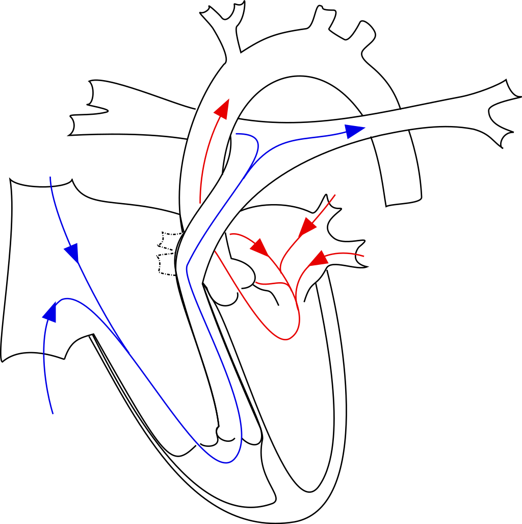 Image Source: Wikimedia Commons (2008). Ebstein’s anomaly, Licensed under a creative commons licence. Available for free at: https://en.wikipedia.org/wiki/Ebstein%27s_anomaly (Accessed: 26 January 2024).
Clinical presentation
The presence of VSD or PFO with severe tricuspid regurgitation leads to right-to-left shunting of blood, resulting in cyanosis. Subtle defects may not be apparent until adulthood, which then results in non-specific symptoms such as decreased exercise tolerance, palpitations, shortness of breath or syncope. Paradoxical embolic events may also occur. Physical examination may reveal cyanosis, clubbing and the pansystolic regurgitant murmur of tricuspid regurgitation.
Investigations
The ECG will show right atrial enlargement with “Himalayan” P waves, prolonged PR interval and right bundle branch block. There may be low volume QRS complexes, pre-excitation in the form of delta waves and arrhythmias including atrial fibrillation and flutter may be present.
The diagnosis can be confirmed with echocardiography showing apical displacement of the septal tricuspid leaflet. The degree of displacement affects the severity of the disease. Other findings include tricuspid regurgitation, right atrium and ventricular dilatation and associated structural defects such as PFO, ASD and VSD.
Management
The treatment and prognosis depend on the severity of the disease. Asymptomatic patients require routine monitoring with surgery indicated for symptomatic adults. Medical therapy is used as a bridge towards surgery or for symptomatic control, which includes diuretics for volume overload, anticoagulation for arrhythmias and standard heart failure treatment. Antibiotic prophylaxis is indicated in cyanotic patients with Ebstein anomaly, patients with previous infective endocarditis; six months after surgical repair or in those with prosthetic material used for repair.
Indications for surgery include progressive RV failure; cyanosis with resting oxygen saturation < 90%, paradoxical embolism (ASD), arrhythmias, progressive cardiomegaly and severe tricuspid regurgitation with NYHA III-IV.
Eisenmenger Syndrome
Eisenmenger syndrome occurs as a result of a chronic process in which an uncorrected left-to-right shunt leads to increased pulmonary blood flow, causing pathological remodelling of the pulmonary vasculature and finally, pulmonary hypertension. It consists of the triad:
- Congenital structural defect causing initial left-to-right shunt
- Subsequent pulmonary hypertension causing shunt reversal from right-to-left
- Cyanosis
Clinical manifestations include clubbing, raised JVP, central cyanosis, right parasternal heave and loud P2. Complications of Eisenmenger syndrome include:
- RV dilatation and dysfunction
- Cyanotic heart disease- erythrocytosis; bleeding
- Intrapulmonary haemorrhage / haemoptysis
- Pulmonary artery aneurysm
- Pulmonary thrombosis
- Stroke/TIA – Paradoxical embolism
- Brain abscess
- Arrhythmias
- Sudden cardiac death
- Infective endocarditis
Investigations
The ECG may reveal atrial tachyarrhythmias with right axis deviation, tall P waves and RBBB. The chest radiograph can show dilatation of central pulmonary arteries and right chamber dilatation. Diagnosis is with an echocardiogram, which can identify and evaluate anatomical defects. Coronary angiography is used to assess pulmonary arterial pressures.
Management
Treatment of pulmonary hypertension in Eisenmenger syndrome is with endothelin antagonists and PDE-V inhibitors. Potential treatments include diuretics, antiarrhythmics and anticoagulation while selected patients may be considered for lung +/- heart transplant for refractory symptoms with one year survival < 50%
Pregnancy with Eisenmenger physiology is considered high risk and is generally contraindicated. For surgical procedures in a patient with Eisenmenger physiology, antibiotic prophylaxis is recommended.
References
- Hoffman JI, Kaplan S. The incidence of congenital heart disease. J Am Coll Cardiol. 2002;39(12):1890-1900. doi:10.1016/s0735-1097(02)01886-7
- Rao PS. Diagnosis and management of cyanotic congenital heart disease: part I. Indian J Pediatr. 2009;76(1):57-70. doi:10.1007/s12098-009-0030-4
- Holst KA, Connolly HM, Dearani JA. Ebstein’s Anomaly. Methodist Debakey Cardiovasc J. 2019;15(2):138-144. doi:10.14797/mdcj-15-2-138
- Basit H, Wallen TJ, Sergent BN. Eisenmenger Syndrome. In: StatPearls. Treasure Island (FL): StatPearls Publishing; February 13, 2023.
- Huang JB, Liang J, Zhou LY. Eisenmenger syndrome: not always inoperable. Respir Care. 2012;57(9):1488-1495. doi:10.4187/respcare.01418
- Baumgartner H, De Backer J, Babu-Narayan SV, et al. 2020 ESC Guidelines for the management of adult congenital heart disease. Eur Heart J. 2021;42(6):563-645. doi:10.1093/eurheartj/ehaa554
- Parker SE, Mai CT, Canfield MA, et al. Updated National Birth Prevalence estimates for selected birth defects in the United States, 2004-2006. Birth Defects Res A Clin Mol Teratol. 2010;88(12):1008-1016. doi:10.1002/bdra.20735
- Hutter PA, Kreb DL, Mantel SF, Hitchcock JF, Meijboom EJ, Bennink GB. Twenty-five years’ experience with the arterial switch operation. J Thorac Cardiovasc Surg. 2002;124(4):790-797. doi:10.1067/mtc.2002.120714
- Stout KK, Daniels CJ, Aboulhosn JA, et al. 2018 AHA/ACC Guideline for the Management of Adults With Congenital Heart Disease: A Report of the American College of Cardiology/American Heart Association Task Force on Clinical Practice Guidelines [published correction appears in J Am Coll Cardiol. 2019 May 14;73(18):2361-2362]. J Am Coll Cardiol. 2019;73(12):e81-e192. doi:10.1016/j.jacc.2018.08.1029
- McMullan DM, Permut LC, Jones TK, Johnston TA, Rubio AE. Modified Blalock-Taussig shunt versus ductal stenting for palliation of cardiac lesions with inadequate pulmonary blood flow. J Thorac Cardiovasc Surg. 2014;147(1):397-401. doi:10.1016/j.jtcvs.2013.07.052
- HEALEY JE Jr. An anatomic survey of anomalous pulmonary veins: their clinical significance. J Thorac Surg. 1952;23(5):433-444.
Coarctation of the Aorta
Introduction
Coarctation of the aorta (CoA) is a congenital stenosis which forms in the region of the ligamentum arteriosum at the descending aorta. It accounts for 4-6% of all congenital cardiovascular defects, with a prevalence of approximately 4 per 10,000 live births. Most cases are sporadic, with a male predominance. The severity of CoA depends on the degree of coarctation and narrowing. As a response to increased left ventricular afterload from left ventricular outflow tract obstruction, remodelling in the form of left ventricular hypertrophy occurs. There is also development of collateral blood supply from the mammary, intercostal and scapular vessels to bypass the stenosis. Amongst patients with CoA, up to 85% of patients have a bicuspid aortic valve (mainly fusion of the left and right coronary cusps).
Up to 35% of patients with Turner’s syndrome have CoA. Other associated abnormalities include:
- Ventricular septal defect
- Patent ductus arteriosus
- Valvular and subvalvular aortic stenosis
- Congenital mitral valve abnormalities
- Aneurysm of the Circle of Willis
- Aortopathies (aortic aneurysm and dissection)
- Collateral circulation within the spinal canal causing spinal subarachnoid haemorrhage
Clinical presentation
Most adults remain asymptomatic unless severe hypertension develops, which may lead to headaches and epistaxis. Lower limb claudication may also develop due to reduced blood flow. Typical findings on examination include hypertension of the upper limbs, diminished blood pressure of the lower limbs and radio-femoral delay. An ejection systolic murmur can be heard anteriorly or posteriorly. A continuous murmur in the left parasternal border may also be heard due to the presence of large collateral vessels.
Investigations
The chest radiograph will show indentation of the aorta at the site of CoA and notching of ribs due to erosion by large collateral arteries.
Echocardiography provides information on the site and extent of CoA, as well as the presence of LVH and associated cardiac defects. Colour Doppler may show increased turbulence in the descending aorta. Cardiac catheterisation studies can demonstrate pressure gradients between the left ventricle and femoral artery. Imaging of the entire aorta is usually performed with CT or CMR.
Management
In adult patients with CoA, transcatheter intervention using covered stents has become the first-choice treatment. The prognosis after CoA repair is generally good, with a >90% 10-year survival rate. It is important to note that CoA repair does not prevent the late recurrence of systemic hypertension. Patients should be reviewed regularly at the clinic and arterial hypertension should be treated according to the 2018 ESC guidelines. Echocardiography should be performed periodically for structural assessment post-repair.
The indications for intervention include radiological evidence of significant collateral flow; heart failure and systemic hypertension secondary to CoA, CoA with ≥50% narrowing relative to the aortic diameter and where the CoA gradient >20mmHg in hypertensive patients.
Antibiotic prophylaxis is not recommended in CoA except for the first 6 months after stenting of CoA or if there has been a history of infective endocarditis.
References
- Abbruzzese PA, Aidala E. Aortic coarctation: an overview. J Cardiovasc Med (Hagerstown). 2007;8(2):123-128. doi:10.2459/01.JCM.0000260215.75535.64
- Baumgartner H, De Backer J, Babu-Narayan SV, et al. 2020 ESC Guidelines for the management of adult congenital heart disease. Eur Heart J. 2021;42(6):563-645. doi:10.1093/eurheartj/ehaa554
- Canniffe C, Ou P, Walsh K, Bonnet D, Celermajer D. Hypertension after repair of aortic coarctation–a systematic review. Int J Cardiol. 2013;167(6):2456-2461. doi:10.1016/j.ijcard.2012.09.084
Complete Guide to Clinical Genetics ST3 Application and Recruitment (2027)
Written by Medibuddy. Last updated: October 2026. Clinical Genetics is

Complete Guide to Clinical Radiology ST3 Application and Recruitment (2027)
Recruitment paused: NHS England is not running Clinical Radiology ST3+ recru

Complete Guide to Paediatric Surgery ST3 Application and Recruitment in United Kingdom (2026)
Written by Medibuddy. Last updated: October 2026. If you’re planning


Take your subscriptions with you
Our mobile app allows you to access your interview and exam question banks wherever you are.

