


 Close
Close
 8th March 2024
Acyanotic Heart Disease
Introduction
Congenital heart disease is the commonest form of congenital anomaly, affecting about 8 in every 1000 live births in the United Kingdom. Diagnostic and management outcomes have significantly improved, which has led to an increasing population of adults with the condition. Adult congenital heart disease (ACHD) can be categorised into acyanotic; cyanotic heart disease and coarctation of the aorta.
Acyanotic heart diseases are congenital cardiac malformations that lead to a left-to-right shunt. Cyanosis is not typically a feature as patients retain a normal level of oxyhaemoglobin saturation in the systemic circulation. However, uncorrected left-to-right shunts may eventually lead to Eisenmenger syndrome. The main types of acyanotic heart defects include:
- Atrial Septal Defect
- Ventricular Septal Defect
- Patent Ductus Arteriosus
Atrial Septal Defect
Atrial septal defect (ASD) is the commonest form of acyanotic heart disease in which a pathological communication is formed that allows shunting of blood between the left and right atrium. It constitutes approximately 5-10% of congenital heart diseases and co-exists with other congenital defects in 30-50% of cases.
A patent foramen ovale (PFO) is a different form of interatrial shunt caused by failure of the foramen ovale to close completely after birth. PFOs are present in 25% of the general population and the majority of adults remain asymptomatic. PFOs may result in paradoxical embolisation of thrombus from the venous circulation into the systemic circulation and the risk increases in the presence of an atrial septal aneurysm.
ASD can be divided into the following subtypes:
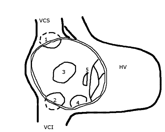
Image Source: Wikimedia Commons (2005). Atrial septal defect, Licensed under a creative commons licence. Available for free at: https://en.wikipedia.org/wiki/Atrial_septal_defect svg (Accessed: 26 January 2024).
Ostium secundum
These constitute 80% of ASDs and are located within, or surrounding, the fossa ovalis. They form as a result of poor development of the septum primum of varying degrees during embryologic development and have associations with other forms of congenital defects, including mitral valve prolapse, pulmonary stenosis and partial anomalous pulmonary venous drainage.
Ostium primum
They account for 15-20% of ASDs and are part of the spectrum of atrioventricular septal defects (AVSD). Primum ASD is caused by failure of the septum primum to fuse with the endocardial cushions at the interatrial septal base and are frequently associated with a cleft in the anterior leaflet of the mitral valve, leading to varying degrees of mitral regurgitation. Partial AVSD includes primum ASD, cleft mitral valve, and ventricular septal defect (VSD). Complete AVSD is characterised by a common AV valve with primum ASD and inlet VSD, often seen in Down’s syndrome.
Sinus venosus defect
Sinus venosus defects constitute 5-10% of ASDs and involve a portion of the atrial wall derived from the sinus venosus but there is no true communication between the right and left atria. Transoesophageal echocardiography (TOE) is generally required for assessment in adults, especially when there is unexplained right chamber dilatation. They can be further subdivided into superior and inferior defects. The commonest of the two is a superior sinus venosus, located below the orifice of the superior vena cava (SVC). The right upper lobe and middle pulmonary veins connect to the junction of the SVC and right atrium or on the SVC itself. An inferior sinus venosus results from the breakdown between the right atrial wall and the inferior right-sided pulmonary veins.
Unroofed coronary sinus
Caused by the absence of the common wall between the coronary sinus and left atrium, this defect accounts for less than 1% of ASDs and is often associated with complete AV septal defect; absence of coronary sinus and a persistent left sided SVC.
Pathophysiology
ASDs cause a left-to-right shunt, which results in volume overload of the RA and RV. The severity depends on the size of the ASD and filling pressures of the ventricles. Factors which heighten the degree of shunting include elevated LV filling pressures in hypertensive cardiomyopathy or increased LA pressure seen in mitral stenosis. In contrast, factors that reduce the degree of left-to-right shunt include pulmonary hypertension or increased RA pressure in tricuspid stenosis.
Clinical presentation
Patients commonly remain asymptomatic until adulthood, where 30% begin to develop breathlessness on exertion by the third decade and more than 75% of patients by the fifth decade. Late complications include supraventricular tachyarrhythmias, severe pulmonary hypertension and right-sided heart failure. Paradoxical embolism causing systemic events may be the first clue to an ASD.
Auscultation will reveal fixed splitting of S2 and a systolic flow murmur at the left second intercostal space. A loud P2 component of the second heart sound indicates pulmonary hypertension. If cyanosis develops, this generally suggests reversal of the shunt (Eisenmenger syndrome).
Investigations
On an ECG, both primum and secundum ASDs display first degree AV block and RSR’ pattern in V1 but can be differentiated by right axis deviation in secundum ASD and left axis deviation in primum ASD.
A chest x-ray may show cardiomegaly and prominent vascular markings.
The first line diagnostic modality is transthoracic echocardiography (TTE), where intravenous contrast studies can be performed to elicit the shunt and colour-flow Doppler can identify intra-arterial flow. The degree of shunting can be assessed using the shunt fraction (Qp/Qs), which is the ratio of pulmonary flow (Qp) to systemic flow (Qs). A left-to-right shunt is considered significant when the ratio of pulmonary to systemic blood flow (Qp/Qs) is >1.5 or when the RV dilates. There may also be functional tricuspid regurgitation, RA and RV enlargement, as well as abnormal septal motion. TOE is optional for further anatomical definition.
Cardiac magnetic resonance (CMR) imaging provides high quality assessment of RV size and function and clear views of the pulmonary veins. It is useful for quantification of pulmonary to systemic flow ratio (Qp:Qs) where either a ratio > 1.5 or evidence of RV dilation confirms a significant shunt.
Cardiac catheterisation is not routinely required for diagnostic purposes except to assess for pulmonary pressures or coronary artery disease. Additional investigations involve oximetry sampling that can demonstrate an oxygen step-up from the vena cava to the right atrium.
Management
Medical management is not typically required as most patients are asymptomatic. Congestive cardiac failure is managed with diuretics and atrial tachyarrhythmias with rate, rhythm, and anticoagulation therapy.
Treatment options include percutaneous and surgical closure and indications for intervention include:
- Dilatation of right chambers without pulmonary artery hypertension (PAH), regardless of symptoms
- Paradoxical embolism
- PAH with marked left-to-right shunt (Qp:Qs>1.5)
A percutaneous approach is the first choice for secundum ASD with adequate margins from AV valves, coronary sinus, origin of vena cava and right pulmonary veins. Surgical repair has a low mortality and a good long-term outcome when performed early during childhood and in the absence of pulmonary hypertension.
ASD closure is contraindicated in irreversible pulmonary hypertension (systolic pulmonary pressure > 2/3 systolic BP) and in Eisenmenger physiology.
Pregnancy
Women with uncomplicated ASD can have uncomplicated pregnancies despite the physiological increase in stroke volume and cardiac output. Venous thromboembolism prophylaxis is important as there exists a risk of paradoxical embolisation.
Antibiotic prophylaxis
The risk of infective endocarditis is higher in those with an ASD than in the general population but the European Society of Cardiology (ESC) guidelines recommend antibiotic prophylaxis in the following:
- Cyanotic congenital heart disease
- Previous infective endocarditis
- Repaired defect with prosthetic material for the first 6 months
- Residual defect after intervention
Ventricular Septal Defect
A ventricular septal defect (VSD) is one of the most common congenital cardiac defects (second to bicuspid aortic valve) with a prevalence of about 20-25% of all congenital heart diseases. Males and females are affected equally. Acquired VSDs, e.g., from myocardial infarction, are not discussed here.
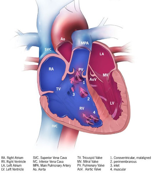
Image Source: Wikimedia Commons (2013). Ventricular septal defect, Licensed under a creative commons licence. Available for free at: https://en.wikipedia.org/wiki/Ventricular_septal_defect (Accessed: 26 January 2024).
There are four subtypes of VSD the commonest of which is membranous VSD, accounting for 80% of defects. They occur within the membranous septum with potential involvement of the tricuspid and aortic valves.
Muscular VSDs are completely surrounded by muscle. They make up to 15-20% of VSDs.
5% of VSDs are outlet/infundibular defects located below the aortic and pulmonary valves. They may be associated with aortic regurgitation.
Inlet VSD, also known as AV canal/AVSD type, typically occur in Down’s syndrome and is found below the AV valves.
Clinical presentation
Most patients with a small VSD will remain asymptomatic and the natural history will depend on the location and size of the defect as these impact the degree of left-to-right shunting.
On auscultation of the heart, a small VSD will produce a holosystolic murmur at the left parasternal border and a palpable systolic thrill. A moderate sized VSD may elicit a mitral diastolic rumble and S3 gallop due to LA and LV dilatation with increased flow across the mitral valve. Large VSD can progress to pulmonary hypertension and Eisenmenger syndrome. Signs of pulmonary hypertension, such as a left parasternal heave and loud P2, may be present. Other findings in large VSD include S4 heart sound, tricuspid regurgitation and Graham Steele murmur.
Complications of VSD include:
- Left heart failure
- Pulmonary hypertension (Cor pulmonale due to pressure and volume overload)
- Tricuspid regurgitation
- Arrhythmias
- Infective endocarditis
- Aortic regurgitation due to leaflet prolapse
- Subaortic stenosis
- Double-chambered right ventricle where a muscle band develops as a result of VSD jet leading to infundibular pulmonary stenosis.
Investigations
The ECG in VSD can show LVH and biphasic QRS complexes in the precordial leads. Echocardiography is useful for colour-flow Doppler, which shows a high-velocity systolic jet across the VSD. If echocardiography provides insufficient data, then CMR is a useful modality. Cardiac catheterisation allows measurement of Qp:Qs, pulmonary pressure and pulmonary vascular resistance.
Management
Asymptomatic patients with small defects do not require intervention apart from routine cardiology follow-up. The following indications warrant intervention with surgical or percutaneous closure:
- Haemodynamically significant VSD in patients with evidence of LV dysfunction
- Prolapse of aortic valve causing moderate aortic regurgitation
- In the presence of severe pulmonary hypertension, closure should only be considered in the case of left-to-right shunt (Qp:Qs ratio >1.5)
- Double chamber RV
- Infective endocarditis
Surgical closure can be performed with low operative mortality and good long-term outcomes. Percutaneous closure can be considered in selected cases, such as muscular and membranous VSDs that are located away from the tricuspid and aortic valves.
Patent Ductus Arteriosus
The patent ductus arteriosus (PDA) is a remnant of the normal embryonic vessel called the ductus arteriosus, which connects the left pulmonary artery to the aorta. If the PDA fails to close after birth, the direction of blood flow leads to a left-to-right shunt. The reported incidence is around 1 in 2000 live births with a 2:1 female predominance. Important risk factors include congenital rubella; premature birth, genetic predisposition and birth at high altitude.

Image Source: Wikimedia Commons (2012). Patent ductus arteriosus, Licensed under a creative commons licence. Available for free at: https://en.wikipedia.org/wiki/Patent_ductus_arteriosus (Accessed: 26 January 2024).
Clinical presentation
Signs and symptoms depend on the size and the length of the PDA (Table 1):
| Silent | Incidental and asymptomatic |
| Small | Qp:Qs <1.5
Asymptomatic with a flow murmur |
| Moderate | Qp:Qs between 1.5 to 2.2
May present with left or right heart failure Continuous machinery murmur at the first left intercostal space +/- thrill |
| Large | Qp:Qs > 2.2
A large PDA initially causes left ventricular overload If uncorrected, severe pulmonary hypertension develops (Eisenmenger physiology) The classic machinery murmur diminishes as pulmonary pressure increases Patients develop cyanosis and clubbing |
Table 1: Clinical presentation of different sized PDA
The complications of PDA include congestive cardiac failure, progressive pulmonary HTN and Eisenmenger syndrome; infective endocarditis, arrhythmias and ductus aneurysms.
Investigations
Echocardiography is important to quantify the degree of LA and LV enlargement; pulmonary artery pressure and size, right heart changes and diastolic flow reversal in the descending aorta. Further evaluation of PDA anatomy can be obtained using CMR. Cardiac catheterisation may be performed to measure pulmonary pressures, pulmonary vascular resistance and flow ratio (Qp:Qs). Oximetry sampling can demonstrate an oxygen step-up at the level of the pulmonary artery.
Management
This depends on the age of the patient, whether they are an infant, child or adult and the size and degree of left-to-right shunting. In preterm infants, pharmacological therapy using cyclo-oxygenase inhibitors such as Indomethacin is used. For adults, treatment is with transcatheter device closure rather than surgical closure, which is reserved for very large PDA, aneurysms or endarteritis.
Indications for PDA closure include small, moderate and large PDAs without Eisenmenger’s physiology and all patients with LV failure and no pulmonary hypertension, regardless of symptoms. Closure is not recommended in severe pulmonary hypertension and Eisenmenger’s physiology with right-to-left shunt due to the risk of life threatening pulmonary oedema.
Cardiac Haemodynamic Assessment
The heart has a dual circulation with deoxygenated blood received via the SVC/IVC into the right atrium, down the AV valve into the RV, and to the lungs via the pulmonary arteries. Once oxygenated, the blood travels to the left atrium via pulmonary veins. This process prevents mixing of oxygen depleted blood from oxygenated blood. The differences in oxygen saturations are shown in the image.

Image Source: Wikimedia Commons (2016). Diagram of the human heart_(cropped, Licensed under a creative commons licence. Available for free at: https://commons.wikimedia.org/wiki/File:Diagram_of_the_human_heart_(cropped).svg (Accessed: 26 January 2024).
Cardiac catheterisation with oxygen studies is useful to identify and localise intracardiac shunts by detecting differences in oxygen saturation values. The normal values for oxygen saturation and pressures at different cardiac sites are presented in Table 2. A left to right shunt is considered if there is a significant increase (>75%) in blood oxygen saturation higher than normal values. Left heart saturations < 94% are suggestive of a right to left shunt.
| Site | O2 saturation | End systolic mmHg | End diastolic mmHg | Mean
mmHg |
|---|---|---|---|---|
| Inferior vena cava | < 75% | – | – | – |
| Superior vena cava | < 75% | – | – | – |
| Right atrium | < 75% | – | – | 1-5 |
| Right ventricle | < 75% | 15-30 | 1-7 | – |
| Pulmonary artery | < 75% | 15-30 | 4-12 | 9-19 |
| Pulmonary capillary wedge pressure | – | – | – | 4-12 |
| Left atrium | 98% | – | – | 2-12 |
| Left ventricle | 98% | 90-140 | 5-12 | – |
| Aorta | 98% | 90-140 | 60-90 | 70-105 |
Table 2: Normal oxygen saturations and pressures at different cardiac sites. Cited from Braunwald’s Heart Disease: A Textbook of Cardiovascular Medicine (Bonow et al, 2022)
Intracardiac shunts can be quantified using the ratio of pulmonary flow (Qp) and total systemic (Qs) blood flow (Table 2).
| Qp/Qs <1.5 | Qp/Qs: 1.5 – 2 | Qp/Qs > 2 |
|---|---|---|
| Small left-to-right shunt | Moderate left-to-right shunt | Large left-to-right shunt |
Table 3. Quantification of intra cardiac shunts using pulmonary blood flow (Qp) and total systemic blood flow (Qs)
References
- Baumgartner H, De Backer J, Babu-Narayan SV, et al. 2020 ESC Guidelines for the management of adult congenital heart disease. Eur Heart J. 2021;42(6):563-645. doi:10.1093/eurheartj/ehaa554
- Hoffman JI, Kaplan S. The incidence of congenital heart disease. J Am Coll Cardiol. 2002;39(12):1890-1900. doi:10.1016/s0735-1097(02)01886-7
- Teshome MK, Najib K, Nwagbara CC, Akinseye OA, Ibebuogu UN. Patent Foramen Ovale: A Comprehensive Review. Curr Probl Cardiol. 2020;45(2):100392. doi:10.1016/j.cpcardiol.2018.08.004
- Geva T, Martins JD, Wald RM. Atrial septal defects. Lancet. 2014;383(9932):1921-1932. doi:10.1016/S0140-6736(13)62145-5
- Bonow Robert O et al. Braunwald’s Heart Disease: A Textbook of Cardiovascular Medicine. Vol. 1. 12th ed. Elsevier 2022.
- Griffin BP. Manual of Cardiovascular Medicine: Wolters Kluwer Health 2018.
Complete Guide to Clinical Genetics ST3 Application and Recruitment (2027)
Written by Medibuddy. Last updated: October 2026. Clinical Genetics is

Complete Guide to Clinical Radiology ST3 Application and Recruitment (2027)
Recruitment paused: NHS England is not running Clinical Radiology ST3+ recru

Complete Guide to Paediatric Surgery ST3 Application and Recruitment in United Kingdom (2026)
Written by Medibuddy. Last updated: October 2026. If you’re planning


Take your subscriptions with you
Our mobile app allows you to access your interview and exam question banks wherever you are.

