


 Close
Close
 8th March 2024
Cyanotic Heart Disease
Introduction
Cyanotic heart disease is the other subdivision of congenital heart disease. They result due to mixed de/oxygenated blood in the systemic circulation or when deoxygenated blood bypasses the pulmonary circulation into the systemic circulation. The occurrence is usually sporadic but may be associated with genetic syndromes including trisomy 21, 13, 18, and Turner syndrome. Environmental risk factors include maternal exposure to drugs/toxins and viral infections during pregnancy. The most commonly occurring congenital cyanotic heart condition is Tetralogy of Fallot, accounting for 7-10% of cyanotic heart disease.
Truncus Arteriosus
This cyanotic birth defect occurs when the embryological truncus arteriosus fails to separate into the pulmonary trunk and aorta. This occurs due to the lack of aortico-pulmonary septum formation and results in a single artery, which arises from both ventricles becoming the aortic and pulmonary vessels. The result is a supply of mixed de/oxygenated blood to the coronary arteries, pulmonary arteries, and systemic circulation.
The condition occurs spontaneously but is also associated with deletions in chromosome 22q11 (DiGeorge syndrome). Cyanosis presents at birth and heart failure may develop in weeks. On cardiac auscultation, a systolic murmur may be heard at the left sternal edge.
Management is with surgical repair to separate the pulmonary artery from the common artery. Once separated, the common artery functions as the aorta.
Transposition of Great Arteries
Transposition of the great arteries (TGA) is the second most common cyanotic heart disease, typically presenting in the first week of birth. It occurs with a prevalence of 20-30 in 100,000 live births and is more common in males (2:1). In TGA, the aorta originates from the morphological RV and the pulmonary artery originates from the morphologic LV.
There are two main classifications: dextro-TGA (D-TGA), where the aorta is anterior and to the right of the pulmonary artery. The aorta arises from the RV and pulmonary artery from the LV.
In levo-TGA (L-TGA), the aorta is anterior but to the left of the pulmonary artery.
Associated abnormalities include VSD, LVOT obstruction, mitral and tricuspid valve abnormalities and coarctation of the aorta, which can be diagnosed using an echocardiogram.
Management
TGA operations include the arterial and atrial switch. Arterial switch surgery is used to correct D-TGA and involves moving the pulmonary artery and aorta into their normal positions to achieve ventriculoarterial concordance.
Where the arterial switch is not possible due to anatomy, the atrial switch is a procedure which involves redirecting the systemic venous return from the vena cava using a conduit (baffle) made from the pericardium to the LV.
Post-surgery lifelong follow up care with cardiology is needed. Without surgical repair, the condition is incompatible with life; therefore, adult patients who present to specialist cardiology clinics would have undergone surgical correction of TGA. Clinical signs include loud A2; left parasternal heave and holosystolic murmur of the systemic AV valve. There may be symptoms of right/left heart failure and arrhythmias can occur.
Tricuspid Atresia
This is the third most common form of cyanotic congenital heart disease and is characterised by an absent tricuspid valve and no direct communication between the RA and RV. The defect occurs during prenatal development and results in an absent or hypoplastic RV. If left untreated, the mortality rate is high (90%) at 1 year of age.
 Image Source: Wikimedia Commons (2009). Tricuspid atresia, Licensed under a creative commons licence. Available for free at: https://en.wikipedia.org/wiki/Tricuspid_atresia (Accessed: 26 January 2024).
In many cases of tricuspid atresia, additional defects are present, which allow the exchange of blood between the systemic and pulmonary circulation. Associated conditions include:
- ASD
- VSD
- Pulmonary outflow obstruction
- Ventricular arterial discordance, including TGA
- Pulmonary artery anomalies
Diagnosis
Most cases of tricuspid atresia are diagnosed during a routine anomaly scan. Central cyanosis is the most noticeable feature on physical examination. There may be tachypnoea in the first two weeks of life and a holosystolic murmur due to VSD. Diagnosis can be confirmed using an echocardiogram.
Management
In neonates, prostaglandin E1 (PGE1) is given initially to maintain patency of the ductus arteriosus. Single-ventricular palliative surgery is then performed to ensure adequate separation between the pulmonary and systemic circulation.
Palliative surgical intervention is usually performed in three stages:
- Modified Blalock-Thomas-Taussig procedure: A Gore-Tex conduit is used to connect the subclavian artery to the pulmonary artery
- Cavo-pulmonary anastomosis (Bi-directional Glenn procedure): This includes redirecting the oxygen-depleted blood from the upper parts of the body to the lungs by connecting the superior vena cava to the right pulmonary artery.
- Fontan procedure: Venous blood is diverted from the IVC to the pulmonary arteries. This creates a system in which the entire venous system is now flowing passively into the pulmonary arteries. The single ventricle pumps oxygenated blood out of the left ventricular outflow tract (LVOT) into the systemic circulation, thus relieving cyanosis.
Tetralogy of Fallot
Tetralogy of Fallot (TOF) is one of the commonest congenital heart diseases requiring intervention in the first year of life. Its prevalence in the UK is approximately 1 in 3600 live births with equal occurrence in males and females. It is characterised by four cardiac defects, which can be remembered by the mnemonic “International House Of Pancakes:”
- I – Interventricular septal defect
- H – Hypertrophic RV
- O – Overriding aorta
- P – Pulmonary stenosis (RVOT obstruction)
TOF may occur in isolation or with genetic conditions, including DiGeorge syndrome (22q11 deletion) and Down syndrome (trisomy 21). It is associated with cardiac features including right aortic arch; persistent left SVC and anomalous origin of the left anterior descending artery (LAD) from the right coronary artery. Pentalogy of Fallot describes TOF with associated (secundum) ASD.
 Image Source: Wikimedia Commons (2006). Tetralogy of Fallot, Licensed under a creative commons licence. Available for free at: https://en.wikipedia.org/wiki/Tetralogy_of_Fallot (Accessed: 26 January 2024).
Pathophysiology
The degree of right ventricular outflow tract (RVOT) obstruction affects the severity of the disease. The VSD is typically large and unrestrictive, which means that the direction of blood flow across the VSD is dependent on the resistance to blood flow out of the right and left ventricular outflow tracts. As the degree of RVOT obstruction increases in TOF, the resistance to blood flow into the pulmonary circulation increases. If this pressure exceeds the systemic vascular resistance, it becomes easier for deoxygenated blood to flow from the RV into the LV across the large VSD, causing cyanosis. One of the pathophysiologic characteristics of TOF is a dynamic RVOT obstruction ranging from minimal obstruction to near-total occlusion, resulting in “tet spells’ (a short period when a child’s skin turns blue).
Clinical presentation
Most adult patients with underlying TOF will have had surgical intervention with or without a prior palliative procedure. Without surgical repair, only 10% of patients survive beyond 20 years of age. Patients with previous palliative repair can present with variable clinical findings, depending on the procedure performed. Those who have undergone total repair are often asymptomatic or may present with late symptoms, which include palpitations, reduced exercise tolerance, shortness of breath, syncope or heart failure. Post repair clinical findings may reveal:
- Raised JVP
- Left parasternal heave – RV hypertrophy
- Soft/absent P2 heart sound
- Soft diastolic murmur suggestive of pulmonary regurgitation
- Pansystolic murmur in residual VSD (due to a leak in the VSD patch)
- Early diastolic murmur suggestive of aortic regurgitation
- Continuous murmurs from aortopulmonary collateral supply
Investigations
Chest radiography, ECG and echocardiography are performed routinely for all patients undergoing paediatric cardiac surgery.
Post correction, the ECG may reveal RBBB and evidence of RVH. A QRS duration of > 180 ms is a predictor of ventricular arrhythmias.
The chest x-ray will show a ‘boot-shaped’ heart due to RVH causing an upturned apex. There may be prominent pulmonary vascular markings and presence of a right sided aortic arch.
Echocardiography is useful in identifying residual VSD and the presence of aortic regurgitation. CMR can help to evaluate the right ventricle, previously placed shunts and aortopulmonary collaterals; quantify pulmonary regurgitation and identify scar tissue as well as associated cardiac defects (as mentioned above).
Invasive cardiac catheterisation can assess levels of RVOT obstruction, pulmonary hypertension and residual shunts in addition to coronary artery anatomy.
Management
Most patients with TOF undergo complete repair by 1 year of age. A small number of neonates require initial palliative repair in the form of shunts or stents due to severe RVOT obstruction. Surgical repair aims to relieve RVOT obstruction, separate the systemic and pulmonary circulations by closure of the VSD, preserve RV function and minimise post-surgery valvular complications.
Ventricular arrhythmias are an important cause of mortality in TOF patients after surgery. Secondary prevention with an implantable cardioverter defibrillator (ICD) is appropriate in patients with documented sustained VT or those who have survived an episode of resuscitated cardiac arrest. The indication for a primary prevention ICD is individualised.
Follow up
The long-term prognosis is generally good in repaired TOF, with studies reporting survival rates exceeding 94% at 25 years.
Routine follow-up with a congenital cardiologist is required to monitor post-operative complications. Echocardiography and ECG are performed yearly until the age of 10 and every two years thereafter. Other investigations, such as an exercise stress test, CMR and Holter monitor, are performed roughly every 3-4 years but sooner if the need arises.
Antibiotic prophylaxis is recommended in the first 6 months after TOF surgery or in those with residual defects after intervention.
Pregnancy is not recommended in women with unrepaired TOF. The decision for pregnancy after surgery should be individualised based on the careful evaluation made by the congenital cardiac team in order to adequately risk-stratify the patient.
Total Anomalous Pulmonary Venous Drainage
Total anomalous pulmonary venous drainage (TAPVD) is a cyanotic heart disease in which all four pulmonary veins fail to drain into the left atrium and instead drain abnormally into the systemic venous circulation via various other pathways. The incidence of TAPVD is between 0.6 to 1.2 per 10,000 live births. Without definitive surgical correction, the mortality rate is approximately 80% by one year of age.
Signs include right ventricular heave, loud S1 and systolic ejection murmur at the left upper sternal border. The ECG will show right axis deviation and the chest radiograph may reveal a Snowman/figure of 8/cottage loaf sign.
Partial anomalous pulmonary venous drainage (PAPVD) occurs when blood from one or more pulmonary veins return abnormally into the right atrium. PAPVD occurs in approximately 0.7% of the population and is commonly associated with ASD and Turner syndrome. Patients can remain asymptomatic with small defects, whereas symptomatic cyanotic patients would require definitive surgical intervention where any associated heart defects such as ASD, VSD, PFO and PDA are surgically closed.
The 5 T’s
One way to remember the five topics presented above is using the method presented below:
- Truncus arteriosus (1 vessel)
- Transposition of great vessels (2 vessels)
- Tricuspid atresia (Tri=3)
- Tetralogy of Fallot (Tetra=4)
- Total anomalous pulmonary venous drainage (5 letters in the name)
Ebstein Anomaly
This congenital abnormality is characterised by the apical displacement of the tricuspid valve with elongated tricuspid valve leaflets, leading to the atrialisation of the RV and dilation of the true tricuspid annulus. The incidence is approximately 1.2 to 5 in 100,000 live births. Although there is an increased risk in mothers taking lithium during early pregnancy, most cases are sporadic.
Associated cardiac defects include PFO, ASD, VSD, pulmonary stenosis, and accessory pathways predisposing patients to tachyarrhythmias and left heart defects such as mitral valve prolapse or bicuspid aortic valve.
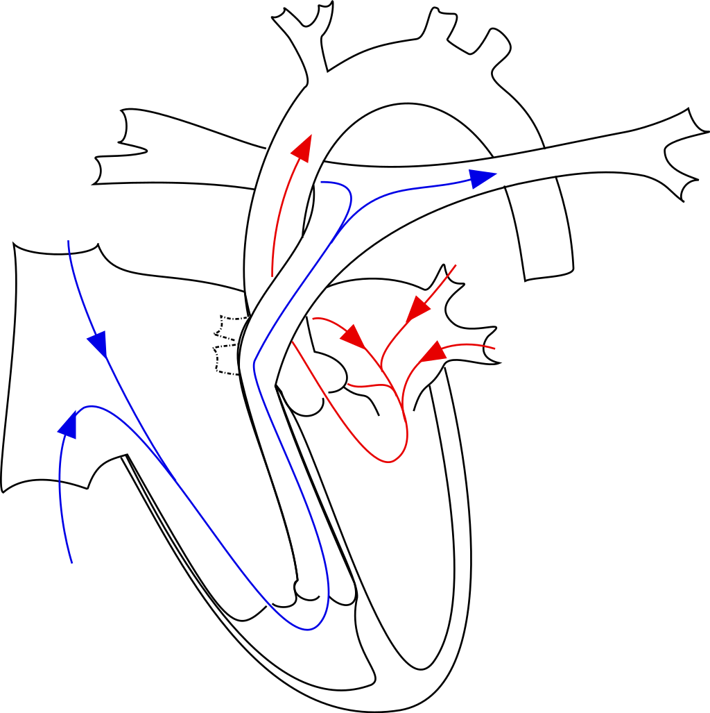 Image Source: Wikimedia Commons (2008). Ebstein’s anomaly, Licensed under a creative commons licence. Available for free at: https://en.wikipedia.org/wiki/Ebstein%27s_anomaly (Accessed: 26 January 2024).
Clinical presentation
The presence of VSD or PFO with severe tricuspid regurgitation leads to right-to-left shunting of blood, resulting in cyanosis. Subtle defects may not be apparent until adulthood, which then results in non-specific symptoms such as decreased exercise tolerance, palpitations, shortness of breath or syncope. Paradoxical embolic events may also occur. Physical examination may reveal cyanosis, clubbing and the pansystolic regurgitant murmur of tricuspid regurgitation.
Investigations
The ECG will show right atrial enlargement with “Himalayan” P waves, prolonged PR interval and right bundle branch block. There may be low volume QRS complexes, pre-excitation in the form of delta waves and arrhythmias including atrial fibrillation and flutter may be present.
The diagnosis can be confirmed with echocardiography showing apical displacement of the septal tricuspid leaflet. The degree of displacement affects the severity of the disease. Other findings include tricuspid regurgitation, right atrium and ventricular dilatation and associated structural defects such as PFO, ASD and VSD.
Management
The treatment and prognosis depend on the severity of the disease. Asymptomatic patients require routine monitoring with surgery indicated for symptomatic adults. Medical therapy is used as a bridge towards surgery or for symptomatic control, which includes diuretics for volume overload, anticoagulation for arrhythmias and standard heart failure treatment. Antibiotic prophylaxis is indicated in cyanotic patients with Ebstein anomaly, patients with previous infective endocarditis; six months after surgical repair or in those with prosthetic material used for repair.
Indications for surgery include progressive RV failure; cyanosis with resting oxygen saturation < 90%, paradoxical embolism (ASD), arrhythmias, progressive cardiomegaly and severe tricuspid regurgitation with NYHA III-IV.
Eisenmenger Syndrome
Eisenmenger syndrome occurs as a result of a chronic process in which an uncorrected left-to-right shunt leads to increased pulmonary blood flow, causing pathological remodelling of the pulmonary vasculature and finally, pulmonary hypertension. It consists of the triad:
- Congenital structural defect causing initial left-to-right shunt
- Subsequent pulmonary hypertension causing shunt reversal from right-to-left
- Cyanosis
Clinical manifestations include clubbing, raised JVP, central cyanosis, right parasternal heave and loud P2. Complications of Eisenmenger syndrome include:
- RV dilatation and dysfunction
- Cyanotic heart disease- erythrocytosis; bleeding
- Intrapulmonary haemorrhage / haemoptysis
- Pulmonary artery aneurysm
- Pulmonary thrombosis
- Stroke/TIA – Paradoxical embolism
- Brain abscess
- Arrhythmias
- Sudden cardiac death
- Infective endocarditis
Investigations
The ECG may reveal atrial tachyarrhythmias with right axis deviation, tall P waves and RBBB. The chest radiograph can show dilatation of central pulmonary arteries and right chamber dilatation. Diagnosis is with an echocardiogram, which can identify and evaluate anatomical defects. Coronary angiography is used to assess pulmonary arterial pressures.
Management
Treatment of pulmonary hypertension in Eisenmenger syndrome is with endothelin antagonists and PDE-V inhibitors. Potential treatments include diuretics, antiarrhythmics and anticoagulation while selected patients may be considered for lung +/- heart transplant for refractory symptoms with one year survival < 50%
Pregnancy with Eisenmenger physiology is considered high risk and is generally contraindicated. For surgical procedures in a patient with Eisenmenger physiology, antibiotic prophylaxis is recommended.
References
- Hoffman JI, Kaplan S. The incidence of congenital heart disease. J Am Coll Cardiol. 2002;39(12):1890-1900. doi:10.1016/s0735-1097(02)01886-7
- Rao PS. Diagnosis and management of cyanotic congenital heart disease: part I. Indian J Pediatr. 2009;76(1):57-70. doi:10.1007/s12098-009-0030-4
- Holst KA, Connolly HM, Dearani JA. Ebstein’s Anomaly. Methodist Debakey Cardiovasc J. 2019;15(2):138-144. doi:10.14797/mdcj-15-2-138
- Basit H, Wallen TJ, Sergent BN. Eisenmenger Syndrome. In: StatPearls. Treasure Island (FL): StatPearls Publishing; February 13, 2023.
- Huang JB, Liang J, Zhou LY. Eisenmenger syndrome: not always inoperable. Respir Care. 2012;57(9):1488-1495. doi:10.4187/respcare.01418
- Baumgartner H, De Backer J, Babu-Narayan SV, et al. 2020 ESC Guidelines for the management of adult congenital heart disease. Eur Heart J. 2021;42(6):563-645. doi:10.1093/eurheartj/ehaa554
- Parker SE, Mai CT, Canfield MA, et al. Updated National Birth Prevalence estimates for selected birth defects in the United States, 2004-2006. Birth Defects Res A Clin Mol Teratol. 2010;88(12):1008-1016. doi:10.1002/bdra.20735
- Hutter PA, Kreb DL, Mantel SF, Hitchcock JF, Meijboom EJ, Bennink GB. Twenty-five years’ experience with the arterial switch operation. J Thorac Cardiovasc Surg. 2002;124(4):790-797. doi:10.1067/mtc.2002.120714
- Stout KK, Daniels CJ, Aboulhosn JA, et al. 2018 AHA/ACC Guideline for the Management of Adults With Congenital Heart Disease: A Report of the American College of Cardiology/American Heart Association Task Force on Clinical Practice Guidelines [published correction appears in J Am Coll Cardiol. 2019 May 14;73(18):2361-2362]. J Am Coll Cardiol. 2019;73(12):e81-e192. doi:10.1016/j.jacc.2018.08.1029
- McMullan DM, Permut LC, Jones TK, Johnston TA, Rubio AE. Modified Blalock-Taussig shunt versus ductal stenting for palliation of cardiac lesions with inadequate pulmonary blood flow. J Thorac Cardiovasc Surg. 2014;147(1):397-401. doi:10.1016/j.jtcvs.2013.07.052
- HEALEY JE Jr. An anatomic survey of anomalous pulmonary veins: their clinical significance. J Thorac Surg. 1952;23(5):433-444.
Complete Guide to Clinical Genetics ST3 Application and Recruitment (2027)
Written by Medibuddy. Last updated: October 2026. Clinical Genetics is

Complete Guide to Clinical Radiology ST3 Application and Recruitment (2027)
Recruitment paused: NHS England is not running Clinical Radiology ST3+ recru

Complete Guide to Paediatric Surgery ST3 Application and Recruitment in United Kingdom (2026)
Written by Medibuddy. Last updated: October 2026. If you’re planning


Take your subscriptions with you
Our mobile app allows you to access your interview and exam question banks wherever you are.

