


 Close
Close
 5th April 2024
Thyroid Disorders
Regulation of thyroid hormones
Physiology
Thyrotropin releasing hormone (TRH) from the hypothalamus stimulates the production of thyroid stimulating hormone (TSH) from the anterior pituitary. TSH, in turn, stimulates the production of T4 (a pro-hormone which is converted to T3 in the liver and kidney) and T3 (an active form which acts on nuclear receptors). Thyroid hormones are mainly protein bound in circulation. The free T4 and T3 exert negative feedback on TRH and TSH, suppressing their release. Where there is thyroid hormone derangement, a good understanding of this negative feedback mechanism will help to determine the likely aetiology (see Table 1).
To summarise the pattern of derangement:
- Raised T3/T4 with low TSH suggests overproduction of thyroid hormones. Causes include: Graves, toxic multinodular goitre, postpartum thyroiditis, subacute thyroiditis, amiodarone induced thyrotoxicosis
- Raised TSH with low T3/T4 suggests a problem with the production of thyroid hormones. Examples listed in table 2
- Low TSH with low T3/T4 implies pathology in the hypothalamic- pituitary axis, that is, lack of production of TRH/TSH leading to low levels of T3/T4. Examples listed in table 2
In addition to the above, physiological and pharmacological factors can affect thyroid hormones. This includes pregnancy, where there is an increase in production and reduced excretion of thyroxine binding globulin (TBG) by oestrogen. In addition, the human chorionic gonadotropin hormone stimulates production of thyroid hormones. This reduces production of TSH through negative feedback. The biochemical picture is raised total T3/T4, normal free T3/T4 and low TSH. These physiological changes rarely cause clinical problems.
Amiodarone can cause both hyper and hypothyroidism. Amiodarone induced thyrotoxicosis (AIT) can either be due to excess iodine load increasing thyroid hormone production (type I AIT) or inflammation (type II AIT). Type I AIT usually presents with a goitre and is associated with an underlying thyroid disorder. Cessation of amiodarone and switch to another anti-arrhythmic should be considered. Generally, type I AIT is treated with antithyroid medication as the underlying process is increased production and type II is treated with steroids as the underlying process is thyroiditis.
Amiodarone induced hypothyroidism is caused by excess iodine inhibiting iodide organification and T4 and T3 synthesis. In such cases, it is not essential to stop amiodarone but levothyroxine can be commenced to achieve an euthyroid state.
Sick euthyroid syndrome
This results from a reduction in binding proteins, reduced peripheral conversion of T4 to T3 and reduced production of TSH. This should be suspected in patients with critical systemic illnesses, deprivation of calories and following major surgery. The biochemical picture is of low T3, low/normal T4 and low/normal TSH. Thyroid function tests should be repeated once the patient has recovered from their illness.
| TSH | T3 | T4 | Likely disorder | Additional features to support diagnosis |
| Low | Raised | Raised | Hyperthyroidism | |
| Grave’s disease | Exophthalmos and/or ophthalmoplegia, pretibial myxoedema and thyroid acropachy | |||
| Toxic multinodular goitre | Goitre with multiple nodules.
Patchy uptake on nuclear scintigraphy |
|||
| Raised | Low | Low | Primary hypothyroidism | |
| Autoimmune | Goitre in Hashimoto’s, no goitre in atrophic | |||
| Riedel’s thyroiditis -70% hypothyroid picture | Hard, fixed goitre, painless. Compression symptoms Retroperitoneal fibrosis. Anti-thyroid peroxidase antibodies | |||
| Low | Low | Low | Secondary hypothyroidism | Features of co-existing hypothalamic/pituitary disease such as hypogonadism or adrenal insufficiency |
| Normal or low | Low | Low or normal | Sick euthyroid syndrome | Features of systemic illness |
| Raised | Normal | Normal | Subclinical hypothyroidism | Often asymptomatic |
| Low | Normal | Normal | Subclinical hyperthyroidism | Often asymptomatic or may have signs/symptoms of underlying diagnosis such as multinodular goitre, Grave’s disease |
References
- National Institute for Health and Care Excellence (UK). Thyroid disease assessment and management. Published 9ᵗʰ November 2019. Available at: https://www.nice.org.uk/guidance/ng145/chapter/recommendations
- UpToDate. Laboratory assessment of thyroid function. Last updated 5ᵗʰ October 2021. Accessed Available at: https://www.uptodate.com/contents/laboratory-assessment-of-thyroid-function?search=thyroid%20function%20tests&source=search_result&selectedTitle=1~150&usage_type=default&display_rank=1
- 2018 European Thyroid Association (ETA) Guidelines for the Management of Amiodarone-associated Thyroid Dysfunction. European thyroid journal. Eur Thyroid J 2018;7:55–66. Available at: https://www.karger.com/Article/Fulltext/486957
- Euthyroid sick syndrome. Stat Pearls. Last updated 30ᵗʰ October 2021. Available at: https://www.ncbi.nlm.nih.gov/books/NBK482219/
Thyroid disorders
Hyperthyroidism/thyrotoxicosis
Affecting females more than males, several disorders can cause thyrotoxicosis, of which Grave’s disease is the commonest cause. This autoimmune disorder occurs where thyroid stimulating antibodies stimulate thyroid gland growth and thyroid hormone production. Clinical features include
- Exophthalmos and/or ophthalmoplegia
- Pretibial myxoedema
- Thyroid acropachy, which consists of digital clubbing, soft tissue swelling of hands and feet and periosteal new bone formation
Investigations
Thyroid function tests will reveal raised T3/T4 and suppressed TSH. TSH receptor antibodies are present in 90% of cases, whilst anti-thyroid peroxidase antibodies are seen in 75%. On radionuclide testing, there is increased homogenous uptake.
Management
Carbimazole is a thioamide drug used to lower thyroid hormone levels by inhibiting thyroid peroxidase. It can cause agranulocytosis and patients are counselled to seek medical advice in the event of developing an unexplained sore throat, to have their FBC checked. There is an increased risk of congenital malformation when carbimazole is administered in the first trimester of pregnancy, where propylthiouracil is the preferred option.
Propranolol can help to control sympathomimetic symptoms of hyperthyroidism. In refractory cases, radioactive iodine or surgery should be considered. Contraindications to radioactive iodine include pregnancy, breastfeeding and uncontrolled thyroid eye disease. Pregnancy should be avoided for up to 6 months after radioactive iodine.
Other causes of hyperthyroidism and distinguishing features include:
- Toxic multinodular goitre, which is more common in elderly women, presenting with a goitre and multiple nodules. There is an absence of eye signs and thyroid acropachy. On the radioiodine uptake scan there is patchy uptake and treatment is with radioactive iodine
- De Quervain’s thyroiditis/subacute thyroiditis occurs secondary to infection. Accompanying features include malaise, fever and pain in the neck. Suppression of uptake is seen in the acute phase on radioiodine scan. Hypothyroidism usually follows
- Solitary toxic adenoma occurs when a single nodule grows on the thyroid gland causing excess thyroid hormone production. It is confirmed by the increased radionuclide uptake in the nodule and a decreased uptake in the surrounding extra nodular thyroid tissue. Management is with radioactive iodine or surgery
- Subclinical hyperthyroidism is a biochemical picture of low TSH but normal T3 and T4. It is associated with an increased risk of atrial fibrillation and decreased bone mineral density. TFTs should be repeated in these patients
Hypothyroidism
Hypothyroidism can be primary as a result of thyroid gland disease or secondary due to dysregulation of the hypothalamic-pituitary axis (Table 2). Co-existing hormonal deficiencies causing hypogonadism or adrenal insufficiency can occur with the latter, making isolated symptoms associated with hypothyroidism difficult to identify.
| Primary hypothyroidism | Secondary hypothyroidism |
|---|---|
| Iron deficiency
Autoimmune thyroiditis Post-ablative therapy or surgery Drugs (carbimazole, propylthiouracil, amiodarone, lithium, interferon alpha, rifampicin) Thyroiditis (subacute, postpartum) Infiltrative disorder (amyloidosis, sarcoidosis, tuberculosis, lymphoma, metastasis) |
Pituitary or hypothalamic dysfunction as a result of:
Tumours (pituitary adenomas or gliomas) Surgery, radiotherapy, trauma Pituitary infarction Sheehan’s syndrome Infiltrative disorder: amyloidosis, sarcoidosis Drugs (cocaine, dopamine, glucocorticoids) |
Long-term sub-optimal hypothyroidism can contribute to hyperlipidaemia and metabolic syndrome, increasing the risk of coronary artery disease and stroke. The commonest cause of primary hypothyroidism in iodine sufficient areas of the world is Hashimoto’s thyroiditis. It is characterised by lymphocytic infiltration and autoimmune destruction of the thyroid gland.
Clinical features
- Goitre, although no goitre is seen in atrophic thyroiditis
- Fatigue
- Constipation, cold intolerance and coarse hair/skin
- Slowing of speech and movement, low mood
- Puffy face, enlarged tongue and voice hoarseness
TFT shows raised TSH with low T3/T4. Anti-thyroid peroxidase (TPO) and anti-thyroglobulin (Tg) antibodies should be checked. Management is with levothyroxine. TSH levels should be checked every 3 months and annually once thyroid function is stable.
Other causes of hypothyroidism and differentiating factors include:
- Iodine, where deficiency or excess can cause hypothyroidism. Iodine deficiency is the leading cause of hypothyroidism (and goitre) world-wide and is associated with iodine intake of <100mcg/day. Excess iodine causes hypothyroidism by inhibiting iodide organification and T4 and T3 synthesis (Wolff-Chaikoff effect)
- Riedel thyroiditis is a fibrotic process associated with mononuclear cell inflammation, which can extend beyond the thyroid gland. 70% present as hypothyroidism with a hard, fixed, painless goitre. Compression can involve parathyroid glands, recurrent laryngeal nerve, trachea and the mediastinum anterior chest wall. TPO antibodies are present in 90% of cases and it is associated with retroperitoneal fibrosis
- De Quervain’s thyroiditis/subacute thyroiditis occurs secondary to infection with accompanying features such as malaise, fever and neck pain. The transient hyperthyroidism is usually followed by hypothyroidism and there is suppression of radionuclide uptake in the acute phase
- Iatrogenic causes include after treatment with radioactive therapy, amiodarone or thyroidectomy
- Subclinical hypothyroidism is usually asymptomatic and does not always require treatment. The risk of progression to overt hypothyroidism is 2-5%. Risk factors associated with progression include positive thyroid autoantibodies, female sex and TSH levels greater than 10mU/L. If TSH is >10mU/L and T4 is within the normal range on two separate occasions more than 3 months apart, then levothyroxine should be considered. If TSH <10 mU/L, levothyroxine is considered in symptomatic patients aged <65 years
Skin manifestations of thyroid disorders
Some general features of skin manifestations in thyroid disease are presented in Table 3.
| Hyperthyroidism | Hypothyroidism |
| Warm, soft, velvety skin
Facial flushing and palmar erythema Non-scarring alopecia Palmoplantar hyperhidrosis |
Coarse, rough, dry skin
Skin pallor Loss of lateral aspect of eyebrows Hair thinning |
Pretibial myxoedema, seen in Grave’s disease, is characterised by bilateral nodules/plaques and non-pitting oedema. It occurs as a result of hydrophilic glycosaminoglycan and osmotic changes. The presence is not related to thyroid function and it can occur before, during or after thyrotoxic state (see image below).
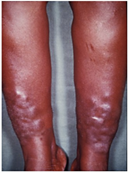
Image Source: Wikimedia Commons (2008). Pretibial_myxedema, Licensed under a creative commons licence. Available for free at: https://en.wikipedia.org/wiki/Pretibial_myxedema (Accessed: 6 February 2024).
Thyroid acropachy is a triad seen in autoimmune thyroid disease consisting of digital clubbing, soft tissue swelling of hands and feet and periosteal new bone formation in metacarpal bones or phalanges.
Thyroid emergencies
Thyroid storm
This is a medical emergency which presents with hyperthermia, altered mental state and severe manifestations of thyrotoxicosis. Triggers include any form of surgery, iodine overload, infection or parturition.
Clinical features include:
- Tachycardia
- Fever
- Hypertension
- Altered mental state
- Signs of heart failure and deranged LFTs (due to enterohepatic recycling of thyroid hormones)
- Hypercalcaemia (secondary to haemoconcentration and enhanced bone resorption)
- Hyperglycaemia (due to catecholamine induced inhibition of insulin release and increased glycogenolysis) may also be seen
Management includes control of adrenergic symptoms with beta blockers, lugol solution to block release of thyroid hormones, carbimazole to block new hormone synthesis and steroids to reduce conversion of T4 to T3.
It is important to check for co-existent Addison’s disease in these patients and this should be addressed first (with intravenous steroids) as this can otherwise exacerbate adrenal crisis.
Myxoedema coma
The manifestation includes severe symptoms of hypothyroidism with reduced mental state and hypothermia. It is more common in elderly women with poorly controlled hypothyroidism. Factors such as heart failure, myocardial infarction, sepsis or stroke can trigger myxoedema coma.
Features include lethargy, bradycardia, hypothermia, seizures and coma.
The principle of management involves re-warming, IV fluids, IV thyroxine, IV steroids.
References
- National Institute for Health and Care Excellence (UK). Hyperthyroidism. Updated January 2021. Available at: https://cks.nice.org.uk/topics/hyperthyroidism/
- National Institute for Health and Care Excellence (UK). Hypothyroidism. Updated May 2021. Available at: https://cks.nice.org.uk/topics/hypothyroidism/
- UptoDate. Infiltrative thyroid disease. Last updated 25ᵗʰ February 2021. Available at: https://www.uptodate.com/contents/infiltrative-thyroid-disease?search=reidel%20thyroiditis&source=search_result&selectedTitle=2~150&usage_type=default&display_rank=2#H2
- UptoDate. Overview of the clinical manifestations of hyperthyroidism in adults. Available at https://www.uptodate.com/contents/overview-of-the-clinical-manifestations-of-hyperthyroidism-in-adults?search=thyroid%20acropachy&topicRef=13682&source=see_link#H2
- StatPearls. Riedel Thyroiditis. Last updated 3ʳᵈ February 2022. Available at: https://www.ncbi.nlm.nih.gov/books/NBK537303
Pregnancy and thyroid diseases
Hyperthyroidism in pregnancy
To confirm biochemical hyperthyroidism during pregnancy, the following levels should be checked:
- Serum TSH
- Free thyroxine (FT4)
- Free triiodothyronine (FT3)
Serum TSH-receptor antibodies should be checked immediately once pregnancy is confirmed if there is current or previous history of Grave’s disease.
Risks associated with untreated hyperthyroidism in pregnancy include:
- Miscarriage
- Premature birth
- Low birthweight
- Preeclampsia
- Thyroid storm
- Congestive heart failure
Management can be difficult requiring specialist input. Propylthiouracil is preferred in early pregnancy as it is less likely to cross the placenta than carbimazole, which is associated with congenital abnormalities. Carbimazole is preferred later on in pregnancy due to the risk of severe hepatotoxicity associated with propylthiouracil. Radioactive iodine and traditional block and replace regimens are contraindicated in pregnancy. It is important to check TSH receptor antibody status at 24-28 weeks to assess the risk of foetal/neonatal hyperthyroidism.
Hypothyroidism in pregnancy
Risks associated with untreated hypothyroidism in pregnancy include the following:
- Increased risk of miscarriage
- Anaemia
- Pre-eclampsia
- Placental abruption
- Postpartum haemorrhage
- Stillbirth
Adverse neonatal outcomes include: preterm delivery, low birthweight, neonatal respiratory distress, congenital abnormalities, congenital hypothyroidism and impaired foetal neurocognitive development.
Management is with levothyroxine by maintaining maternal free thyroxine levels in the upper third of normal reference ranges. Thyroxine is safe in pregnancy and breastfeeding. TSH should be checked 4-6 weeks postpartum.
Postpartum thyroid dysfunction
Postpartum thyroiditis (PPT) is a transient autoimmune lymphocytic thyroiditis which can result in thyrotoxicosis, hypothyroidism or thyrotoxicosis followed by hypothyroidism (Table 4). It usually resolves within a year but there is an increased risk of developing permanent hypothyroidism. Anti-thyroid peroxidase antibodies can be found in 90% of people with postpartum thyroiditis.
| Postpartum thyrotoxicosis | Postpartum hypothyroid |
| Differentiating from Grave’s: PPT has a high T4:T3 ratio, negative thyroid receptor antibody test and no clinical signs of Grave’s disease.
TFTs to be checked 4-8 weeks after resolution of thyrotoxic phase to screen for hypothyroid phase Referral to endocrinology particularly if suspicion of Grave’s disease. Treatment is usually not required, beta-blockers can be considered for symptomatic management. |
Most symptoms resolve within 1 year postpartum. Treatment is recommended if TSH levels are >10 mlU/L, bothersome symptoms or considering further pregnancy.
Levels should be reassessed at 4-8 weeks in untreated cases. |
References:
- National Institute for Health and Care Excellence (UK). Postpartum. Updated May 2021. Available at: https://cks.nice.org.uk/topics/hypothyroidism/management/postpartum/
- Patient UK. Thyroid disease in pregnancy. Last updated 26ᵗʰ June 2016. Available at: https://patient.info/doctor/thyroid-disease-in-pregnancy
Thyroid cancer
Background
Thyroid cancers can be broadly divided into those arising from thyroid follicular epithelium (papillary, follicular and anaplastic) or calcitonin producing C cells (medullary cancer). Thyroid hormone levels are very rarely deranged with thyroid cancers and hence are not associated with symptoms of hypo or hyperthyroidism. Instead, they secrete thyroglobulin which acts as a tumour marker post treatment. Certain cancers metastasise to the thyroid gland. These include renal, colon, breast, and melanoma.
Thyroid cancer subtypes
- Papillary is the most common subtype of thyroid cancer, occurring mainly in young people, with female preponderance. Presentation is with a nodule and associated with lymph node metastasis. Histologically, there is an encapsulated papillae consisting of one or two layers of tumour cells surrounding a well-defined fibrovascular core. Management involves surgery and radioiodine. Papillary cancer has a good prognosis and can be monitored by measurement of thyroglobulin levels
- Follicular cancer also presents as a thyroid nodule and is associated with capsular and vascular invasion. It affects middle aged people and is more common in women. The management is similar to papillary cancer
- Hürthle/Oxyphil cell thyroid cancer is usually classified as a type of follicular thyroid cancer but it is distinct in that it grows more aggressively. Tumours can be benign or malignant
- Anaplastic cancer mainly affects the elderly and can present as a mass effect secondary to local invasion. This subtype carries a poor prognosis
- Medullary thyroid cancer is associated with raised calcitonin levels. It may be sporadic or inherited as a genetic condition called MEN2. The increased risk of malignancy is a consequence of the RET oncogene
- Lymphoma of the thyroid gland is rare and associated with Hashimoto’s. The majority of the cases are B cell lymphoma
References
Thyroid lymphoma. Last updated 8ᵗʰ September 2021. Available at: https://www.ncbi.nlm.nih.gov/books/NBK544282
Complete Guide to Clinical Genetics ST3 Application and Recruitment (2027)
Written by Medibuddy. Last updated: October 2026. Clinical Genetics is

Complete Guide to Clinical Radiology ST3 Application and Recruitment (2027)
Recruitment paused: NHS England is not running Clinical Radiology ST3+ recru

Complete Guide to Paediatric Surgery ST3 Application and Recruitment in United Kingdom (2026)
Written by Medibuddy. Last updated: October 2026. If you’re planning


Take your subscriptions with you
Our mobile app allows you to access your interview and exam question banks wherever you are.

