


 Close
Close
 21st July 2024
Anaemia
Anaemia
Overview of anaemia
Anaemia is a common condition defined as haemoglobin (Hb) more than two standard deviations below normal:
| Men: Hb < 130 g/L | Non-pregnant women: Hb < 120 g/L |
Anaemia is not a diagnosis on its own and merits assessment and investigation into the cause. Broadly, this can be split into blood loss, deficiency in erythropoiesis and haemolysis.
Anaemia can also be classified by morphology based on the mean corpuscular volume (MCV):
| Microcytic <80 fL | Normocytic 80-100 fL | Macrocytic >100 fL |
General symptoms include fatigue, palpitations, dyspnoea, angina, dizziness and tinnitus.
Signs include pallor (including conjunctival) and hyperdynamic circulatory changes (tachycardia, flow murmurs, cardiomegaly) when severe. There may be evidence of the underlying cause.
Some patients are asymptomatic even with severe anaemia.
Microcytic anaemia
Iron-deficiency anaemia (IDA)
IDA is the most common form of anaemia worldwide. Causes can be classified into:
- Dietary deficiency
- Malabsorption, e.g. inflammatory bowel disease, coeliac disease, H. pylori infection, gastrectomy
- Increased blood loss, e.g. chronic bleeding (gastrointestinal, GI, or urogenital)
- Increased requirement, e.g. in pregnancy there is a physiological increase in iron requirements
- Miscellaneous, e.g. blood donation, self harm, secondary to medication
Signs specific to iron deficiency include atrophic glossitis, dry skin and damaged hair, angular cheilitis, and koilonychia (spoon-shaped nails).
Assuming a full blood count (FBC) showing microcytic anaemia has already been performed, it is important to assess iron status. Typical findings in IDA are:
- Serum iron: low or normal (shows diurnal variation)
- Total iron binding capacity (TIBC): high – a measure of the amount of iron that transferrin can bind
- Transferrin saturation: low – when <16% of the iron supply limits production of RBCs
- Ferritin: low – a measure of iron stores, although this is also an acute phase reactant
A blood film may show microcytic, hypochromic red cells and pencil cells (Figure 1).
Further investigation to determine the cause varies on the patient’s age, gender and presentation, however may include coeliac serology (anti-tissue transglutaminase, TTG, antibodies), urine dipstick, gastroscopy and colonoscopy, and stool for ova and parasites. Faecal occult blood testing is not recommended as it has a poor sensitivity. If there is a convincing history of GI bleeding with recurrent negative endoscopies and colonoscopies then a capsule endoscopy may be indicated to investigate for small bowel angiodysplasia – associations include von Willebrand disease and aortic stenosis (Heyde syndrome).
For those with IDA who are aged over 60 or have features of a GI cancer (change in bowel habit, weight loss, PR [per rectum] bleeding) a 2-Week-Wait referral is required.
Oral replacement of iron with ferrous sulphate, fumarate or gluconate is usually sufficient, with monitoring of Hb within 4 weeks, then 4 months and, once normal, continuation of replacement for 3 months. If patients do not tolerate treatment (GI side effects are common), alternate-day dosing or parenteral replacement can be of benefit. Food (especially calcium and phosphate) and reduced gastric acidity can reduce iron absorption, so oral iron should not be taken with food or within 4 hours of taking a PPI (proton pump inhibitor) or antacid.
Oral iron can also impact absorption of other medications, commonly reducing absorption of levothyroxine, bisphosphonates and certain antibiotics (tetracyclines and quinolones).
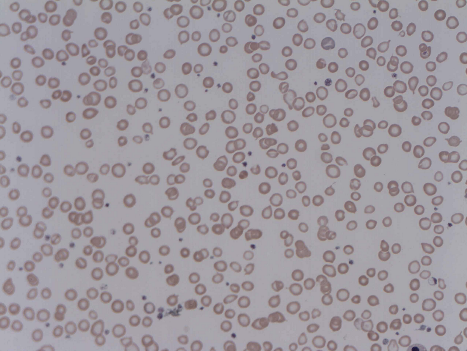
Image Source: Wikimedia Commons (2014). Iron deficiency Anaemia, Licensed under a creative commons licence. Available for free at: https://upload.wikimedia.org/wikipedia/commons/3/32/Iron_deficiency_Anaemia.jpg (Accessed: 16 January 2024)
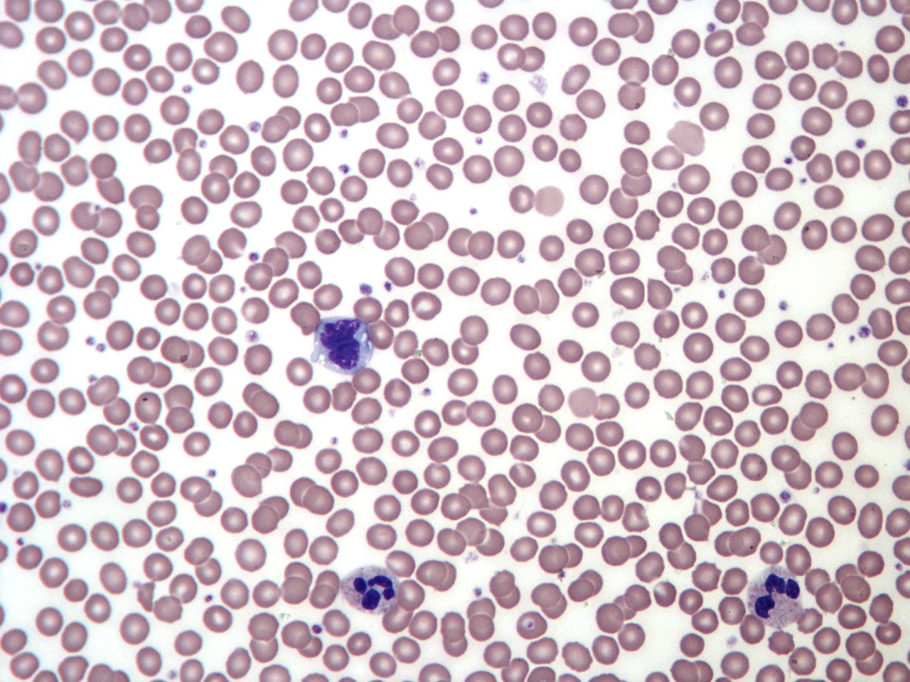
Image Source: Wikimedia Commons (2011). Normal Adult Blood Smear, Licensed under a creative commons licence. Available for free at: https://commons.wikimedia.org/wiki/File:Normal_Adult_Blood_Smear.JPG (Accessed: 16 January 2024)
Sideroblastic anaemia
This is a disorder of erythropoiesis due to failure to incorporate iron into haemoglobin. Iron instead accumulates in mitochondria and gives rise to ring sideroblasts in the bone marrow.
It can be congenital (often X-linked) or acquired. Acquired causes include myelodysplastic disorders, alcohol, lead excess, anti-tuberculosis (TB) medication, and chemotherapy.
Investigation findings may include increased ferritin, a hypochromic microcytic blood film, and ring sideroblasts in the marrow (seen with Prussian blue stain). There may also be evidence of haemosiderosis – haemosiderin deposition, which may cause cirrhosis, cardiomyopathy and pancreatic dysfunction leading to diabetes melitus (DM).
Treatments include removing the cause, if known, pyridoxine and red blood cell (RBC) transfusion.
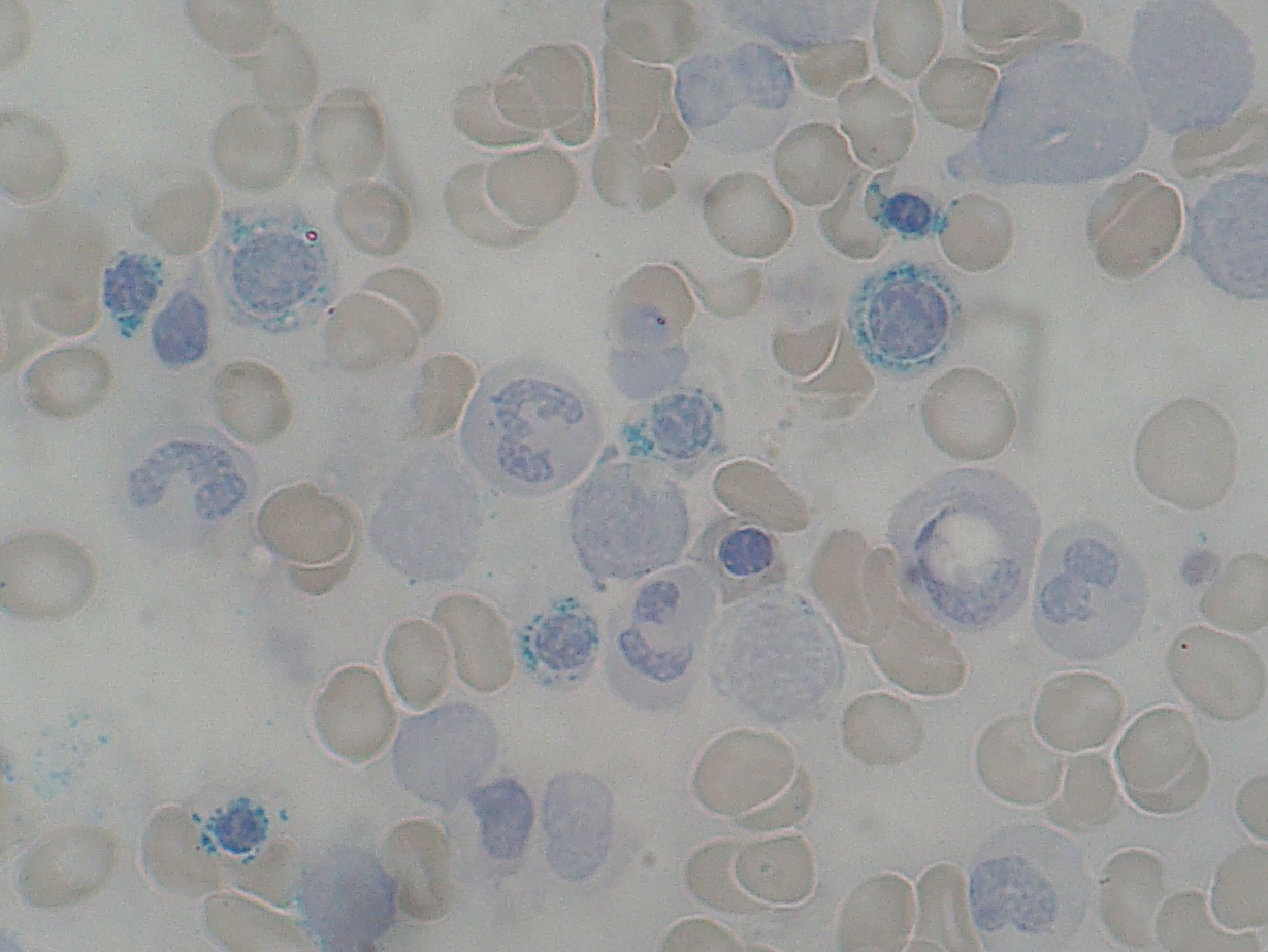
Image Source: Wikimedia Commons (2010). Ring Sideroblast smear 2010-01-13, Licensed under a creative commons licence. Available for free at: https://commons.wikimedia.org/wiki/File:Ring_Sideroblast_smear_2010-01-13.JPG (Accessed: 16 January 2024)
Normocytic anaemia
Anaemia of chronic disease
This is the most common form of anaemia seen in hospital inpatients.
It is due to inflammation-mediated reduced RBC production (decreased production of and response to erythropoietin), often coupled with shortened RBC survival (active cytokines) and dysregulated iron homeostasis. Causes include chronic infection, autoimmune disorders, malignancy and chronic disease (e.g. heart or renal failure).
Often MCV is normal, however it can present as a microcytic anaemia. Ferritin is often raised with low iron and other haematinics normal.
Management is by treating the underlying disease. Iron replacement, erythropoietin and RBC transfusions may also be required.
Other causes of normocytic anaemia
Other aetiologies that can give rise to a normocytic anaemia include:
- Nutrient deficiencies – either in the early stages or mixed, e.g. both iron and B12 deficiency – red cell distribution width (RDW) measures variation in RBC size and when significantly raised, can signify the presence of both microcytic and macrocytic cells (which may average out to a normal MCV)
- Acute blood loss
- Haemolysis
- Myelodysplastic syndrome
- Cancers with bone marrow infiltration
Macrocytic anaemia
This can be divided into megaloblastic and non-megaloblastic causes.
Megaloblastic macrocytic anaemia
Megaloblastic anaemia is caused by delayed nuclear maturation relative to the cytoplasm, often due to impaired DNA (deoxyribonucleic acid) synthesis. The blood film may show oval-shaped macrocytes and hypersegmented neutrophils (Figure 4). The most common causes are B12 and folate deficiency, as well as a range of drugs (often chemotherapy or immunosuppressants).
If a patient is nutrient deficient, the aetiology should be considered. Causes include malabsorption, such as in coeliac disease (anti-TTG antibodies will be present) or pernicious anaemia (intrinsic factor antibodies are diagnostic; parietal cell antibodies can be found in the unaffected population), and dietary factors. If the cause is not found, then a bone marrow biopsy should be considered.
In cases of severe B12 deficiency there is a risk of subacute combined degeneration (SCD) of the cord, which can present with a progressive, distal, symmetrical peripheral sensory neuropathy first affecting the legs. This has a classic triad of extensor plantar responses (upper motor neuron, UMN, sign) and absent knee and ankle reflexes (both lower motor neuron, LMN). In concomitant B12 and folate deficiency, B12 replacement should be instigated first, as giving folate whilst B12 deficient can precipitate or worsen SCD.
Non-megaloblastic macrocytic anaemia
Non-megaloblastic causes of macrocytic anaemia include alcohol excess and liver disease, conditions causing increased reticulocyte production (e.g. haemolysis), hypothyroidism, pregnancy, myelodysplasia and aplastic anaemia.
It is, therefore, important to check TFTs (thyroid function tests) and LFTs (liver function tests), including GGT (gamma-glutamyl transferase).

Image Source: Wikimedia Commons (2009). Hypersegmented neutrophil, Licensed under a creative commons licence. Available for free at: https://commons.wikimedia.org/w/index.php?curid=5837715 (Accessed: 16 January 2024).
References
- Auerbach M. Treatment of iron deficiency anemia in adults [Internet]. Mentzer W, Means R, Tirnauer J, Kunins L, editors. UpToDate. 2022 [cited 2022 May 24]. Available from: https://www.uptodate.com/contents/treatment-of-iron-deficiency-anemia-in-adults
- Camaschella C, Brugnara C. Microcytosis/Microcytic anemia [Internet]. Mentzer W, Means R, Tirnauer J, editors. UpToDate. 2022 [cited 2022 May 24]. Available from: https://www.uptodate.com/contents/microcytosis-microcytic-anemia
- Joint Formulary Committee. BNF – Interactions [Internet]. British National Formulary (online). London: BMJ Group and Pharmaceutical Press; 2022 [cited 2022 May 24]. Available from: https://bnf.nice.org.uk/interaction/iron.html
- Means R, Brodsky R. UpToDate [Internet]. Mentzer W, editor. UpToDate. 2021 [cited 2021 Dec 11]. Available from: https://www.uptodate.com/contents/diagnostic-approach-to-anemia-in-adults
- National Institute for Health and Care Excellence. Anaemia – iron deficiency [Internet]. NICE. 2021 [cited 2021 Dec 13]. Available from: https://cks.nice.org.uk/topics/anaemia-iron-deficiency/
- National Institute for Health and Care Excellence. Gastrointestinal tract (lower) cancers – recognition and referral [Internet]. NICE. 2021 [cited 2021 Dec 13]. Available from: https://cks.nice.org.uk/topics/gastrointestinal-tract-lower-cancers-recognition-referral/management/referral-for-suspected-gastrointestinal-tract-lower-cancer/
- Provan D, Baglin T, Dokal I, De Vos J. Oxford Handbook of Clinical Haematology [Internet]. 4th ed. Oxford: Oxford University Press; 2015 [cited 2021 Dec 17]. Available from: https://oxfordmedicine.com/view/10.1093/med/9780199683307.001.0001/med-9780199683307
- Saltzman J. Angiodysplasia of the gastrointestinal tract [Internet]. Friedman L, Travis A, editors. UpToDate. 2022 [cited 2022 May 24]. Available from: https://www.uptodate.com/contents/angiodysplasia-of-the-gastrointestinal-tract
- Wilkinson I, Raine T, Wiles K, Goodhart A, Hall C, O’Neill H. Haematology. In: Oxford Handbook of Clinical Medicine (10 ed) [Internet]. Oxford University Press; 2017 [cited 2021 Dec 11]. Available from: https://oxfordmedicine.com/view/10.1093/med/9780199689903.001.0001/med-9780199689903-chapter-8
- Zaiden R. Assessment of anaemia – Differential diagnosis of symptoms [Internet]. BMJ Best Practice. 2022 [cited 2021 Dec 11]. Available from: https://bestpractice.bmj.com/topics/en-gb/93
Haemolytic anaemia
Overview of haemolytic anaemia
This is anaemia secondary to premature destruction of RBCs. It can range in severity and be acute or chronic. There are various underlying causes, but useful distinctions include whether or not the process is immune-system driven and whether it occurs in intra- or extravascular space(s), the latter including the spleen, liver and bone marrow. If haemolysis is not compensated for by increased RBC production, then it is likely to cause symptomatic anaemia with fatigue and jaundice.
Results indicating a haemolytic process include raised reticulocytes, low haptoglobin (this binds to free haem released from lysed RBCs), raised LDH (lactate dehydrogenase) and raised unconjugated bilirubin.
Intravascular haemolysis is suggested by positive urinary tests:
- Haemoglobinuria: free haemoglobin rather than whole RBCs
- Haemosiderinuria: free haemoglobin which has been filtered and stored as haemosiderin in the renal tubules is then excreted in the urine; this can stay positive for ~1 week
Splenomegaly is associated with extravascular haemolysis, and patients may have a history of recurrent pigment gallstones due to excess unconjugated bilirubin.
Hereditary haemolysis
RBC membrane defects
Hereditary spherocytosis
An autosomal dominant condition most prevalent in people descended from Northern Europe. There is a deficiency of cell membrane proteins leading to a more spherical RBC compared to the normal biconcave shape, with a raised mean corpuscular haemoglobin concentration (MCHC) (typically >36 g/dL). The abnormal RBCs, called spherocytes (Figure 5) have a reduced lifespan as well as reduced mechanical stability and deformability, which can cause membrane loss and phagocytosis within the spleen. In turn, this leads to an even more spherical shape and reduced mobility through the circulation leading to haemolysis.
A diagnosis can be made clinically and patients often have a family history as well as bloods consistent with a chronic haemolytic anaemia. If there is uncertainty, the specific EMA (eosin-5′-maleimide) binding test can be used. Osmotic fragility testing is possible but has poor sensitivity and specificity. Acute exacerbations can occur secondary to infection, nutritional deficiencies or pregnancy, due to either increased haemolysis or inadequate RBC production.
Treatment aims to minimise complications and may include folic acid supplementation, RBC transfusions, erythropoietin, splenectomy and cholecystectomy (patients are at risk of pigment gallstones made up of bilirubin). Following splenectomy patients will have signs of reduced splenic function, for example Howell-Jolly bodies (nuclear remnants within RBCs) and are more at risk of infection from encapsulated bacteria.
Hereditary elliptocytosis
An autosomal dominant condition with a similar pathophysiology to hereditary spherocytosis, but is rarer and often asymptomatic or causes only mild disease.

Image Source: Wikimedia Commons (2010). Hereditary Spherocytosis smear 2010-03-17, Licensed under a creative commons licence. Available for free at: https://upload.wikimedia.org/wikipedia/commons/6/69/Hereditary_Spherocytosis_smear_2010-03-17.JPG (Accessed: 16 January 2024)
Enzyme deficiencies
Glucose-6-phosphate dehydrogenase (G6PD) deficiency
G6PD deficiency is an X-linked recessive condition, which therefore generally affects males, often of Mediterranean, African or middle and far Eastern descent. There is mainly intravascular haemolysis.
It can present in the newborn, commonly with neonatal jaundice, or with episodes of acute haemolysis secondary to triggers including drugs (primaquine, sulfonamides, henna dye), fava beans and intercurrent illness, or with chronic haemolytic anaemia.
Investigations include those mentioned previously to assess for haemolysis. The blood film may show bite cells (semicircular indent on RBC) and blister cells (vacuole on the edge of the RBC membrane), and Heinz bodies. A G6PD enzyme assay should be performed. This needs to be done 8-12 weeks after an acute haemolytic crisis to avoid a falsely elevated result, as reticulocytes have high levels of the enzyme, but levels decrease as RBCs mature.
Treatments include RBC transfusion if symptomatic anaemia or severe haemolysis and avoidance of triggers.
Pyruvate kinase (PK) deficiency
This is an autosomal recessive condition which often presents with neonatal jaundice, followed by unexplained chronic anaemia later in life with splenomegaly ± gallstones. There is mainly extravascular haemolysis.
Investigations include an enzyme assay to assess PK activity as well as genetic testing.
Treatment is often supportive with folic acid and RBC transfusions if needed. Splenectomy can help reduce haemolysis if there is severe disease.
Haemoglobinopathies
These include thalassaemia and sickle cell anaemia.
Acquired haemolysis
Autoimmune haemolytic anaemia (AIHA)
In AIHA, extravascular haemolysis occurs due to the presence of autoantibodies against RBCs. This is categorised as either warm or cold AIHA according to the temperature at which this occurs.
Haemolytic markers are raised, and a positive Coombs test is expected.
| Warm AIHA | Cold agglutinin disease (most common cold AIHA) |
| Mediated by IgG – binds to RBC antigens at ~37°C | Mediated by IgM – bind to RBC antigens at <4°C |
| Commonly idiopathic or can be secondary to lymphoproliferative or autoimmune disease | Can be idiopathic or secondary to lymphoproliferative disease or infection |
| May present with symptomatic anaemia
Chronic compensated haemolysis, jaundice and splenomegaly are common |
Often presents with symptoms induced by exposure to cold temperatures, e.g. acrocyanosis
Chronic compensated haemolysis |
| Spherocytes are seen due to RBC membrane destruction by splenic phagocytes | No significant spherocytosis as the haemolysis is due to complement activation, which leads to phagocytosis of the whole RBC |
| First-line treatment is oral steroids (prednisolone 1 mg/kg) and consideration of rituximab
Second-line treatment includes other immunosuppressants (e.g. azathioprine or cyclophosphamide) or IVIg Splenectomy can be considered in resistant cases Treatment of the underlying cause can improve symptoms but may take some time to have an effect |
First-line treatment is rituximab and warm RBC transfusion if severe anaemia
Other treatments include avoidance of cold conditions, treatment of the underlying disorder or plasmapheresis in severe cases |
Drug-induced haemolysis
Many drugs have been found to cause haemolysis, and these can occur by immune (i.e. AIHA) or non-immune mechanisms. Examples include penicillins, cephalosporins, NSAIDs, sulfasalazine, quinine and rifampicin.
Microangiopathic haemolytic anaemia (MAHA)
This is a type of intravascular haemolysis where RBCs are damaged and haemolyse due to mechanical damage whilst circulating. This can be caused by endothelial abnormalities within blood vessels leading to microthrombi, which causes trauma to the RBCs.
Causes include disseminated intravascular coagulation (DIC), haemolytic uraemic syndrome (HUS), thrombotic thrombocytopaenic purpura (TTP), eclampsia/HELLP (haemolysis, elevated liver enzymes and low platelets) syndrome, mechanical valves and vasculitides.
The blood film will show schistocytes (a fragmented part of an RBC) (Figure 6) and there is marked reticulocytosis.
Clinically, it can present differently depending on the primary cause, and treatment will depend on the diagnosis.

Image Source: Wikimedia Commons (2014). Microangiopathic haemolytic anaemia_(Schistocytes_and_spherocytes, Licensed under a creative commons licence. Available for free at: https://commons.wikimedia.org/wiki/File:Microangiopathic_haemolytic_anaemia_(Schistocytes_and_spherocytes).jpg (Accessed: 16 January 2024)
Thrombotic thrombocytopaenic purpura
This is often thought of as a pentad of clinical features: haemolytic anaemia, thrombocytopaenia, renal injury, fever and neurological symptoms.
Reduced functional levels of ADAMTS13 (the enzyme that cleaves large multimers of von Willebrand factor, vWF, into smaller molecules) leads to platelet aggregation on the endothelium of small vessels and, therefore, thrombosis. This causes thrombocytopaenia due to increased platelet consumption. This can be acquired (with autoantibodies directed at ADAMTS13) or a hereditary deficiency of the enzyme. Levels can be checked, however treatment will need to be instigated urgently before results are available.
First-line treatment is plasma exchange, and medical treatment can include glucocorticoids, rituximab and caplacizumab (anti-vWF).
Haemolytic uraemic syndrome
This is defined by the triad of microangiopathic haemolytic anaemia, thrombocytopaenia and acute kidney injury.
It is more commonly found in children than in adults and is predominantly caused by shiga toxin-producing E. coli (STEC) 0157:H7, and mostly associated with diarrhoea, although this usually resolves prior to the development of other features. This can be confirmed with a stool culture or PCR for STEC and a stool assay for shiga toxin.
The infection is self resolving, and treatment is mostly supportive with IV fluid replacement, RBC or platelet transfusion and dialysis if required. In severe or atypical cases other treatments may be required, depending on the underlying cause. HUS is a notifiable disease.
Infections
A variety of infections can cause haemolytic anaemia. These include malaria (most commonly falciparum), babesiosis, bartonellosis and viral infections (e.g. dengue, yellow fever, Epstein-Barr virus, EBV).
Paroxysmal nocturnal haemoglobinuria (PNH)
PNH is a rare, acquired clonal abnormality in haematopoietic stem cells, which have reduced complement inhibitors (CD55 and CD 59) on their surface. This leaves cells derived from these stem cells prone to haemolysis through the complement system.
Patients can present with a wide range of symptoms, classically with more noticeable dark urine in the morning (although haemolysis does occur at lower levels throughout the day as well), symptomatic anaemia, jaundice, and unusual sites of venous thrombosis (e.g. abdominal or cerebral). It can also be associated with bone marrow failure and, therefore, symptoms of pancytopaenia such as recurrent infection, bleeding or bruising.
Specific investigations for PNH include sending blood for flow cytometry to detect reductions in CD55 and CD59 and bone marrow biopsy to evaluate any degree of bone marrow failure.
Treatment options include eculizumab (anti-complement monoclonal antibody) and anticoagulation and in patients with co-existing bone marrow failure, stem cell transplant is a curative option.
References
- Andreoli S, Khalid M. Haemolytic uraemic syndrome [Internet]. Connor R, editor. BMJ Best Practice. 2019 [cited 2022 May 26]. Available from: https://bestpractice.bmj.com/topics/en-gb/470
- Brodsky R. Clinical manifestations and diagnosis of paroxysmal nocturnal hemoglobinuria [Internet]. Larson R, Rosmarin A, editors. UpToDate. 2021 [cited 2021 Dec 20]. Available from: https://www.uptodate.com/contents/clinical-manifestations-and-diagnosis-of-paroxysmal-nocturnal-hemoglobinuria
- Brodsky R. Pathogenesis of paroxysmal nocturnal hemoglobinuria [Internet]. Larson R, Rosmarin A, editors. UpToDate. 2021 [cited 2022 May 25]. Available from: https://www.uptodate.com/contents/pathogenesis-of-paroxysmal-nocturnal-hemoglobinuria
- Brodsky R. Treatment and prognosis of paroxysmal nocturnal hemoglobinuria [Internet]. Larson R, Rosmarin A, editors. UpToDate. 2021 [cited 2021 Dec 20]. Available from: https://www.uptodate.com/contents/treatment-and-prognosis-of-paroxysmal-nocturnal-hemoglobinuria
- Brodsky R. Diagnosis of hemolytic anemia in adults [Internet]. Mentzer W, editor. UpToDate. 2021 [cited 2021 Dec 17]. Available from: https://www.uptodate.com/contents/diagnosis-of-hemolytic-anemia-in-adults
- Brugnara C, Brodsky R. Warm autoimmune hemolytic anemia (AIHA) in adults [Internet]. Mentzer W, Means R, Tirnauer J, editors. UpToDate. 2022 [cited 2022 Jan 3]. Available from: https://www.uptodate.com/contents/warm-autoimmune-hemolytic-anemia-aiha-in-adults
- Brugnara C, Bertentsen S. Cold agglutinin disease [Internet]. Mentzer W, Tirnauer J, editors. UpToDate. 2021 [cited 2022 Jan 3]. Available from: https://www.uptodate.com/contents/cold-agglutinin-disease
- Densmore J. Haemolytic anaemia [Internet]. BMJ Best Practice. 2020 [cited 2021 Dec 17]. Available from: https://bestpractice.bmj.com/topics/en-gb/98
- George J, Cuker A. Diagnosis of immune TTP [Internet]. Leung L, Tirnauer J, editors. UpToDate. 2021 [cited 2022 May 26]. Available from: https://www.uptodate.com/contents/diagnosis-of-immune-ttp
- George J, Cuker A. Pathophysiology of TTP and other primary thrombotic microangiopathies (TMAs) [Internet]. Leung L, Tirnauer J, editors. UpToDate. 2022 [cited 2022 May 26]. Available from: https://www.uptodate.com/contents/pathophysiology-of-ttp-and-other-primary-thrombotic-microangiopathies-tmas
- George J, Nester C. Diagnostic approach to suspected TTP, HUS, or other thrombotic microangiopathy (TMA) [Internet]. Leung L, Tiranuer J, editors. UpToDate. 2022 [cited 2022 May 26]. Available from: https://www.uptodate.com/contents/diagnostic-approach-to-suspected-ttp-hus-or-other-thrombotic-microangiopathy-tma
- George J, Nester C. Thrombotic microangiopathies (TMAs) with acute kidney injury (AKI) in adults: CM-TMA and ST-HUS [Internet]. Brodsky R, Tirnauer J, editors. UpToDate. 2021 [cited 2022 May 26]. Available from: https://www.uptodate.com/contents/thrombotic-microangiopathies-tmas-with-acute-kidney-injury-aki-in-adults-cm-tma-and-st-hus
- Glader B. Diagnosis and management of glucose-6-phosphate dehydrogenase (G6PD) deficiency [Internet]. Means R, editor. UpToDate. 2021 [cited 2021 Dec 18]. Available from: https://www.uptodate.com/contents/diagnosis-and-management-of-glucose-6-phosphate-dehydrogenase-g6pd-deficiency
- Niaudet P, Boyer O. Overview of hemolytic uremic syndrome in children [Internet]. Mattoo T, Wilkie L, editors. UpToDate. 2021 [cited 2022 May 26]. Available from: https://www.uptodate.com/contents/overview-of-hemolytic-uremic-syndrome-in-children
- Niaudet P, Boyer O. Treatment and prognosis of Shiga toxin-producing Escherichia coli (STEC) hemolytic uremic syndrome (HUS) in children [Internet]. Mattoo T, Wilkie L, editors. UpToDate. 2021 [cited 2022 May 26]. Available from: https://www.uptodate.com/contents/treatment-and-prognosis-of-shiga-toxin-producing-escherichia-coli-stec-hemolytic-uremic-syndrome-hus-in-children
- Provan D, Baglin T, Dokal I, De Vos J. Oxford Handbook of Clinical Haematology [Internet]. 4th ed. Oxford: Oxford University Press; 2015 [cited 2021 Dec 17]. Available from: https://oxfordmedicine.com/view/10.1093/med/9780199683307.001.0001/med-9780199683307
- Wilkinson I, Raine T, Wiles K, Goodhart A, Hall C, O’Neill H. Haematology. In: Oxford Handbook of Clinical Medicine (10 ed) [Internet]. Oxford University Press; 2017 [cited 2021 Dec 11]. Available from: https://oxfordmedicine.com/view/10.1093/med/9780199689903.001.0001/med-9780199689903-chapter-8
Haemoglobinopathies
Sickle cell anaemia (SCA)
Overview of sickle cell anaemia
SCA is an autosomal recessive disorder affecting the Hb beta chain, causing HbS to be produced rather than HbA. It is most commonly seen in people of African ethnicity or descent. Sickle trait (HbAS) is protective against malaria.
In the UK, screening is available for pregnant women in high-risk groups, as well as neonatal screening. Children usually only become symptomatic after around 4 months, when HbS production takes over from foetal Hb (HbF). When exposed to reduced oxygen levels HbS deforms and forms sickle cells, which have poorer flow through vessels and can cause occlusion and sickle crises.
Investigations may show chronic haemolysis, anaemia, raised reticulocytes and bilirubin. The blood film shows sickle cells, nucleated RBCs and Howell-Jolly bodies (indicate hyposplenism). A sickle solubility test will be positive; however, electrophoresis is required to confirm the diagnosis and differentiate between HbSS and HbAS.
Long-term complications include anaemia, iron overload (secondary to transfusions), pigment gallstones, avascular necrosis, chronic kidney disease, pulmonary hypertension, hyposplenism, retinopathy and impaired growth.

Image Source: Wikimedia Commons (2011). Stomach blood supply, Licensed under a creative commons licence. Available for free at: https://upload.wikimedia.org/wikipedia/commons/7/70/Sickle_Cell_Blood_Smear.JPG (Accessed: 16 January 2024)
Management of chronic sickle cell anaemia
Long-term management of sickle cell disease includes hydroxyurea to reduce HbS levels and increase HbF, prophylactic antibiotics (usually started in childhood), and vaccinations (pneumococcal and influenza) because of functional asplenism (due to repeated splenic infarction) that occurs whilst patients are young and increases the risk of severe infections from encapsulated bacteria.
Management of sickle cell crises
There are multiple acute manifestations of sickle cell anaemia. In general, these crises occur when HbS RBCs deform due to hypoxia, acidosis, dehydration, hypothermia or infection. This produces sickle-shaped cells, which can occlude vessels and are more prone to haemolysis. Patients are often well outside of crises.
A general approach to a patient with sickle cell disease presenting acutely to hospital will aim to identify any potential triggers or early signs of a sickle cell crisis and reduce the risk of patients developing a crisis:
- Supportive measures: adequate hydration, target sats >94%, keep warm
- Send a group and save and consider transfusion if the patient is anaemic below their baseline
- Check blood tests for signs of increased haemolysis
- Consider sending a HbS%
- Adequate analgesia
- Screen for infection and have a low threshold to perform a septic screen and start empirical antibiotics
Key clinical facts about specific types of sickle cell crises are as follows:
| Acute chest crisis | Episodes can occur with no specific trigger, however they are commonly triggered by infection, vaso-occlusive pain, fat embolism and asthma exacerbations.
It presents with chest pain, shortness of breath, fever, cough and wheeze, and can lead to multiorgan failure if not treated rapidly. Pulmonary infiltrates on chest X-ray are classical but initially may not be present. Acute management includes analgesia, oxygen, incentive spirometry to prevent further atelectasis, IV antibiotics (typically cefotaxime and azithromycin), RBC transfusion or exchange transfusion to reduce HbS% to <30, and possible early escalation for consideration of non-invasive (NIV) or mechanical ventilation. |
| Vaso-occlusive (VOC) or painful crisis | Microvascular occlusion often triggered by cold, dehydration, hypoxia or infection. Generally affects bones containing marrow, but other sites include mesenteric ischaemia, central nervous system (CNS) (strokes, cognitive impairment or seizures), avascular necrosis of the femoral head, leg ulcers and priapism (urological aspiration and irrigation may be required).
It is important to use the patient’s and their relative’s reports to assess pain. Analgesia should be administered within 30 minutes, and patients often have a personalised treatment plan, however IV morphine can be used if not. Reassessment following this is important as patients may need additional doses. Adequate hydration helps to improve symptoms, and exchange transfusion can be used to treat neurological complications, including an acute stroke, due to a VOC. |
| Aplastic crisis | Bone marrow suppression due to parvovirus B19 infection causing reduction in RBCs and reticulocytes. This is self limiting and usually resolves within 14 days, but supportive RBC transfusion may be needed. |
| Sequestration crisis | Pooling of blood, often in the spleen or liver, which worsens anaemia and can cause hypovolaemic shock. This often occurs in children whose spleens have not yet infarcted and become fibrotic. Viral infections can be a trigger.
Management includes fluid resuscitation and cautious RBC transfusion (sequestered blood may re-enter the circulation and lead to hyperviscosity). |
| Haemolytic crisis | A rapid fall in Hb due to haemolysis, which can be associated with infections and other sickle crises. Management involves RBC transfusions and treatment of the underlying cause. |
Thalassaemia
Thalassaemias are disorders of haemoglobin production with a reduced production of one type of globin chain (alpha or beta). This leads to an excess of the unaffected chain, which ultimately damages RBCs in the bone marrow and causes haemolysis. Thalassaemia is more common in patients from the Mediterranean, northern Africa and parts of the Middle and Far East.
FBC will show a microcytic anaemia, with blood film findings including tear drop cells, target cells and nucleated RBCs. Raised bilirubin, LDH and reticulocytes indicate haemolysis.
Beta thalassaemia
Mutations of the beta globin gene on chromosome 11 lead to β⁺ (reduced beta chain production) or β⁰ (absence of beta chain production). Various combinations of mutations are possible leading to different phenotypes.
Beta thalassaemia major (two defective genes)
This presents in early childhood with impaired growth, abdominal distention (hepatosplenomegaly), frontal bossing of the skull (extramedullary haematopoesis) and severe anaemia.
Hb electrophoresis shows significantly reduced or absent HbA (adult haemoglobin, the most common haemoglobin tetramer), with elevated HbF and HbA₂. X-rays of the skull can show the ‘hair-on-end’ sign due to bone marrow hyperplasia and widening of the medullary cavity in order to aid haematopoesis (this can also be seen, but generally to a lesser extent, in other causes of bone marrow hyperplasia, e.g. hereditary spherocytosis and sickle cell disease). Genetic testing is used to confirm the diagnosis and evaluate the nature of the mutation and can also be used to assess for carrier status in relatives.
Patients need lifelong RBC transfusions (2-4 weekly) to reduce extramedullary haematopoeisis and, therefore, need to be monitored for iron overload with 3-monthly ferritin levels. If iron chelation is indicated (criteria include ferritin >1000 ug/L, liver iron concentration >3 mg/g of dry weight on MRI, cardiac T2* <20 ms), subcutaneous (SC) desferrioxamine is first line, and patients should undergo 3-monthly monitoring of side effects including visual and auditory neurotoxicity, growth impairment, renal and liver failure and increased susceptibility to Yersinia infections.
Splenectomy is indicated if there are increasing transfusion requirements. Hydroxycarbamide can increase HbF and improve symptoms. Bone marrow transplantation offers a cure but is rarely done due to the associated risks.
Beta thalassaemia intermedia (mild homozygous mutations or heterozygotes with another haemoglobinopathy)
This causes moderate anaemia but patients are not usually transfusion dependent.
Beta thalassaemia minor or trait (heterozygous with one functioning gene)
Patients are usually asymptomatic, although Hb can drop in pregnancy. They are likely to have mild anaemia, with a similar picture to iron deficiency but with significantly low MCV (< 75). Electrophoresis shows HbA2 >3.5% with a mildly raised HbF.
Alpha thalassaemia
Alpha globin chains are coded for by two separate genes on each copy of chromosome 16 (normal alleles are designated αα/αα). There are 4 distinct phenotypes of alpha thalassaemia:
- Silent carrier (1 affected gene): asymptomatic with normal Hb
- Alpha thalassaemia trait (2 affected genes): asymptomatic with mild microcytic anaemia
- HbH disease (3 affected genes): moderate anaemia and haemolysis with jaundice and hepatosplenomegaly, and failure to thrive in infancy
- Hb Bart or hydrops fetalis syndrome (deletion of all 4 alpha globin genes): death in utero
References
- Benz E, Angelucci E. Diagnosis of thalassemia (adults and children) [Internet]. Vichinsky E, Tirnauer J, editors. UpToDate. 2022 [cited 2022 May 30]. Available from: https://www.uptodate.com/contents/management-of-thalassemia
- Benz E, Angelucci E. Management of thalassemia [Internet]. Vinchinsky E, Tirnauer J, editors. UpToDate. 2022 [cited 2022 May 30]. Available from: https://www.uptodate.com/contents/management-of-thalassemia
- DeBaun M. Acute vaso-occlusive pain management in sickle cell disease [Internet]. Vichinsky E, Tirnauer J, editors. UpToDate. 2022 [cited 2022 May 28]. Available from: https://www.uptodate.com/contents/acute-vaso-occlusive-pain-management-in-sickle-cell-disease
- Field J, Willen S. Acute chest syndrome (ACS) in sickle cell disease (adults and children) [Internet]. Vichinsky E, DeBaun M, Tirnauer J, editors. UpToDate. 2022 [cited 2022 May 28]. Available from: https://www.uptodate.com/contents/acute-chest-syndrome-acs-in-sickle-cell-disease-adults-and-children
- Field J, Vichinsky E. Overview of the management and prognosis of sickle cell disease [Internet]. DeBaun M, Tirnauer J, editors. UpToDate. 2022 [cited 2022 Jan 7]. Available from: https://www.uptodate.com/contents/overview-of-the-management-and-prognosis-of-sickle-cell-disease
- Lanzkron S. Sickle cell anaemia [Internet]. BMJ Best Practice. 2022 [cited 2022 Jan 5]. Available from: https://bestpractice.bmj.com/topics/en-gb/100
- Provan D, Baglin T, Dokal I, De Vos J. Oxford Handbook of Clinical Haematology [Internet]. 4th ed. Oxford: Oxford University Press; 2015 [cited 2022 Jan 5]. Available from: https://oxfordmedicine.com/view/10.1093/med/9780199683307.001.0001/med-9780199683307
- Sheth S. Beta-thalassaemia [Internet]. BMJ Best Practice. 2021 [cited 2022 Jan 7]. Available from: https://bestpractice.bmj.com/topics/en-gb/251
- Vichinsky E. Overview of the clinical manifestations of sickle cell disease [Internet]. DeBaun M, Tirnauer J, editors. UpToDate. 2020 [cited 2022 Jan 5]. Available from: https://www.uptodate.com/contents/overview-of-the-clinical-manifestations-of-sickle-cell-disease
- Wilkinson I, Raine T, Wiles K, Goodhart A, Hall C, O’Neill H. Haematology. In: Oxford Handbook of Clinical Medicine (10 ed) [Internet]. Oxford University Press; 2017 [cited 2022 Jan 5]. Available from: https://oxfordmedicine.com/view/10.1093/med/9780199689903.001.0001/med-9780199689903-chapter-8
Bone marrow failure
Overview of bone marrow failure
Production of RBCs, WBCs and platelets occurs in the bone marrow, but in certain conditions where demand is increased there can be haematopoiesis in the liver and spleen. Different cells arise from different lineages, and issues can arise in any or all parts of cell production and differentiation.
Pancytopenia
Pancytopenia is a reduction in all peripheral blood cell lineages and usually diagnosed when the three main cell lines – haemoglobin, platelets and neutrophils – are below the normal ranges. Patients may present with symptoms associated with cytopenias (infections, fatigue, dyspnoea, bruising or bleeding) or the underlying disease process.
Blood tests including FBC, reticulocytes, urea and electrolytes (U&E), clotting screen and blood film should be sent, and the patient may subsequently undergo a bone marrow biopsy.
There are many different causes of pancytopenia, which can be broadly grouped into: bone marrow infiltration, bone marrow aplasia and blood cell destruction or sequestration.
| Congenital | Gaucher’s – lysosomal storage disorder with bone marrow infiltration and splenomegaly
Fanconi’s anaemia – aplastic anaemia, developmental delay, microcephaly, café-au-lait lesions and increased risk of AML (acute myeloid leukaemia) Wiskott-Aldrich syndrome Haemophagocytic lymphohistiocytosis (HLH) |
|
| Bone marrow infiltration | Malignant | Leukaemia, myelodysplastic syndromes, multiple myeloma
Metastatic cancer |
| Non-malignant | Myelofibrosis
Infections (e.g. TB, fungal) |
|
| Bone marrow failure | Immune destruction or suppression | Aplastic anaemia, paroxysmal nocturnal haemoglobinuria
Drugs (e.g. cytotoxic drugs, sulfonamides) Autoimmune (e.g. SLE, rheumatoid arthritis [RA], sarcoidosis) Infections (e.g. HIV [human immunodeficiency virus], EBV, hepatitis) |
| Destruction or sequestration | Consumption | Disseminated intravascular coagulation |
| Splenomegaly | Liver disease
Infections (e.g. EBV) Autoimmune (e.g. SLE, RA) Malignancy (e.g. lymphoma, leukaemia) Storage disorders (e.g. Gaucher’s disease) |
|
Aplastic anaemia
This is a disorder whereby the bone marrow ceases to produce stem cells, leading to pancytopenia. It can present with symptoms related to reduction in any of the cell lineages, for example anaemia, infection or bleeding.
An FBC shows evidence of cytopenia (at least 2 cytopenias needed for diagnosis: Hb <100 g/L, platelet <50 10⁹/L, neutrophils <1.5 10⁹/L). Reticulocytes are low as the bone marrow is hypo-productive. Bone marrow biopsy confirms the diagnosis with a hypocellular marrow but no abnormal cell populations or fibrosis (suggestive of leukaemia and myelofibrosis respectively).
Acquired aplastic anaemia is commonly idiopathic, but causes include drugs (chloramphenicol, NSAIDs – indomethacin, gold), viral illness, PNH and SLE.
Treatment varies depending on the severity. Asymptomatic patients may only need observation or supportive transfusions. Severe disease (reticulocytes <50 x10⁹/L, platelets <20 x 10⁹/L and neutrophils <0.5 x10⁹/L) can be cured with a stem cell transplant, however this is reserved for those under the age of 40 who are medically fit, as otherwise prognosis is poor. For those who are likely not able to tolerate stem cell transplantation or those with more moderate disease, treatment with triple therapy (eltrombopag, horse antithymocyte globulin and cyclosporine) is another option. Supportive care is considered for frailer patients (performance status >2).
Agranulocytosis
This is a reduction in granulocytes (includes neutrophils, basophils and eosinophils), which increases the risk of infection. Patients will often present with recurrent infections or oral ulcerations, although it can be asymptomatic.
It is often secondary to drugs (carbimazole, propylthiouracil, clozapine, sulfonamides, gold or dapsone), and patients on these medications need to be monitored and safety netted to present for medical attention if any signs of a fever.
Treatment includes stopping the offending medication and consideration of granulocyte colony-stimulating factor (GCSF). Bone marrow biopsy may not be needed if the presentation is likely secondary to a high-risk drug.
References
- Berliner N. Approach to the adult with pancytopenia [Internet]. Newburger P, Rosmarin A, editors. UpToDate. 2021 [cited 2022 Jan 22]. Available from: https://www.uptodate.com/contents/approach-to-the-adult-with-pancytopenia
- Coates T. Drug-induced neutropenia and agranulocytosis [Internet]. Newburger P, Rosmarin A, editors. UpToDate. 2022 [cited 2022 May 31]. Available from: https://www.uptodate.com/contents/drug-induced-neutropenia-and-agranulocytosis
- Lipton J. Assessment of pancytopenia [Internet]. BMJ Best Practice. 2021 [cited 2022 Jan 17]. Available from: https://bestpractice.bmj.com/topics/en-gb/1024
- Marsh J, Kulasekararaj A. Aplastic anaemia in adults [Internet]. BMJ Best Practice. 2018 [cited 2022 Jan 15]. Available from: https://bestpractice.bmj.com/topics/en-gb/96
- Olson T. Aplastic anemia: Pathogenesis, clinical manifestations, and diagnosis [Internet]. Mentzer W, Rosmarin A, editors. UpToDate. 2021 [cited 2022 Jan 22]. Available from: https://www.uptodate.com/contents/aplastic-anemia-pathogenesis-clinical-manifestations-and-diagnosis
- Provan D, Baglin T, Dokal I, De Vos J. Oxford Handbook of Clinical Haematology [Internet]. 4th ed. Oxford: Oxford University Press; 2015 [cited 2021 Dec 17]. Available from: https://oxfordmedicine.com/view/10.1093/med/9780199683307.001.0001/med-9780199683307
- Olson T, Dunbar C. Treatment of aplastic anemia in adults [Internet]. Larson R, Rosmarin A, editors. UpToDate. 2022 [cited 2022 May 31]. Available from: https://www.uptodate.com/contents/treatment-of-aplastic-anemia-in-adults
- Wilkinson I, Raine T, Wiles K, Goodhart A, Hall C, O’Neill H. Haematology. In: Oxford Handbook of Clinical Medicine (10 ed) [Internet]. Oxford University Press; 2017 [cited 2021 Dec 11]. Available from: https://oxfordmedicine.com/view/10.1093/med/9780199689903.001.0001/med-9780199689903-chapter-8
Blood transfusion
Red blood cell transfusion
Guidelines do not give a fixed Hb level for transfusion and advise using clinical judgement. It is generally suggested to transfuse if Hb <70 g/L, and to target 70-90 g/L post-transfusion. For those with cardiac disease, a higher threshold is used, Hb <80 g/L, with a target of 80-100 g/L. These targets do not apply in cases of significant bleeding, symptomatic anaemia, acute coronary syndrome (ACS) or transfusion-dependent anaemia.
The blood grouping system commonly refers to ABO and rhesus D antigens, however there are other clinically significant antigens present. Patients will have antibodies against antigens missing from their own RBCs and it is important to ensure compatible blood is given – otherwise there is risk of a haemolytic reaction.
Alternatives to blood transfusion
It is generally recommended to use alternative methods to optimise patients instead of transfusing where possible. This includes iron replacement, which can be IV in an acute setting or oral in the community, and intraoperative cell salvage and tranexamic acid, which should be considered in all patients expected to have high blood loss.
Transfusion reactions
| Acute haemolytic reaction | A life-threatening reaction caused by ABO incompatibility or other mismatched antigens. Presents early in the transfusion with fever, chills, back/flank pain, hypotension, DIC (bleeding or oozing from lines) or haemoglobinuria.
Management: stop transfusion, IV fluids to aim for urine output > 100 ml/hour, confirm patient identity and the name on the blood product. A full set of bloods, including a group and save, Coombs test and haemolysis screen, should be sent. |
| Anaphylaxis | Angioedema, bronchospasm/wheeze, cyanosis, hypotension, flushing, and urticaria.
Management: stop transfusion, treat anaphylaxis (adrenaline, consider antihistamine and hydrocortisone, and supportive measures as required, including airway). |
| Bacterial infection | Can be caused by a blood product contaminated with a microorganism. Clinical features include fever >39ºC (or increase >2ºC following transfusion), rigors, tachycardia and hypotension – often occurring at least 30 minutes after the transfusion has been completed.
Management: stop transfusion, then manage as sepsis, including sending cultures. Blood bank needs to be informed and the blood bag sent to microbiology for culture. |
| Transfusion-related acute lung injury (TRALI) | Acute lung injury or acute respiratory distress syndrome (ARDS) within 6 hours of transfusion. Symptoms often occur within the first hours of transfusion: hypoxia, hypotension and pulmonary infiltrates on CXR.
Management: stop transfusion, otherwise mostly supportive – oxygen supplementation, consideration of NIV. Early ITU discussions are needed, as patients may need mechanical ventilation ± circulatory support. Must be reported to the blood bank for recall of donors for HLA (human leukocyte antigens) antibody testing. |
| Transfusion-associated circulatory overload (TACO) | Fluid overload within 12 hours of last transfusion (secondary to transfusion). Presents with dyspnoea, hypoxia, elevated jugular venous pressure (JVP) and bibasal crepitations. Often hypertensive – can help differentiate from TRALI.
Management: slow or stop transfusion depending on severity and indication for the transfusion. Supplemental oxygen and a diuretic can be given if required. |
| Non-haemolytic febrile reaction | A diagnosis of exclusion – first rule out other reactions. Fever and shivering, often within 1 hour of start of transfusion, but with no other systemic features.
Management: slow or stop transfusion, give antipyretics. |
| Allergic reaction | A common reaction associated with itching and urticaria.
Management: slow or stop transfusion, give antihistamines (e.g. chlorphenamine). |
| Transfusion-associated graft-versus-host disease (ta-GVHD) | Often fatal. Can develop 4-30 days post-transfusion. Due to proliferation of T-lymphocytes from the donor’s blood, which attack host HLA-expressing cells. Presents with fever, pancytopenia, erythroderma, deranged LFTs and diarrhoea.
Risk factors:
Gamma irradiation of blood products inactivates T cells, thus reducing the risk of ta-GVHD; patients who are defined as high risk need irradiated blood. Treatment is often ineffective and includes stem cell transplantation (limited time to find a suitable donor) and immunosuppression with steroids, IVIg or cyclosporin. |
Platelet transfusion
Usual thresholds for transfusion in non-bleeding patients are platelets <10 x10⁹/L or <20 x10⁹/L in the context of sepsis. In bleeding patients, this threshold is increased to <30 x10⁹/L or higher for severe bleeding or high-risk sites, e.g. CNS bleeding. The peak increment can be measured 10-60 minutes post transfusion, and then reduces over the next 72 hours, with the expected increase to be around 30 x10⁹/L per pool of platelets transfused.
Complications include all of the transfusion reactions associated with blood transfusions (Table 4), however the most common reactions with platelet transfusion include infection (platelets are stored at room temperature so bacteria can divide rapidly) and alloimmunisation (production of anti-HLA antibodies against the donor platelets, leading to no significant increment in platelet count).
References
- Foukaneli T, Kerr P, Bolton‐Maggs PHB, Cardigan R, Coles A, Gennery A, et al. Guidelines on the use of irradiated blood components. British Journal of Haematology. 2020 Aug 18;191(5):704–24. [cited 2022 Jan 25]. Available from: https://onlinelibrary.wiley.com/doi/full/10.1111/bjh.17015
- Kaufman R. Refractoriness to platelet transfusion [Internet]. Kleinman S, Tirnauer J, editors. UpToDate. 2022 [cited 2022 Jun 19]. Available from: https://www.uptodate.com/contents/refractoriness-to-platelet-transfusion
- National Institute for Health and Care Excellence. Blood transfusion [Internet]. NICE. 2015 [cited 2022 Jan 25]. Available from: https://www.nice.org.uk/guidance/ng24
- Provan D, Baglin T, Dokal I, De Vos J. Oxford Handbook of Clinical Haematology [Internet]. 4th ed. Oxford: Oxford University Press; 2015 [cited 2022 Jan 25]. Available from: https://oxfordmedicine.com/view/10.1093/med/9780199683307.001.0001/med-9780199683307
- Tobian A. Transfusion-associated graft-versus-host disease [Internet]. Kleinman S, Tirnauer J, editors. UpToDate. 2022 [cited 2022 Jan 25]. Available from: https://www.uptodate.com/contents/transfusion-associated-graft-versus-host-disease
- Tobian A. Approach to the patient with a suspected acute transfusion reaction [Internet]. Kleinman S, Tirnauer J, editors. UpToDate. 2022 [cited 2022 Jan 25]. Available from: https://www.uptodate.com/contents/approach-to-the-patient-with-a-suspected-acute-transfusion-reaction
- Tobian A. Hemolytic transfusion reactions [Internet]. Kleinman S, Tirnauer J, editors. UpToDate. 2022 [cited 2022 Jan 26]. Available from: https://www.uptodate.com/contents/hemolytic-transfusion-reactions
- Weinberg J. Transfusion reaction [Internet]. BMJ Best Practice. 2019 [cited 2022 Feb 25]. Available from: https://bestpractice.bmj.com/topics/en-gb/1048
- Wilkinson I, Raine T, Wiles K, Goodhart A, Hall C, O’Neill H. Haematology. In: Oxford Handbook of Clinical Medicine (10 ed) [Internet]. Oxford University Press; 2017 [cited 2022 Jan 25]. Available from: https://oxfordmedicine.com/view/10.1093/med/9780199689903.001.0001/med-9780199689903-chapter-8
- Yuan S. Platelet transfusion: Indications, ordering, and associated risks [Internet]. Kleinman S, Tirnauer J, editors. UpToDate. 2022 [cited 2022 Jun 19]. Available from: https://www.uptodate.com/contents/platelet-transfusion-indications-ordering-and-associated-risks
Complete Guide to Clinical Genetics ST3 Application and Recruitment (2027)
Written by Medibuddy. Last updated: October 2026. Clinical Genetics is

Complete Guide to Clinical Radiology ST3 Application and Recruitment (2027)
Recruitment paused: NHS England is not running Clinical Radiology ST3+ recru

Complete Guide to Paediatric Surgery ST3 Application and Recruitment in United Kingdom (2026)
Written by Medibuddy. Last updated: October 2026. If you’re planning


Take your subscriptions with you
Our mobile app allows you to access your interview and exam question banks wherever you are.

