


 Close
Close
 23rd July 2024
Myeloproliferative neoplasms and myelodysplastic syndromes
Overview of myeloproliferative neoplasms (MPN)
MPNs can be split according to Philadelphia chromosome presence:
| Philadelphia chromosome-negative MPNs | Polycythaemia vera (PV) |
| Essential thrombocythaemia (ET) | |
| Primary myelofibrosis (PMF) | |
| Philadelphia chromosome-positive MPN | Chronic myeloid leukaemia |
The differences between MPN, myelodysplastic syndromes (MDS) and acute leukaemias can be grossly summarised as below; there is a degree of overlap between them:

Polycythaemia vera (PV)
Background of PV
PV is a true primary erythrocytosis from clonal proliferation of haematopoietic stem cells, as opposed to a secondary erythrocytosis such as from hypoxia or a relative erythrocytosis from reduced plasma volume.
Median age at diagnosis is ~60 years with a slight male predominance.
JAK2 mutations are associated with almost all cases of PV. The majority of these exhibit the V617F mutation on exon 14 of the JAK2 gene; more rarely there are mutations on exon 12.
Presentation and diagnosis of PV
Most patients are identified incidentally with an elevated haemoglobin or haematocrit.
Common presenting complaints include aquagenic pruritus (pruritus after a warm bath or shower) and erythromelalgia (red, painful extremities). Arterial or venous thrombosis (stroke, myocardial infarction, deep vein thrombosis) and haemorrhage (intracranial or gastrointestinal) can occur. Hyperviscosity symptoms may be present (headache, visual disturbance, fatigue).
Signs include facial plethora (ruddy complexion), gouty arthritis, splenomegaly and hypertension.
Diagnosis:
- Bloods: full blood count (erythrocytosis, raised haematocrit and red cell mass, thrombocytosis and neutrophilia are common)
- JAK2 mutation analysis
If haematocrit (or red cell mass) is raised and JAK2 mutations are present, this is sufficient for diagnosis. If the patient is JAK2 mutation negative, then further investigations including serum erythropoietin (normal or low in PV, raised in patients with secondary polycythaemia), bone marrow biopsy and ultrasound imaging may be necessary.
Management and prognosis of PV
Treatment is not curative; aims are to reduce plasma viscosity, aiming for a haematocrit <0.45, and reduce thrombosis risk. All patients should be given advice on managing other cardiovascular risk factors.
Plasma viscosity is reduced with regular venesection and cytoreductive therapy (e.g. hydroxycarbamide).
Thrombosis risk is reduced with regular low-dose aspirin.
PV may progress to PMF (spent-phase PV, 10-15% after 10 years) or transform to MDS or AML. Occurrence of these confers a poor prognosis.
Median survival in untreated symptomatic patients is ~18 months. Treatment extends median survival to >13 years.
Essential thrombocythaemia (ET)
Background of ET
ET is a true primary thrombocytosis that is not a reactive erythrocytosis or due to another MPN or MDS.
Median age at diagnosis is ~60 years with a female predominance.
The precise aetiology is unknown. JAK2 V617F mutations are found in ~50% of ET. Mutually exclusive JAK2, CALR or MPL mutations are found in ~80-90% of ET.
Presentation and diagnosis of ET
Patients are asymptomatic and diagnosis is incidental in ~50% of cases.
Vasomotor symptoms may be the presenting complaint, including headaches, lightheadedness, atypical chest pain, paraesthesia, livedo reticularis and erythromelalgia. Arterial or venous thrombosis and bleeding (mucosal or gastrointestinal) can occur. Aquagenic pruritus is very uncommon.
Splenomegaly may be present on examination.
Diagnostic criteria for ET include:
- Platelets >450 x10⁹/L (with blood film showing marked thrombocytosis with platelet anisocytosis)
- Bone marrow biopsy showing proliferation mainly of the megakaryocytic lineage
- Exclusion of reactive thrombocytosis and other MPNs and MDSs
- Demonstration of JAK2, CALR or MPL mutations
Management and prognosis of ET
Treatment is not curative; aims are to reduce symptoms and risk of complications. Treatment is determined by risk of thrombosis. All patients should be given advice on managing other cardiovascular risk factors.
High risk patients (≥60 years old or history of arterial or venous thrombosis) receive cytoreductive therapy with hydroxycarbamide. Patients with previous venous thrombosis receive anticoagulation and patients with previous arterial thrombosis receive low-dose aspirin.
Intermediate risk patients receive low-dose aspirin ± hydroxycarbamide.
Low risk patients can be monitored with no therapy or receive low-dose aspirin.
Generally, ET is an indolent disease with most patients having a normal life expectancy. Transformation to AML or primary myelofibrosis can occur but is uncommon.
Primary myelofibrosis (PMF)
Background of PMF
PMF is an MPN characterised by bone marrow fibrosis, extramedullary haematopoiesis (causing splenomegaly) and leucoerythroblastic blood films. It primarily causes proliferation of cells of megakaryocyte and granulocyte lineage. PMF refers to only de novo myelofibrosis. Myelofibrosis from another cause is called secondary or reactive myelofibrosis. Myelofibrosis occurring from PV or ET are termed post-PV and post-ET myelofibrosis respectively.
PMF is the least common MPN. Median age of diagnosis is ~65 years with a slight male predominance.
Precise aetiology is unknown. The JAK2 V617F mutation is found in up to 58% of patients. Mutually exclusive JAK2, CALR or MPL mutations are found in ~90% of PMF.
Presentation and diagnosis of PMF
Fatigue is the most common presenting complaint. Weight loss, fevers and night sweats are also common and are due to an overexpression of inflammatory cytokines, resulting in a hypercatabolic state.
Marked splenomegaly is a hallmark of PMF. It is present in almost all patients, and they may present with symptoms from this such as left upper quadrant discomfort and early satiety.
A significant minority are asymptomatic at diagnosis, incidentally being identified with abnormal bloods or splenomegaly.
Diagnosis is multimodal:
- Bloods: usually show anaemia; platelets and white cells may be low, normal or high (thrombocytosis>thrombocytopenia)
- Blood film: teardrop-shaped red blood cells and a leucoerythroblastic picture (nucleated red cells, immature white cells) are disease hallmarks
- Bone marrow aspirate: usually unsuccessful, ‘dry tap’
- Bone marrow biopsy: extensive fibrosis, may be hypercellular
Further immunophenotyping and mutation analysis are required to ensure secondary causes of myelofibrosis are ruled out.
Management and prognosis of PMF
If eligible, patients should undergo allogeneic SCT. This can prolong survival and has curative potential.
If ineligible for transplant, treatment is for symptomatic relief. Ruxolitinib and fedratinib (JAK2 inhibitors) are effective at improving symptoms (reducing organomegaly and improving blood counts). Hydroxycarbamide can be used if JAK2 inhibitors are ineffective but it is less efficacious. Splenectomy and red cell transfusion are also used to manage symptoms. Symptomatic treatments do not affect prognosis.
Median survival is ~6 years, ranging from <1 to >30 years.
Death is usually from leukaemic transformation or from bone marrow failure complications (infection, haemorrhage).
Myelodysplastic syndromes (MDS)
Background of MDS
MDSs are a heterogenous group of conditions characterised by ineffective haematopoiesis, causing impaired cell maturation and proliferation.
Median age of diagnosis is 70 years with a male predominance.
Around 80% of cases are primary. Common chromosomal abnormalities include deletions of chromosomes 5, 7, 11, 12, 13 and 20.
Secondary MDSs can arise from risk factors including previous exposure to chemotherapy (alkylating agents and topoisomerase II inhibitors), previous SCT and environmental factors (benzene and tobacco).
Presentation and diagnosis of MDS
Patients may be asymptomatic at diagnosis, being identified with abnormal blood tests.
They may present with signs and symptoms of anaemia, neutropenia and thrombocytopenia. Autoimmune conditions are more common in patients with MDS, most commonly rheumatic heart disease, rheumatoid arthritis and pernicious anaemia.
Diagnosis relies upon:
- Bloods: anaemia with variable thrombocytopenia and neutropenia
- Blood film: red cell dysplasia (e.g. oval macrocytic cells, basophilic stippling) and white cell dysplasia (e.g. pseudo-Pelger-Huet abnormality) (Figures 11 and 12)
- Bone marrow: hypercellular with single or multilineage dysplasia; further investigation of the sample with cytochemistry, immunocytochemistry, flow cytometry and cytogenetic analysis for diagnosis and prognosis
MDSs are classified based on the number of cell lineages demonstrating dysplasia, percentage of blast cells, presence of Auer rods, percentage of ringed sideroblasts and cytogenetics. Classification is important for treatment and prognosis.
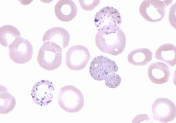
Image Source: Wikimedia Commons (2014). Basophilic stippling 2, Licensed under a creative commons licence. Available for free at: https://en.wikipedia.org/wiki/Basophilic_stippling#/media/File:Basophilic_stippling_2.jpg (Accessed: 16 January 2024)

Image Source: Wikimedia Commons (2008). Hypogranular neutrophil with a pseudo-Pelger-Huet nucleus in MDS, Licensed under a creative commons licence. Available for free at: https://commons.wikimedia.org/wiki/File:Hypogranular_neutrophil_with_a_pseudo-Pelger-Huet_nucleus_in_MDS.jpg (Accessed: 16 January 2024)
Management and prognosis of MDS
Risk is assessed using percentage of bone marrow blasts, cytogenetics and number and degree of cytopenias.
For lower-risk asymptomatic patients, active surveillance is used. Symptomatic lower-risk or higher-risk patients who are medically fit and willing may undergo intensive therapy (e.g. induction chemotherapy with allogeneic SCT).
If unable or unwilling to undergo an intensive regime, patients can have a lower intensity regime alongside supportive therapy (e.g. transfusions and growth factors).
Median overall survival ranges from 8.8 years in the lowest risk group to 9 months in the highest risk group.
References
- Tefferi A. Clinical manifestations and diagnosis of polycythaemia vera [Internet]. Larson RA, Rosmarin AG, editors. UpToDate. 2020 [cited 2022 Jan 02]. Available from: https://www.uptodate.com/contents/clinical-manifestations-and-diagnosis-of-polycythemia-vera
- Bose P, Verstovsek S. Polycythaemia vera [Internet]. BMJ Best Practice. 2021 [cited 2022 Jan 02]. Available from: https://bestpractice.bmj.com/topics/en-gb/178
- McMullin MF, Harrison CN, Ali S, Cargo C, Chen F, Ewing J et al. A guideline for the diagnosis and management of polycythaemia vera. A British Society for Haematology Guideline. British Journal of Haematology. 2019;184(2):176-191. Available from: doi: 10.1111/bjh.15648
- Tefferi A. Prognosis and treatment of polycythaemia vera [Internet]. Larson RA, Rosmarin AG, editors. UpToDate. 2021 [cited 2022 Jan 02]. Available from: https://www.uptodate.com/contents/prognosis-and-treatment-of-polycythemia-vera
- Mukherjee D. Essential thrombocytosis [Internet]. BMJ Best Practice. 2021 [cited 2022 Jan 02]. Available from: https://bestpractice.bmj.com/topics/en-gb/896
- Provan D, Baglin T, Dokal I, de Vos J. Oxford Handbook of Clinical Haematology. 4th ed. Oxford: Oxford University Press; 2015
- Tefferi A. Diagnosis and clinical manifestations of essential thrombocythaemia [Internet]. Larson RA, Rosmarin AG, editors. UpToDate. 2021 [cited 2022 Jan 02]. Available from: https://www.uptodate.com/contents/diagnosis-and-clinical-manifestations-of-essential-thrombocythemia
- Tefferi A. Prognosis and treatment of essential thrombocythaemia [Internet]. Larson RA, Rosmarin AG, editors. UpToDate. 2021 [cited 2022 Jan 02]. Available from: https://www.uptodate.com/contents/prognosis-and-treatment-of-essential-thrombocythemia
- Spivak JL. Myelofibrosis [Internet]. BMJ Best Practice. 2018 [cited 2022 Jan 03]. Available from: https://bestpractice.bmj.com/topics/en-gb/1132
- Tefferi A. Clinical manifestations and diagnosis of primary myelofibrosis [Internet]. Larson RA, Rosmarin AG, editors. UpToDate. 2020 [cited 2022 Jan 03]. Available from: https://www.uptodate.com/contents/clinical-manifestations-and-diagnosis-of-primary-myelofibrosis
- Tefferi A. Management of primary myelofibrosis [Internet]. Larson RA, Rosmarin AG, editors. UpToDate. 2021 [cited 2022 Jan 03]. Available from: https://www.uptodate.com/contents/management-of-primary-myelofibrosis
- Tefferi A. Prognosis of primary myelofibrosis [Internet]. Larson RA, Rosmarin AG, editors. UpToDate. 2021 [cited 2022 Jan 03]. Available from: https://www.uptodate.com/contents/prognosis-of-primary-myelofibrosis
- Ganti AP, Marr A. Myelodysplastic syndrome [Internet]. BMJ Best Practice. 2020 [cited 2022 Jan 03]. Available from: https://bestpractice.bmj.com/topics/en-gb/377
- Aster JC, Stone RM. Clinical manifestations and diagnosis of myelodysplastic syndromes (MDS) [Internet]. Larson RA, Rosmarin AG, editors. UpToDate. 2021 [cited 2022 Jan 03]. Available from: https://www.uptodate.com/contents/clinical-manifestations-and-diagnosis-of-myelodysplastic-syndromes-mds
- Sekeres MA. Overview of the treatment of myelodysplastic syndromes [Internet]. Larson RA, Rosmarin AG, editors. UpToDate. 2021 [cited 2022 Jan 03]. Available from: https://www.uptodate.com/contents/overview-of-the-treatment-of-myelodysplastic-syndromes
- Sekeres MA. Prognosis of the myelodysplastic syndromes in adults [Internet]. Larson RA, Rosmarin AG, editors. UpToDate. 2021 [cited 2022 Jan 03]. Available from: https://www.uptodate.com/contents/prognosis-of-the-myelodysplastic-syndromes-in-adults
Complete Guide to Clinical Genetics ST3 Application and Recruitment (2027)
Written by Medibuddy. Last updated: October 2026. Clinical Genetics is

Complete Guide to Clinical Radiology ST3 Application and Recruitment (2027)
Recruitment paused: NHS England is not running Clinical Radiology ST3+ recru

Complete Guide to Paediatric Surgery ST3 Application and Recruitment in United Kingdom (2026)
Written by Medibuddy. Last updated: October 2026. If you’re planning


Take your subscriptions with you
Our mobile app allows you to access your interview and exam question banks wherever you are.

