


 Close
Close
 5th August 2024
Seizures and the Unconscious State
The unconscious patient
Loss, or change in level, of consciousness is a common reason for presentation to healthcare providers, with a wide range of differentials including a number of serious pathologies and requires detailed evaluation and investigation.
This chapter will focus primarily on neurological causes, but initial attention will be paid to the general approach.
Causes of altered consciousness level
The table that follows is not an exhaustive list but touches upon the major differential diagnoses encountered in day-to-date clinical practice, as well as diagnoses favoured in the MRCP exams.
| System | Aetiology | Important special considerations |
|---|---|---|
| Neurological | Seizures | |
| Intracranial haemorrhage | Different sites of haemorrhage provoke distinct clinical syndromes, e.g. the ‘lucid interval’ of extradural haemorrhage
Vasospasm is a common cause of delayed deterioration post-SAH; nimodipine is given to reduce the risk of this |
|
| Infarction (site dependent) | Major infarction can present with altered consciousness; patients with vascular risk factors and a total coma should be urgently evaluated for locked in syndrome (basilar artery thrombus) | |
| Space-occupying lesions and mass effect | ||
| Meningitis and encephalitis | ||
| Cardiovascular | Vasovagal syncope | May present with twitching of arms and legs due to anoxia, and so may be confused with seizure |
| Arrhythmia | ||
| Extremes of blood pressure | ||
| Structural or electrical heart disease | ||
| Cardiac arrest | Neurological prognostication following return of spontaneous circulation can only take place after 72 h of optimal post-ROSC (return of spontaneous circulation) care; after this, lack of motor response is the most sensitive parameter | |
| Respiratory | Hypoxia | |
| Hypercapnia | ||
| Carbon monoxide poisoning | Classically taught ‘cherry-red’ colouration of mucous membranes is rare in practice – assess based on history and blood gas | |
| Renal and metabolic | Hypoglycaemia | |
| DKA and HHS | ||
| Uraemia | May occur if there is an acute deterioration into ESRF and may manifest with fatigue, nausea, itching, encephalopathy, pericarditis | |
| Hepatic encephalopathy | Requires screening for multiple potential precipitants: constipation, electrolyte imbalance, GI bleeding, underlying infection (especially SBP, spontaneous bacterial peritonitis) | |
| Gland failure: thyroid, adrenals, pituitary | All may present with alteration of conscious level – clinical examination may reveal signs suggestive of co-existent autoimmune diseases
Treat with hydrocortisone 200 mg ± glucose until adrenal insufficiency ruled out, IV/NG thyroid hormone replacement if indicated |
|
| Haematological | Thrombotic thrombocytopaenic purpura | Classical pentad: anaemia, thrombocytopaenia, fever, renal and CNS involvement
Testing will demonstrate reduced ADAMTS-13 activity; treatment is with urgent plasma exchange and high dose steroids |
| Psychiatric | Psychogenic non-epileptic seizures | |
| Catatonia | ||
| Delirium | ||
| Other | Trauma | A multiply injured patient with reduced consciousness level should be assessed in the usual trauma ABCDE approach, which may reveal non-neurological causes for CNS dysfunction |
| Drugs and toxins | ||
| Infection and sepsis |
Assessment and management of the unconscious patient
Given the wide range of potential causes, it is necessary to cast a wide net when assessing unconscious patients. If the patient is unable to give a history, collateral is essential. It is important to capture the events leading up to, during, and, if applicable, after the episode.
All approaches should begin with ABCDE, with urgent input from critical care teams if appropriate. Intubation and ventilation may be required if there is the potential for airway compromise or evidence of inadequate ventilation, and there may be a need to support blood pressure with vasoactive drugs.
Examination should include a complete neurological examination, in particular assessing for:
- Meningism – the triad of photophobia, neck stiffness, and headache may guide towards a diagnosis
- Cranial nerve signs – especially those that can be passively detected, e.g. pupillary asymmetry
- Signs of head injury
- Evidence of raised intracranial pressure – pupillary or fundal abnormality, Cushing’s reflex (bradycardia, hypotension, and altered respiration), though keeping in mind that these are late signs
- Alteration of respiratory pattern – although if found, this may indicate either a ‘peripheral’ or central cause
A complete systems examination is essential and may indicate a potential aetiology; significant findings may include murmurs, jaundice, petechiae, and injection sites. Occupational and environmental causes may also reveal themselves on examination, for example, signs of lead or carbon monoxide poisoning.
Important baseline investigations include:
- FBC, U&Es, LFTs, blood gas analysis – for rapid assessment of Hb, lactate and carboxyhaemoglobin
- Blood glucose – this may give an early diagnosis, especially if presenting with seizures
- ECG – this may reveal an underlying rhythm disturbance or structural heart disease
- CT head – for identification of infarction, haemorrhage, infection, or mass effect
- Toxicology screening of blood and urine – if there is suggestive history or features on examination
There may be insufficient findings on preliminary investigations to identify a cause for altered consciousness levels. In these cases, more focused investigations should be carried out to identify a cause:
- Lumbar puncture – to diagnose infective and inflammatory causes of unconscious state, or if CT-negative subarachnoid haemorrhage is suspected
- EEG – this may reveal a diagnosis of seizure disorder, including non-convulsive status epilepticus
- MRI head – if CT imaging does not explain the clinical picture or if follow up imaging is required
Management of altered conscious level depends on the cause. Supportive management should be instigated and may include supporting the airway, ventilation and blood pressure, correction of metabolic and electrolyte abnormalities, maintenance of normothermia and management of seizure states. Often, it may be necessary to commence empirical treatments, for example for suspected meningoencephalitis, but once the patient is stabilised the underlying cause should be rapidly identified and targeted treatment started.
It may be useful to become familiar with the DVLA (Driver and Vehicle Licensing Agency) rules on driving following a loss of consciousness, available in the references.
References
- Blanchard G. Assessment of altered mental status. BMJ Best Practice [Internet]. 2020 [cited 29 November 2021]. Available from: https://bestpractice.bmj.com/topics/en-gb/843
- Dankiewicz J, Cronberg T, Lilja G, Jakobsen J, Levin H, Ullén S et al. Hypothermia versus Normothermia after Out-of-Hospital Cardiac Arrest. New England Journal of Medicine. 2021;384(24):2283-2294
- Driver and Vehicle Licensing Agency. Assessing fitness to drive – a guide for medical professionals [Internet]. London: HM Government; 2021 [cited 1 December 2021]. Available from: https://www.gov.uk/government/publications/assessing-fitness-to-drive-a-guide-for-medical-professionals
- Manji H, Connolly S, Kitchen N, Lambert C, Mehta A. Oxford Handbook of Neurology [Internet]. 2nd ed. Oxford: Oxford University Press; 2014 [cited 29 November 2021]. Available from: https://oxfordmedicine.com/view/10.1093/med/9780199601172.001.0001/med-9780199601172
- Young G. Stupor and coma in adults. UpToDate [Internet]. Eds: Aminoff M, Hockberger R, Wilterdink J. 2021 [cited 29 November 2021]. Available from: https://www.uptodate.com/contents/stupor-and-coma-in-adults
Seizures and seizure-like activity
Overview of seizures and seizure-like activity
Seizures are defined as transiently occurring events, classically behavioural and/or motor disturbances, secondary to abnormal excessive or synchronised electrical activity within neuronal networks. They may occur at any age and may be secondary to a systemic or brain-specific insult, epilepsy, or have an unknown aetiology.
A single seizure does not constitute a diagnosis of epilepsy – this is only diagnosed when two unprovoked seizures occur less than 24 hours apart, one unprovoked seizure occurs with a high risk of recurrence (e.g. with a focal lesion), or an epilepsy syndrome is diagnosed.
Classification
When assessing patients presenting with seizures or seizure-like activity, an important step to establishing possible causes is to classify the nature of the seizure:
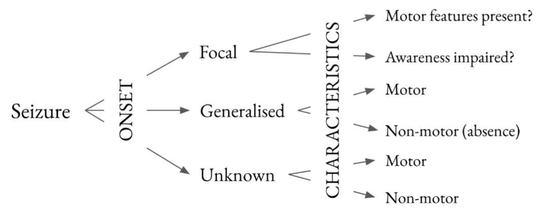
Adapted from Scheffer I, Berkovic S, Capovilla G, Connolly M, French J, Guilhoto L et al. ILAE classification of the epilepsies: Position paper of the ILAE Commission for Classification and Terminology. Epilepsia. 2017;58(4):512-521
Overview of causes of seizures
| Primary neurological | Epilepsy (including secondary to focal lesions, genetic syndromes, and degenerative processes)
Intracranial haemorrhage Cerebral venous sinus thrombosis Acute stroke Hypoxic-ischaemic brain injury Traumatic brain injury |
| Secondary causes | Hypoxia
Malignant hypertension Eclampsia Extremes of: glucose, Na⁺, Ca²⁺, Mg²⁺ (consider effects of medications, e.g. PPIs on Na⁺, Mg²⁺) Hyperthyroidism Uraemia Drug and alcohol intoxication/withdrawal Intercurrent illness in those with known seizure disorders Rare causes: Acute intermittent porphyria Thrombotic thrombocytopaenic purpura Posterior reversible encephalopathy syndrome (PRES) |
Diagnosis and management
Clinical features of seizures
It is important to elicit features before, during, and after the episode being reported. Premorbid state and any prodromal illness are also key features and may guide towards identification of a precipitant, as well as helping to differentiate a true seizure from an alternative explanation. Collateral history is, once again, essential.
| Focal | Generalised | |
|---|---|---|
| Clinical features | Aura
Automatism: lip smacking, chewing, fidgeting Sensory disturbance (e.g. visual) Speech difficulty Post-ictal period |
Aura
Absence Motor features: tonic-clonic, myoclonic Tongue biting Incontinence Post-ictal period |
| Important syndromes | Jacksonian march: focal epilepsy syndrome affecting primary motor cortex (PMC) resulting in progression of focal features as abnormal activity spreads through PMC
Todd’s paresis: post-ictal weakness classically seen in contralateral limb to focus of seizure; also associated with generalised seizures |
Juvenile myoclonic epilepsy: manifests in teenage years with myoclonus (which can be confused with clumsiness) and progresses to generalised epilepsy in most
May be associated with developmental and inherited syndromes, particularly errors of metabolism and mitochondrial disease |
Investigation of seizures
The approach should be as per that to a patient with altered conscious level. It is important to rule out potential precipitants with bedside investigations (e.g. glucose, electrolytes). Potential effects of existing illnesses or medication must be considered, e.g. hypertension, immunosuppression or renal failure may suggest PRES; disturbance to sodium and/or magnesium as a result of PPI use etc.
As with all acute neurological presentations, first-line imaging is CT brain. This may demonstrate a precipitating lesion (e.g. tumour, haemorrhage), but MRI provides the best resolution to demonstrate epileptogenic foci (e.g. in mesial temporal lobe or hippocampus).
EEG is an important facet in the diagnosis of epilepsy. It may be normal but can demonstrate abnormalities or epileptogenic discharges if taken during an event so should be combined with imaging studies. In cases where a precipitating factor is suspected, EEG can be carried out with provocation, e.g. for photosensitive epilepsy.
Management of seizures
Anti-epileptic drugs are not usually indicated after a first seizure unless there is thought to be a high risk of recurrence, e.g. if a precipitating lesion is identified.
Be aware of drug-drug interactions impairing the effectiveness of AEDs – many are metabolised via the cytochrome P450 pathway. This should be considered carefully when a patient with previously well-controlled epilepsy presents with a new seizure.
| Focal | Generalised |
|---|---|
| First line if male or woman beyond childbearing years: lamotrigine or carbamazepine
If woman of childbearing age: lamotrigine Second line: levetiracetam, oxcarbazepine |
Benzodiazepines to terminate seizure with later consideration of anti-epileptic drugs
Cause specific, e.g. MgSO₄ for eclampsia, correction of Na⁺ (hypertonic saline if causing seizures)/Ca²⁺/Mg²⁺/glucose First line if male or woman beyond childbearing years: sodium valproate If woman of childbearing age: lamotrigine Second line: carbamazepine, oxcarbazepine |
It is important to be aware of the impact of a seizure on a patient’s eligibility to drive following the event. In broad summary: patients must not drive a car or motorcycle for 6 months after an isolated or provoked seizure (unless there is a factor that is likely to drive recurrence, i.e. a precipitating lesion) or for 12 months after the most recent recurrent seizure. This is much more stringent for bus and lorry driving: patients must not drive for 5 and 10 years for isolated and recurrent seizures respectively, and may not be reliant on anti-epileptic drugs.
Status epilepticus
Status epilepticus is a medical emergency defined as a seizure lasting more than 5 minutes or recurrent seizures without recovery in between.
Patients should be treated with a benzodiazepine (IV preferred, buccal or rectal if no IV route available) after the first 5 minutes and then re-treated after 5 minutes if the seizure does not terminate. If this is ineffective, second line agents (anti-epileptic drugs: levetiracetam, phenytoin, or valproate) should be given. Patients for whom this is ineffective will require general anaesthesia with intubation and ventilation.
Status epilepticus may be non-convulsive – the patient may appear to be simply comatose with no other explanation. There may be limited or subtle features to point towards this diagnosis, though examination may reveal, for example, subtle twitching or eye movement abnormalities. EEG confirmation is required.
Psychogenic non-epileptic seizures
These are involuntary behavioural and/or motor responses to triggers, which may clinically resemble a seizure. The aetiology is not entirely understood, but there is thought to be a likely psychological mechanism. Psychosocial stressors may trigger these events.
Key features that differentiate these from true seizure events include:
- Unusual seizure-type movements – especially side-to-side movement of the head
- Eye closure with resistance to eye opening
- Normal respiratory and heart rates, lack of cyanosis
- Rapid recovery of consciousness following the event
- Long duration
- Normal prolactin and CK after the event (though sensitivity is variable)
Video EEG may be helpful and will demonstrate normal electrical activity during episodes. Neuroimaging is typically recommended to exclude an epileptogenic lesion.
One of the mainstays of management is patiently and supportively explaining the diagnosis to the patient, usually with psychotherapeutic support.
References
- Duncan R. Psychogenic non-epilectic seizures: etiology, clinical features, and diagnosis. UpToDate [Internet]. Eds: Garcia P, Dashe J. 2021 [cited 1 December 2021]. Available from: https://www.uptodate.com/contents/psychogenic-nonepileptic-seizures-etiology-clinical-features-and-diagnosis
- International League Against Epilepsy. Diagnostic Manual [Internet]. Epilepsydiagnosis.org. 2020 [cited 1 December 2021]. Available from: https://www.epilepsydiagnosis.org/index.html
- Jirsch J, Hirsch L. Non-convulsive status epilepticus: classification, clinical features, and diagnosis. UpToDate [Internet]. Eds: Garcia P, Edlow J, Dashe J. 2021 [cited 1 December 2021]. Available from: https://www.uptodate.com/contents/nonconvulsive-status-epilepticus-classification-clinical-features-and-diagnosis
- Manji H, Connolly S, Kitchen N, Lambert C, Mehta A. Oxford Handbook of Neurology [Internet]. 2nd ed. Oxford: Oxford University Press; 2014 [cited 29 November 2021]. Available from: https://oxfordmedicine.com/view/10.1093/med/9780199601172.001.0001/med-9780199601172
- National Institute for Health and Clinical Excellence. CG137: Epilepsies: diagnosis and management [Internet]. 2012 [cited 1 December 2021]. Available from: https://www.nice.org.uk/guidance/cg137/chapter/1-Guidance
- Schachter S. Evaluation and management of the first seizure in adults. UpToDate [Internet]. Eds: Garcia P, Edlow J, Dashe J. 2021 [cited 1 December 2021]. Available from: https://www.uptodate.com/contents/evaluation-and-management-of-the-first-seizure-in-adults
- Stone J. Functional (Dissociative) Seizures – Functional Neurological Disorder (FND) [Internet]. Neurosymptoms.org. 2021 [cited 1 December 2021]. Available from: https://www.neurosymptoms.org/en_GB/symptoms/fnd-symptoms/dissociative-non-epileptic-attacks-blackouts/
Infective and inflammatory causes
Overview of infective and inflammatory causes
Infective and inflammatory processes within the CNS are notable causes of alteration in consciousness level or seizures and may be accompanied by other symptoms, including fever, headache, focal neurology and behavioural change.
The majorly encountered causes, both in clinical practice and MRCP exams, include the following:
| Infective | Inflammatory (and other) |
|---|---|
| Meningitis and encephalitis | Autoimmune encephalitis (including acute disseminated encephalomyelitis) |
| Abscess of brain or surrounding tissue | Paraneoplastic encephalitis |
| Cerebral toxoplasmosis | Cerebral vasculitis |
| Cerebral malaria | Neurosarcoidosis |
| Drug-induced meningitis | |
| Metastatic spread | |
| Space-occupying lesions |
Infective pathologies
A wide range of pathogens can cause CNS infection – this section will focus on a selection of viral and bacterial agents which commonly cause meningitis, encephalitis, and abscesses. Some of these are more classically associated with diseases in the immunocompromised or with a returning traveller, for example cerebral toxoplasmosis or malaria respectively. They remain, however, important differentials and so should be kept in mind when considering patients with the appropriate risk factors – it is important to elicit a broad history, including travel, sexual, and immunosuppressive risk, when assessing these patients.
A broad, but by no means exhaustive, sample of commonly encountered infective causes is given in the table below. Microscopy findings are indicated for common pathogens identifiable in this way (though it is important to note that stain microscopy is not a definitive test for the presence of organisms in the CSF).
| Viral | Bacterial | Fungal and parasitic |
|---|---|---|
| Herpes simplex | Streptococcus pneumoniae
|
Cryptococcus spp.
|
| Enteroviruses | Listeria monocytogenes
|
Histoplasma capsulatum |
| West Nile (and other Arboviruses) | Neisseria meningitidis
|
Toxoplasma gondii |
| Rabies | Haemophilus influenzae
|
Plasmodium falciparum |
| HIV | Mycobacterium tuberculosis | |
| Measles | Bartonella henselae | |
| Mumps | Treponema pallidum | |
| Borrelia burgdorferi | ||
| Leptospira spp. | ||
| Staphylococcus aureus and viridans streptococci (most common in abscesses) |
Encephalitis and meningitis
Encephalitis occurs when there is inflammation of the brain parenchyma, whereas meningitis refers specifically to inflammation of the meninges. There may be overlap in the clinical syndrome, and they may indeed coexist (meningoencephalitis). A brief comparison is shown below.
| Encephalitis | Meningitis | |
|---|---|---|
| Common causes | Primarily viral (e.g. HSV type 1, VZV (Varicella zoster virus), West Nile, rabies)
Bacterial, fungal, parasitic, and autoimmune aetiologies much rarer |
Bacterial: L. monocytogenes (<1 and >60 years), N. meningitidis, S. pneumoniae (most common)
Viral: enteroviruses (most common), HSV type 1, HIV (rare) |
| Classical presenting features | Fever
Headache Seizures Altered conscious level or mental state |
Fever
Headache Nausea Neck stiffness Photophobia Seizures (rarer) Altered conscious level or mental state |
| Examination findings | Focal neurological deficits
Flaccid paralysis (West Nile virus) Rash or eruption |
Meningism
Purpura or petechiae (N. meningitidis) Focal neurological deficits (rarer) |
| Investigation findings | Radiology
CT frequently normal MRI may show focal involvement (e.g. temporal lobes in HSV) Classical LP findings WCC ↑ (usually lymphocytes) Protein ↑ Glucose ↔ (↓ if mumps) EEG (electroencephalogram) Can show focal abnormality (e.g. temporal discharges) but findings are frequently non-specific |
Radiology
Not typically indicated unless signs of ↑ICP (intracranial pressure) MRI may show meningeal enhancement Classical LP findings WCC ↑↑ if bacterial, ↑ if viral or TB (all can be lymphocytic in early infection) Protein ↑↑ if bacterial, ↑ if viral, TB, syphilis or Lyme disease Glucose generally ↓ but can be ↓↓ especially if TB/fungal Values may not be as raised in early bacterial meningitis |
| Management strategies | If acute viral encephalitis suspected, aciclovir 10 mg/kg IV TDS (may require renal adjustment) until causative organism confirmed, which may require adjustment of therapy
If meningeal irritation, also cover with ceftriaxone 2 g IV BD |
If bacterial meningitis suspected, ceftriaxone 2 g IV BD and dexamethasone 10 mg IV QDS until causative organism identified, which may require adjustment of therapy or a switch to e.g. anti-TB or antifungal agents
Viral meningitis must be confirmed prior to switching to supportive management – analgesia, fluids, antipyretics |
Special considerations
Some patients presenting with a clinical picture of meningitis and/or encephalitis may have features in the history requiring further assessment, as well as follow on actions that may need to be considered:
- Immunosuppression: those with HIV and other immunodeficiencies are at risk of opportunistic infections with atypical organisms and if they present with features of meningitis and/or encephalitis, should be assessed for these. Cryptococcus neoformans is an invasive fungus for which having a CD4 count of <100 is a risk factor, and may present initially with pulmonary disease with progression to the CNS. Classically, Indian ink staining has been used to detect it in the CSF, but cryptococcal antigen is more sensitive. If suspected, it should be empirically treated until ruled out. Patients with a relative immunodeficiency, for example as a result of age or diabetes, are more at risk of less common organisms, including Listeria, for which cover with ampicillin is required.
- Travel history: patients with recent travel or migration who present with altered consciousness levels and/or seizures should be evaluated for potential imported or rare pathologies. TB meningitis can occur even in the absence of active pulmonary TB and should be considered in patients from a high prevalence area with suggestive symptoms and signs. Those who have travelled to areas with mosquitos must have malaria considered and have blood smears carried out for this. More temperate areas with mosquitos, especially in the US, may have a higher incidence of West Nile virus. Potential tick exposure should prompt consideration of tick-borne encephalitis or Lyme disease. If there is a history of travel to areas with an increased prevalence of penicillin-resistant pneumococci, then rifampicin should be added to the initial treatment regimen.
- Malignancy: some solid malignancies, namely melanoma, non-small cell lung cancer, and breast cancer, as well as haematological malignancies including lymphomas, can spread to the leptomeninges, causing a range of symptoms. This is usually as a result of haematogenous spread but may also be direct invasion or (rarely) a primary tumour. Patients may complain of headache, nausea, or lower limb weakness, but signs may be subtle, including cerebellar dysfunction and meningeal irritation. There may also be signs and symptoms of an obstructive hydrocephalus. Investigation is via cytology for malignant cells in the CSF, which may also have a raised protein.
- Other epidemiological factors: unusual causes can be associated with exposure to carriers of pathogens, human or animal, in those susceptible to severe disease. For example, cat scratch disease, caused by Bartonella henselae, can cause encephalitis in a small minority of cases. Those unvaccinated against measles, mumps and rubella (MMR) may be at risk of encephalitis from these causes if exposed.
- Contacts of confirmed meningococcal disease: meningitis and encephalitis are notifiable diseases in the UK. Those who are contacts of patients with confirmed N. meningitidis meningitis should be offered prophylaxis: single dose ciprofloxacin or rifampicin for 2 days as second line. For non-B serotypes, an ACWY vaccine should be offered.
Autoimmune and paraneoplastic encephalitis
These are rarer but clinically extremely significant causes of encephalitis. Paraneoplastic encephalitis refers to an autoimmune process that is associated specifically with a malignancy, whilst autoimmune encephalitis may happen in the absence of a malignant process. Several important causes are summarised below.
Anti-NMDA receptor encephalitis
This is a rapidly progressing encephalitis that affects predominantly young women and presents with a prominent psychiatric component, motor features, seizures, autonomic dysfunction, and occasionally severe hypoventilation. It can be preceded by a mild viral-type illness. It has a strong association with ovarian teratomas and should prompt a search for such a tumour. It may also be associated with small-cell lung cancer and testicular teratoma. Formal diagnosis is by anti-NMDA receptor antibodies in blood and CSF (with the latter being more specific), though these may take time, so diagnosis is frequently made on clinical grounds. Treatment is with immunomodulation (e.g. methylprednisolone ± IVIg or rituximab), as well as management of any neoplasm found.
Paraneoplastic encephalitis
Some malignancies are associated with autoimmune processes that target the central and/or peripheral nervous system. Classically, this is a limbic encephalitis, presenting with prominent behavioural or mood components but also with seizures and cognitive dysfunction. There may also be involvement of the brainstem or descending CNS tracts. One of the most frequent associations is with small-cell lung cancer by way of anti-Hu antibodies, though there is a wide range of autoantibodies and cancer types associated with CNS involvement. Treatment, again, utilises immunomodulatory agents in combination with management of the underlying malignancy.
References
- Dalmau J, Rosenfeld M. Paraneoplastic and autoimmune encephalitis. UpToDate [Internet]. Eds: Wen P, Eichler A. 2021 [cited 6 January 2022]. Available from: https://www.uptodate.com/contents/paraneoplastic-and-autoimmune-encephalitis
- Demopoulos A. Clinical features and diagnosis of leptomeningeal metastases from solid tumours. UpToDate [Internet]. Eds: Wen P, Loeffler J, Eichler A. 2020 [cited 13 April 2022). Available from: https://www.uptodate.com/contents/clinical-features-and-diagnosis-of-leptomeningeal-metastases-from-solid-tumors
- Gluckmann S. Viral encephalitis in adults. UpToDate [Internet]. Eds: Hirsch M, Mitty J. 2021 [cited 6 January 2022]. Available from: https://www.uptodate.com/contents/viral-encephalitis-in-adults
- Hasbun R. Clinical features and diagnosis of bacterial meningitis in adults. UpToDate [Internet]. Eds: Tunkel A, Mitty J. 2021 [cited 6 January 2022]. Available from: https://www.uptodate.com/contents/clinical-features-and-diagnosis-of-acute-bacterial-meningitis-in-adults
- Manji H, Connolly S, Kitchen N, Lambert C, Mehta A. Oxford Handbook of Neurology [Internet]. 2nd ed. Oxford: Oxford University Press; 2014 [cited 29 November 2021]. Available from: https://oxfordmedicine.com/view/10.1093/med/9780199601172.001.0001/med-9780199601172
- Patel P. Encephalitis. BMJ Best Practice [Internet]. 2020 [cited 6 January 2022]. Available from: https://bestpractice.bmj.com/topics/en-gb/436
- Tunkel A. Aseptic meningitis in adults. UpToDate [Internet]. Eds: Hirsch M, Mitty J. 2021 [cited 6 January 2022]. Available from: https://www.uptodate.com/contents/aseptic-meningitis-in-adults
Space-occupying lesions
Overview of space-occupying lesions
Space-occupying lesions within the central nervous system can be diagnosed following presentation with either focal or generalised neurological features. The most common presenting feature is headache, although in 25% of patients, seizures are the initial presentation. Other classic findings include nausea and vomiting, sensory changes, aphasia, focal weakness, depressed level of consciousness, or cognitive and behavioural changes.
There are a number of possible aetiologies of space-occupying lesions, and these are broadly summarised below.
| Tumour | Vascular | Infectious | Inflammatory |
|---|---|---|---|
| Primary | Anomalous vasculature | Abscess (classically bacterial or fungal) | Granulomatous disease (e.g. sarcoidosis) |
| Metastatic | Haemorrhage | Tuberculoma | Vasculitis |
| Infarct | Parasitic cyst formation | Tumefactive MS (multiple sclerosis) lesions | |
| Progressive multifocal leukoencephalopathy (PML) |
Primary and metastatic tumours
Brain tumours are the ninth most common neoplasm diagnosed in the UK and the second most common in children. The majority of primary brain tumours arise from the meninges and the most common of these by far are meningiomas, which are more common in women and likely to be benign.
Malignant brain tumours primarily arise from neuroepithelial tissue and are generally termed gliomas. The most common of these in adults is glioblastoma multiforme (GBM), a highly aggressive tumour of astrocyte origin. Their incidence is similar in men and women and is associated with advancing age. Less aggressive tumours (low-grade gliomas) are more common in children and young people and carry a better prognosis.
Other primary brain tumours include pituitary adenomas, haemangiomas, nerve sheath tumours, such as vestibular Schwannoma, and primary CNS lymphoma. When considering brain tumours, it is especially important to consider metastatic disease – metastatic deposits in the brain are significantly more common than primary brain tumours. Typical primary sites are: lung, breast, kidney, lower GI tract, and skin.
Investigation and management
Initial diagnosis is with contrast CT brain, though the preferred form of imaging is MRI with gadolinium contrast for better resolution and ability to identify tissue types. If metastatic disease is suspected, staging CT imaging should be arranged. If there are no concerns about raised intracranial pressure, and especially if there is concern of a CNS lymphoma (e.g. in the patient with HIV), a lumbar puncture should be performed for flow cytometry.
Management should be led by neurosurgical and oncology teams, but, in general, for lesions with mass effect and surrounding oedema, high-dose steroids should be given, unless there is a suspicion for CNS lymphoma. In this case, steroids should ideally be withheld until a tissue diagnosis is obtained. If the presentation was with seizures, anti-seizure medication should be started.
General principles for ongoing management are usually combination therapy – surgical resection, radiotherapy (including radiosurgery, e.g. Gamma Knife), and chemotherapy in select cases (temozolomide is standard for GBM).
Spinal cord lesions
These are relatively infrequently encountered compared to other CNS tumours but are an important cause of focal neurology, typically in conjunction with unremitting back pain. They are classified as intramedullary (within the substance of the cord), intradural but extramedullary (such as meningiomas or nerve sheath tumours), and extradural (usually metastases).
Like brain tumours, the majority of intramedullary spinal cord tumours are gliomas, with ependymomas predominating. Intradural but extramedullary lesions are most frequently meningiomas, but approximately a quarter are nerve sheath tumours – either Schwannomas or neurofibromas. Extradural lesions most frequently occur within the vertebral bodies and are most commonly caused by metastatic disease, typically from a breast, lung or prostate primary. They classically manifest with spinal cord or cauda equina compression but can cause intradural invasion.
Investigation of spinal cord lesions is primarily via MRI or CT myelogram for those unable to undergo MRI scanning. Management is usually a combination of surgery and radiotherapy.
Infectious and inflammatory causes
When considering an infectious or inflammatory cause of a space-occupying lesion, it is essential to ascertain any risks for immunosuppression, recent travel history, and the patient’s social context. Presentation is classically with headache and fever, potentially in combination with cranial nerve signs, meningeal irritation, seizures, or change in mental state or conscious level.
Inflammatory causes may present insidiously without a pre-existing diagnosis, for example in neurological manifestations of systemic lupus erythematosus. They may result in a range of symptoms, ranging from subtle focal neurology to seizures and coma.
Important things to establish when considering a patient with a potential infectious or inflammatory brain lesion:
- Is there underlying immune suppression? HIV, diabetes, and end-stage renal disease receiving haemodialysis are all risk factors for brain abscess formation, and it is important to consider opportunistic pathogens such as Toxoplasma gondii, the risk of which is increased by a low CD4 count (<100). Some medications, especially immunomodulatory agents, have been associated with PML.
- Has there been recent operative management of the brain or head and neck, including dental procedures?
- Is there a recent history of sinusitis, otitis or dental infection? The majority of brain abscesses form by direct spread, and there are characteristic organisms for different sites of origin:
| Sinuses | Streptococcus spp., Haemophilus spp., Bacteroides spp., Fusobacterium spp. |
| Ears | Streptococcus spp. (acute), P. aeruginosa (chronic), Proteus mirabilis |
| Dental | Streptococcus spp., Fusobacterium spp., Bacteroides spp. |
| Neurosurgical | Staphylococcus spp., Streptococcus spp., P. aeruginosa, Enterobacter spp. |
- Has the patient recently travelled to an area where certain parasites are endemic? Taenia solium, for example, is known to be endemic in many regions of Central and South America and is the causative agent for neurocysticercosis.
- Are there risk factors for TB?
- Is there a history suggestive of an underlying systemic disorder? A careful systems review and examination is essential, especially if considering vasculitis or granulomatous disease, or MS-related lesions.
Initial investigation is via CT brain imaging with contrast, although once again MRI with gadolinium contrast provides best resolution; these may demonstrate ring-enhancing lesions. Lumbar puncture is frequently contraindicated, and it may be that direct biopsy is most diagnostic. It is also essential to perform blood cultures, and HIV serology may be helpful.
If there is suspicion of a brain abscess, broad-spectrum antibiotic therapy with good aerobic and anaerobic cover should be instituted urgently – this may include vancomycin, ceftriaxone, or metronidazole. Once a causative organism or underlying condition is identified, targeted therapy should be initiated, though neurosurgical intervention may be required. Steroids should be given if there is vasogenic oedema and mass effect, and prophylactic anticonvulsant therapy may be indicated.
References
- Cancer Research UK. Brain, other CNS and intracranial tumours statistics [Internet]. Cancer Research UK. 2021 [cited 11 Feb 2022]. Available from: https://www.cancerresearchuk.org/health-professional/cancer-statistics/statistics-by-cancer-type/brain-other-cns-and-intracranial-tumours/
- Hall W. Brain abscess. BMJ Best Practice [Internet]. 2021 [cited 11 Feb 2022]. Available from: https://bestpractice.bmj.com/topics/en-gb/925/
- Manji H, Connolly S, Kitchen N, Lambert C, Mehta A. Oxford Handbook of Neurology [Internet]. 2nd ed. Oxford: Oxford University Press; 2014 [cited 29 November 2021]. Available from: https://oxfordmedicine.com/view/10.1093/med/9780199601172.001.0001/med-9780199601172
- Southwick F. Pathogenesis, clinical manifestations, and diagnosis of brain abscess. UpToDate [Internet]. Eds: Tunkel A, Hall K. 2021 [cited 11 Feb 2022]. Available from: https://www.uptodate.com/contents/pathogenesis-clinical-manifestations-and-diagnosis-of-brain-abscess
- Welch W, Schiff D, Gerszten P. Spinal cord tumours. UpToDate [Internet]. Eds: Loeffler J, Wen P, Eichler A. 2021. [cited 11 Feb 2022]. Available from: https://www.uptodate.com/contents/spinal-cord-tumors
- Wong E, Wu J. Overview of the clinical features and diagnosis of brain tumours in adults. UpToDate [Internet]. Eds: Eds: Loeffler J, Wen P, Eichler A. 2021. [cited 11 Feb 2022]. Available from: https://www.uptodate.com/contents/overview-of-the-clinical-features-and-diagnosis-of-brain-tumors-in-adults
Complete Guide to Clinical Genetics ST3 Application and Recruitment (2027)
Written by Medibuddy. Last updated: October 2026. Clinical Genetics is

Complete Guide to Clinical Radiology ST3 Application and Recruitment (2027)
Recruitment paused: NHS England is not running Clinical Radiology ST3+ recru

Complete Guide to Paediatric Surgery ST3 Application and Recruitment in United Kingdom (2026)
Written by Medibuddy. Last updated: October 2026. If you’re planning


Take your subscriptions with you
Our mobile app allows you to access your interview and exam question banks wherever you are.

