


 Close
Close
 11th August 2024
Human prion disease – Creutzfeldt-Jakob disease
Creutzfeldt-Jakob disease (CJD) is a rare and inevitably fatal cause of progressive dementia with neurological symptoms. Intracellular human prion proteins are misfolded from an alpha-helical structure into a beta-pleated form, which in turn causes adjacent proteins to undergo this transformation. These ‘infectious proteins’ drive the pathophysiology of neuronal injury and eventual cell death as a result of accumulation of abnormal prion proteins in the CNS.
There are three main types of CJD:
- Sporadic (sCJD) – accounts for the majority of cases, up to 90% of CJD
- Acquired – include iatrogenic CJD (iCJD) (transmission of prion protein from contaminated surgical equipment or cadaveric tissue grafts) and variant CJD (vCJD) (ingestion of CJD-contaminated beef or blood transfusion)
- Familial – mutations in the gene coding for PRNP (endogenous prion protein) on chromosome 20; usually autosomal dominant with 100% penetrance, however, clinical presentation can be variable and less aggressive than sCJD and vCJD
| sCJD | Familial | vCJD | iCJD | |
|---|---|---|---|---|
| Onset | 60s | Younger onset than sCJD | Young adults – teenagers, 20s | Variable incubation period – also related to timing of inoculation event |
| Features
(in order of presentation) |
Cognitive symptoms – memory loss, aphasia and impaired executive functioning with progressive dementia
Ataxia Rigidity Limb myoclonus Familial can have a variable rate of progression, although it can present almost identically to sCJD |
Psychiatric symptoms – often severe depression
Dementia (initially mild cognitive impairment) Ataxia Painful sensory symptoms Involuntary movements – dystonia, chorea, myoclonus |
For neurosurgical inoculation – similar presentation to sCJD | |
Diagnosis of CJD
CSF examination – can be bland with mildly raised protein. Elevated 14-3-3 protein has high sensitivity and specificity for sCJD but is not useful for vCJD; real-time quaking-induced conversion for abnormal prions has high sensitivity and specificity.
EEG – periodic triphasic spikes
Imaging – MRI brain with FLAIR (fluid attenuated inversion recovery) and diffusion-weighted imaging (DWI) best demonstrates affected areas:
- sCJD – hyperintensities in the basal ganglia (caudate and putamen) and ‘cortical ribboning’ on DWI
- vCJD – bilateral periventricular thalamic hyperintensities, ‘hockey stick sign’
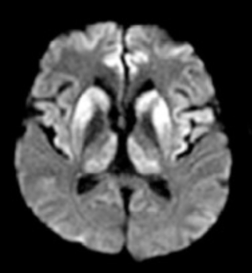
Image Source: ncbi.nlm.nih.gov 2025, PMC5619326, Licensed under a creative commons licence. Available for free at: https://www.ncbi.nlm.nih.gov/pmc/articles/PMC5619326/pdf/dn-09-04-0424.pdf (Accessed: 17 July 2024)
![MRCP Part 2 - [L] Axial T2 FLAIR MRI showing typical bilateral basal ganglia hyperintensity in sCJD [R] DWI MRI showing ‘cortical ribboning’ in sCJD](https://medibuddy.co.uk/wp-content/uploads/2024/08/Screenshot-at-Aug-08-17-55-24.png)
Image Source: ncbi.nlm.nih.gov 2025, PMC5619326, Licensed under a creative commons licence. Available for free at: https://www.ncbi.nlm.nih.gov/pmc/articles/PMC5619326/pdf/dn-09-04-0424.pdf (Accessed: 17 July 2024)
Complete Guide to Clinical Genetics ST3 Application and Recruitment (2027)
Written by Medibuddy. Last updated: October 2026. Clinical Genetics is

Complete Guide to Clinical Radiology ST3 Application and Recruitment (2027)
Recruitment paused: NHS England is not running Clinical Radiology ST3+ recru

Complete Guide to Paediatric Surgery ST3 Application and Recruitment in United Kingdom (2026)
Written by Medibuddy. Last updated: October 2026. If you’re planning


Take your subscriptions with you
Our mobile app allows you to access your interview and exam question banks wherever you are.

