


 Close
Close
 11th August 2024
Myopathies
Overview of myopathies
Myopathies may be inherited or acquired.
Inherited forms include muscular dystrophies, mitochondrial diseases, metabolic and other congenital myopathies (e.g. McArdles or Von Gierke’s), and channelopathies.
Acquired forms have a variety of aetiologies including drug-induced, endocrine (Addison’s, Cushing’s, hypo- or hyperthyroidism, DM, hyperaldosteronism), metabolic (glycogen storage disease, lipid metabolism), toxic or inflammatory.
Muscular dystrophies
Myotonic dystrophy
This is the most common muscular dystrophy. It is an autosomal dominant disorder that generally develops in the 2nd-4th decades and is characterised by respiratory and skeletal muscle weakness (distal limb and facial/neck most commonly), cataracts, frontal balding, cardiac conduction problems and myotonia (delayed relaxation of muscles following contraction).
Duchenne (DMD) and Becker muscular dystrophy (BMD)
DMD and BMD are both caused by mutations of the dystrophin gene on the X chromosome. Males, therefore, are most affected, though female carriers can display mild symptoms.
DMD is the more severe and more common form (prevalence 3/100,000) and typically presents in the first years of life. Features include proximal muscle weakness (with walking problems including toe walking or waddling gait), cardiomyopathy or conduction issues, and scoliosis/bony fractures. The prognosis is poor, with most wheelchair bound by the early teens and death usually from respiratory compromise or cardiomyopathy, in the teens or twenties.
BMD, the rarer form (0.3/100,000), presents with milder symptoms in the teens or twenties. Many survive into their thirties.
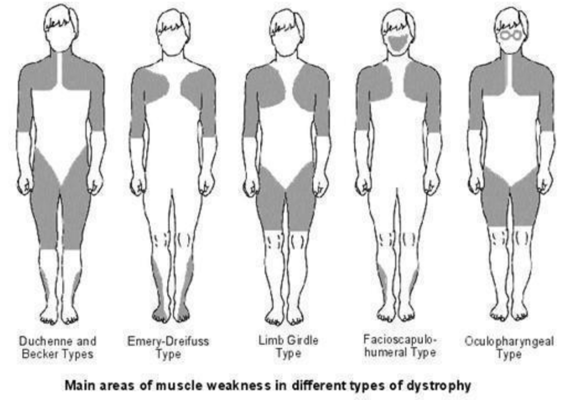
Image Source: Egton Medical Information Systems Limited. All rights reserved (2023). Bones joints muscles/muscular-dystrophies, Licensed under a creative commons licence. Available for free at: https://patient.info/bones-joints-muscles/muscular-dystrophies (Accessed: 17 January 2024)
Facioscapulohumeral muscular dystrophy (FSHD)
FSHD is an autosomal dominant condition with prevalence of 1-2/100,000. Patients generally present in their teens with weakness of facial muscles, scapula (upwards displacement), upper arm (with deltoid sparing), and lower leg. Weakness is generally asymmetric. Genetic testing confirms the diagnosis identifying mutations leading to inappropriate DUX4 gene expression.
Emery-Dreifuss muscular dystrophy
This has autosomal dominant or X-linked inheritance. Clinical features include scapula, humerus and peroneal weakness, and cardiac conduction defects often requiring pacing. Patients may also develop contractures of the cervical extensors, biceps and long finger extensors.
Mitochondrial disorders
MERRF: Myoclonic epilepsy with ragged red fibres
The principal clinical features of MERRF are myoclonus, epilepsy, myopathy and ataxia, with neuropathy, visual problems, lipomas, spasticity, deafness and Wolff-Parkinson-White (WPW) syndrome. The characteristic ragged red fibres are found on muscle biopsy.
MELAS: Mitochondrial encephalomyopathy, lactic acidosis and stroke-like episodes
Clinical features include stroke-like episodes, seizures, migraine-like headaches, deafness, short stature and myopathy.
MIDD: Maternally inherited diabetes and deafness
MIDD is characterised by insulin-dependent diabetes and sensorineural deafness, often alongside myopathy, visual problems, short stature, and cardiac and renal issues (focal segmental glomerulosclerosis).
MNGIE: Mitochondrial neurogastrointestinal encephalomyopathy
Clinical features include gastrointestinal dysmotility (secondary to visceral myopathy), ptosis, ophthalmoplegia, polyneuropathy and distal myopathy. Onset is from childhood to the 4th decade.
CPEO: Chronic progressive external ophthalmoplegia and KSS: Kearns-Sayre syndrome
Patients with CPEO develop ophthalmoplegia and ptosis. KSS has similar features with the addition of retinitis pigmentosa. Onset of KSS, generally in childhood, is earlier than that of CPEO (usually 4th decade).
Glycogen storage diseases (GSD)
The glycogen storage diseases are inborn errors of metabolism that lead to issues with glucose processing and energy release.
Muscle symptoms include pain and stiffness following exercise, elevated CK (creatine kinase) and muscle transaminases and myoglobinuria. In McArdle’s, the ‘second wind’ phenomenon may be noted in which after an initial barrier (with fatigue, tachycardia and breathlessness) is surmounted, exercise tolerance improves. This is due to activation of other energy sources (e.g. fatty acids, hepatic glucose production) to supplement ineffective muscle energy release due to myophosphorylase deficiency.
Dermatomyositis and polymyositis (DM, PM)
Dermatomyositis and polymyositis both present with symmetrical proximal muscle weakness and evidence of inflammation on muscle biopsy (fibre necrosis, degeneration and regeneration, and inflammatory cell infiltrates) which is the gold standard for diagnosis.
Dermatomyositis has additional dermatologic features which may include: Gottron’s papules (scaly purple/red knuckles), periorbital heliotrope rash, shawl-sign (an erythematous rash over the neck and chest), nailfold erythema and periungual telangiectasia, subcutaneous calcification (calcinosis cutis), and ‘mechanic’s hands’ (rough, cracked, darkened areas). Interstitial lung disease occurs in 30-40% of patients.
Both are treated with immunosuppression, usually initially with prednisolone.
Both PM and dermatomyositis may be associated with malignancy via a paraneoplastic mechanism. Cervical, lung, ovarian, pancreatic, bladder and gastric adenocarcinomas are most strongly linked.
Inclusion-body myositis (IBM)
IBM causes a specific pattern of muscle wasting, with weakness of the quadriceps and forearm muscles. Swallow function may also be affected. Onset is generally in the 4th to 6th decade. There is no current treatment.
Muscle enzymes and CK are often mildly elevated. Diagnosis is on the basis of typical examination findings, lab tests and muscle biopsy which may show the characteristic rimmed-vacuoles, along with mononuclear cell inflammatory infiltrates and non-necrotic fibres.
Drug-induced myopathies
Common drug causes of myopathies include:
| Steroids | Alcohol |
| Statins | Cocaine |
| Penicillamine | Antipsychotics |
| Colchicine | Antiretrovirals – AZT (azidothymidine) and other NRTIs (nucleoside reverse transcriptase inhibitor) |
References
- Manji H. Oxford Handbook of Neurology. Oxford Medical Publications; 2020
- Stone WL, Basit H, Adil A. Glycogen Storage Disease [Internet]. PubMed. Treasure Island (FL): StatPearls Publishing; 2020. [Accessed 2022 Jan 22] Available from: https://www.ncbi.nlm.nih.gov/books/NBK459277/
- Darras BT. Duchenne and Becker Muscular Dystrophy: Clinical Features and Diagnosis [Internet]. Patterson MC (Ed), Firth HV (Ed). UpToDate. 2020 [cited 2022 Feb 14]. Available from: https://www.uptodate.com/contents/duchenne-and-becker-muscular-dystrophy-clinical-features-and-diagnosis
- Darras BT. Limb-girdle muscular dystrophy [Internet]. Nordli DR (Ed), Shefner JM (Ed). UpToDate. 2022 [cited 2022 Feb 14]. Available from: https://www.uptodate.com/contents/limb-girdle-muscular-dystrophy
- Darras BT. Emery-Dreifuss muscular dystrophy [Internet]. Patterson MC (Ed). UpToDate. 2020 [cited 2022 Feb 14]. Available from: https://www.uptodate.com/contents/emery-dreifuss-muscular-dystrophy
- Darras BT. Facioscapulohumeral muscular dystrophy [Internet]. Nordli DR (Ed), Shefner JM (Ed). UpToDate. 2022 [cited 2022 Feb 14]. Available from: https://www.uptodate.com/contents/facioscapulohumeral-muscular-dystrophy
- Darras BT. Myotonic dystrophy: Aetiology, clinical features, and diagnosis [Internet]. Shefner JM (Ed), Nordli DR (Ed). UpToDate. 2021 [cited 2022 Feb 14]. Available from: https://www.uptodate.com/contents/myotonic-dystrophy-etiology-clinical-features-and-diagnosis
- Vydra DG, Rayi A. Myotonic Dystrophy [Internet]. PubMed. Treasure Island (FL): StatPearls Publishing; 2020 [accessed 2022 Jan 22]. Available from: https://www.ncbi.nlm.nih.gov/books/NBK557446/
- Craigen WJ. Overview of inherited disorders of glucose and glycogen metabolisms [Internet]. Hahn S (Ed). UpToDate. 2021 [cited 2022 Feb 14]. Available from: https://www.uptodate.com/contents/overview-of-inherited-disorders-of-glucose-and-glycogen-metabolism
Complete Guide to Clinical Genetics ST3 Application and Recruitment (2027)
Written by Medibuddy. Last updated: October 2026. Clinical Genetics is

Complete Guide to Clinical Radiology ST3 Application and Recruitment (2027)
Recruitment paused: NHS England is not running Clinical Radiology ST3+ recru

Complete Guide to Paediatric Surgery ST3 Application and Recruitment in United Kingdom (2026)
Written by Medibuddy. Last updated: October 2026. If you’re planning


Take your subscriptions with you
Our mobile app allows you to access your interview and exam question banks wherever you are.

