


 Close
Close
 13th February 2025
Red eye
Red eye
Overview of red eye
Red eye is a common ophthalmological presentation, with urgent causes requiring immediate referral to ophthalmology. The following aetiologies will be discussed:
| Urgent causes of red eye | Non-urgent causes of red eye |
|---|---|
| Acute closed-angle glaucoma (see later section) | Blepharitis |
| Orbital cellulitis | Conjunctivitis |
| Infectious keratitis | Episcleritis |
| Anterior uveitis (see later section) | Subconjunctival haemorrhage |
| Scleritis |
Table 1 Aetiologies of red eye
Urgent causes of red eye
Orbital cellulitis
Infection of the subcutaneous tissues surrounding the eye may involve the orbit (orbital cellulitis), or tissues anterior to the orbital septum only (periorbital, or preseptal cellulitis). Orbital cellulitis often arises from local spread of paranasal sinus infection or spread of periorbital cellulitis. It is an emergency which can lead to permanent vision loss or even brain abscess and death and, therefore, requires an emergency ophthalmology and/or ENT (Ear, nose and throat) referral.
The eyelid will be inflamed (Figure 1). Ocular signs may include a swollen, red and proptosed eye, with painful and reduced eye movements. Optic nerve dysfunction may be present in severe cases, manifesting as reduced visual acuity, impaired colour vision or relative afferent pupillary defect. The patient may be systemically unwell and pyrexial.
High-resolution CT of the orbit, sinuses and brain is key for diagnosis and to rule out an underlying abscess. Treatment is with IV broad-spectrum antibiotics but sinus or abscess drainage by ENT may also be required.
In contrast to orbital cellulitis, periorbital cellulitis may present with a swollen and red eyelid, with or without pyrexia, but does not usually cause any proptosis, decrease in eye movements or worsening visual acuity. It can usually be treated with oral antibiotics, though due to high risk of orbital extension, especially in children, urgent ophthalmology review is necessary.
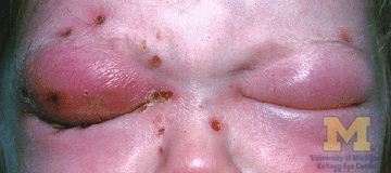
Image Source: Wikimedia Commons (2011). Orbital cellulitis, Licensed under a creative commons licence. Available for free at: https://commons.wikimedia.org/wiki/File:Orbital_cellulitis.jpg#/media/File:Orbital_cellulitis.jpg (Accessed: 18 January 2024).
Infectious keratitis
Infectious keratitis is an infection of the cornea; it is most commonly caused by bacteria but can also have a range of viral, fungal and parasitic aetiologies. All suspected corneal infections or ulcers should be referred to ophthalmology urgently due to risk of potentially sight-threatening complications.
Bacterial
Risk factors include anything that may disrupt the epithelium; the most notable in younger patients is contact lens wear, particularly overnight. Others include trauma, surgery or underlying corneal pathologies such as severe dry eye or bullous keratopathy. Causative organisms are most commonly Staphylococcus aureus, Streptococci species, and Pseudomonas aeruginosa. It should be noted that some organisms are able to penetrate intact corneal epithelium, including Shigella, Neisseria, Listeria and corynebacterium species.
The patient often complains of a foreign body sensation. They may have photophobia and be unable to keep the eye open. On fluorescein staining, an epithelial defect may be visible, with conjunctival injection surrounding the cornea. A corneal opacity may also be seen. Mucopurulent discharge may be present, and in severe cases a hypopyon (pus in the anterior chamber). Treatment is usually with topical antibiotics. Corneal scraping and conjunctival swabs may be carried out for culture and sensitivity to guide further choice of antibiotic.
Herpes zoster ophthalmicus (ophthalmic shingles)
Varicella zoster virus that becomes dormant in the dorsal root ganglia after chickenpox may reactivate in later years to cause herpes zoster (shingles). This can affect the ophthalmic division of the trigeminal nerve (CN V1), causing herpes zoster ophthalmicus (HZO). It is most common in the 6th and 7th decades of life, with more severe disease seen in immunosuppressed patients. There is usually a prodrome lasting around 3-5 days, during which the patient is systemically unwell, with symptoms such as fever, malaise and a headache. This is followed by tingling, burning, or pain which may be mild or severe, and a vesicular rash, both respecting the CN V1 dermatomal distribution (anterior forehead and nose), not crossing the midline (Figure 2).
Ocular manifestations can include conjunctivitis, keratitis, uveitis or episcleritis. Dendritic lesions may be seen on fluorescein staining, which have tapered ends (pseudodendrites) in comparison to the terminal bulbs seen on dendrites in herpes simplex infection. A good predictor of eye disease with shingles is the presence of skin lesions at the side or tip of the nose, indicating involvement of the nasociliary nerve; this is called Hutchinson’s sign.
Oral antivirals are indicated within 72 hours of the rash appearing and reduce the duration of symptoms, severity and risk of chronic pain after rash resolution (post-herpetic neuralgia). Intravenous acyclovir may be required in severe cases or in immunocompromised patients. Antibiotics may also be given to prevent secondary bacterial infection of the eye.
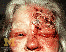
Image Source: Wikimedia Commons (2011). Trigeminal herpes with uveitis and keratitis, Licensed under a creative commons licence. Available for free at: https://commons.wikimedia.org/wiki/File:Trigeminal_herpes_with_uveitis_and_keratitis.jpg#/media/File:Trigeminal_herpes_with_uveitis_and_keratitis.jpg (Accessed: 18 January 2024).
Herpes simplex keratitis
This is most commonly secondary to infection with herpes simplex virus 1 (HSV1). The patient may have a foreign body sensation, photophobia, blurred vision and watering. Examination with fluorescein shows a dendritic ulcer, which is a linear, branching lesion with terminal bulbs, usually in the centre of the cornea (Figure 3). Contrast this to the tapered ends of the pseudodendrites seen in HZO. HSV keratitis is usually treatable with topical antivirals.
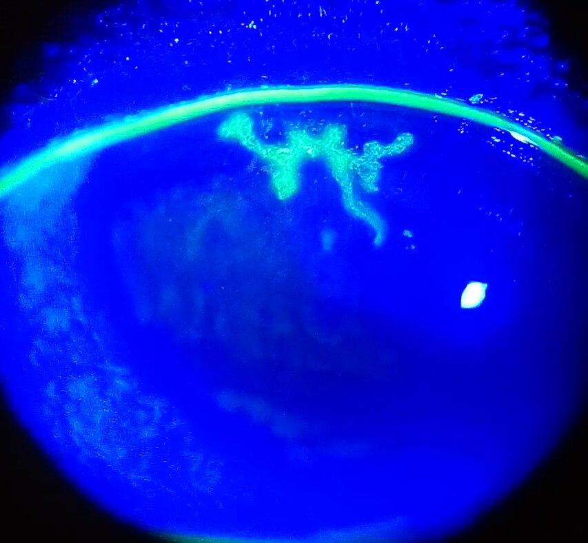
Image Source: Wikimedia Commons (2017). Dendritic corneal ulcer, Licensed under a creative commons licence. Available for free at: https://commons.wikimedia.org/w/index.php?curid=58964337#/media/File:Dendritic_corneal_ulcer.jpg (Accessed: 18 January 2024).
Acanthamoeba keratitis
Vigilance for infection with the rare but potentially sight-threatening Acanthamoeba spp should be maintained, especially in patients who have had limited response to antibiotics. It is a protozoan usually found in soil or fresh water and is associated with poor contact lens hygiene such as swimming or showering in contact lenses. Patients often present with unilateral eye pain that is characteristically out of proportion to clinical findings. Grey-coloured irregular epithelial corneal infiltrates may be seen and in later stages, a grey ring-shaped stromal infiltrate. Infiltrates along corneal nerves (radial keratoneuritis) are pathognomonic.
Scleritis
The sclera is the outer, fibrous layer making up the ‘white’ of the eyes and consists of a thin outermost episclera, containing a rich blood supply, a more dense stromal layer, and a layer continuous with the uveal tract. Scleritis is the inflammation of the stromal layer and can rapidly lead to deterioration and permanent loss of vision due to tissue destruction. Importantly, scleritis is most commonly immune mediated and can represent a host of underlying inflammatory conditions; rheumatoid arthritis is the most common association. Scleritis may also have infective causes, such as herpes zoster, syphilis or tuberculosis. Urgent ophthalmology review is imperative in these cases, in addition to diagnosis and treatment of underlying disease.
The patient may complain of a gradually worsening constant eye pain which becomes severe and keeps them up at night. The pain is often poorly responsive to analgesia and may radiate to the jaw or temple. Photophobia may be present.
In diffuse anterior scleritis, there is a red sclera due to dilatation of the deep vessels, with scleral oedema. Scleritis may also be nodular in pattern (tender single or multiple nodules). Necrotising disease, which can involve an extremely tender sclera and can present with patches of non-perfused overlying episclera, occurs in vaso-occlusive disease. There may also be a blue tinge to the sclera.
Due to disease affecting the deeper scleral vasculature, instillation of 2.5-10% phenylephrine drops will not blanch the redness. This is in contrast to the less concerning diagnosis of episcleritis.
Immune-mediated non-necrotising scleritis is treated with NSAIDs, although often steroids or other immunosuppression are required where this is inadequate (or in necrotising disease).
Non-urgent causes of red eye
Blepharitis
Blepharitis is a chronic condition involving inflammation of the eyelid margins. Anterior blepharitis involves the base of the eyelashes and is most commonly caused by bacteria or seborrhoeic dermatitis. Posterior blepharitis is due to dysfunction of the meibomian glands (that secrete the lipid layer of the tear film) on the posterior lid margin. Symptoms include a foreign body sensation, burning and itching. Lids are erythematous with crusting around the eyelashes. First-line treatment is with warm compresses and eyelid scrubs. Refractory cases may be given antibiotics or topical steroids, dependent on the cause.
Conjunctivitis
Bacterial
Usually presents as a red eye (unilateral or bilateral) with a sticky, purulent discharge (Figure 4). The patient feels irritation rather than pain. Lid erythema may be present. The most common pathogens are Staphylococcus aureus, Streptococcus pneumoniae and Haemophilus influenzae. It is usually self limiting, but broad-spectrum topical antibiotic drops or ointment can be given, e.g. chloramphenicol.
Trachoma is conjunctivitis caused by Chlamydia trachomatis and is the leading infectious cause of blindness worldwide. Active disease is characterised by follicular conjunctivitis (large conjunctival follicles located at the upper margin of the cornea may leave depressions known as ‘Herbert’s pits’, which are pathognomonic for trachoma), with repeated infection leading to inflammation and conjunctival scarring causing ingrowth of eyelashes towards the cornea (entropion) and subsequent corneal opacities due to recurrent abrasions. As part of the World Health Organisation strategy, management includes surgery, antibiotics (such as a single dose of oral azithromycin), facial cleanliness and environmental changes.
Cases of trachoma and Neisseria gonorrhoeae conjunctival infection must be referred to an ophthalmologist.
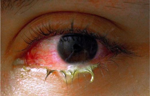
Image Source: Wikimedia Commons (2008). Swollen eye with conjunctivitis, Licensed under a creative commons licence. Available for free at: https://en.m.wikipedia.org/wiki/File:Swollen_eye_with_conjunctivitis.jpg (Accessed: 18 January 2024)
Viral
Viral conjunctivitis is most commonly caused by adenovirus (90% of cases). It may start unilaterally and spread to the other eye or be bilateral throughout its duration. It presents with watery rather than purulent discharge. The patient usually describes an irritation or grittiness. Preauricular lymphadenopathy may be present. Associated upper respiratory tract infection, pharyngitis and fever may be seen in some serotypes (pharyngoconjunctival fever). Most cases are self limiting within 2-3 weeks, but in severe cases steroids may be required. In all cases, stringent hygiene measures should be taken to prevent transmission, and lubricating eye drops may be used for symptom relief.
Allergic
Acute IgE-mediated conjunctival reaction is often to pollen or other environmental allergens. The predominant symptom is itching, which may be associated with conjunctival swelling (chemosis), conjunctival injection, lacrimation and rhinitis. Most cases can be treated with cold compresses and artificial tears, with use of antihistamines or mast cell stabilisers if no improvement.
Episcleritis
Episcleritis involves the outermost layer of the sclera in contrast to the deeper stromal inflammation seen in scleritis and is limited to the superficial episcleral plexus, causing either diffuse or nodular redness (Figure 5). Disease associations include rheumatoid arthritis, inflammatory bowel disease, spondyloarthropathies and vasculitides. Patients often have no pain or mild discomfort, but this can be variable, although when pain is more severe, scleritis should be considered. Instillation of 10% phenylephrine drops will blanch the vessels engorged in episcleritis, but will not blanch the deeper vessels involved in scleritis. Usually, it is self limiting, lasting a few weeks. Simple measures such as cool compresses or artificial tears may be used, in addition to NSAIDs for symptomatic relief.
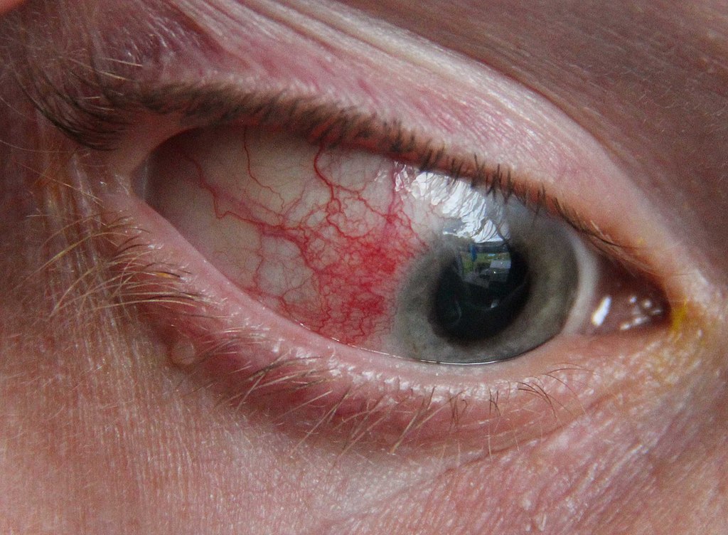
Image Source: Wikimedia Commons (2013). Episcleritiseye, Licensed under a creative commons licence. Available for free at: https://en.wikipedia.org/wiki/Episcleritis#/media/File:Episcleritiseye.jpg (Accessed: 18 January 2024).
Subconjunctival haemorrhage
Subconjunctival haemorrhage is bleeding into the subconjunctival space (Figure 6). It is largely asymptomatic, but occasionally, patients may report a transient pain or ‘pop’ sensation. It is often spontaneous and idiopathic, perhaps precipitated by vomiting, sneezing or coughing, but may also result from trauma, surgery or ocular disease. Hypertension, vascular disease and coagulopathies may be an underlying cause, more so in the older population. No specific treatment is required as this is usually self limiting.
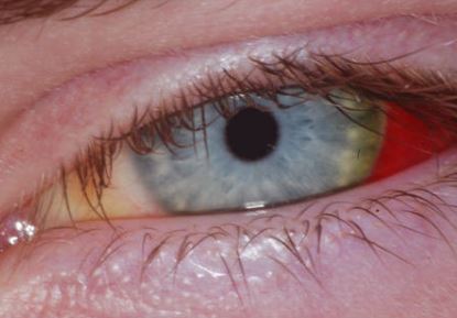
Image Source: Wikimedia Commons (2017). Subconjunctival hemorrhage before after (cropped4weeks), Licensed under a creative commons licence. Available for free at: https://en.wikipedia.org/wiki/File:Subconjunctival_hemorrhage_before_after_%28cropped4weeks%29.jpg (Accessed: 18 January 2024).
References
- Albrecht MR, Levin MJ. Epidemiology, clinical manifestations, and diagnosis of herpes zoster [Internet]. Hirsch MS, editor. UpToDate. 2021 [cited 2021 Nov 13]. Available from: https://www.uptodate.com/contents/epidemiology-clinical-manifestations-and-diagnosis-of-herpes-zoster
- Bowling B. Kanski’s Clinical Ophthalmology, A Systematic Approach. 8th ed. Elsevier Limited. 2016
- Denniston A, Murray P, editors. Oxford handbook of ophthalmology [Internet]. 4th ed. Oxford, United Kingdom: Oxford University Press; 2018 [cited 2021 Nov 13]. Available from: https://oxfordmedicine.com/view/10.1093/med/9780198804550.001.0001/med-9780198804550
- Jacobs D. The red eye: Evaluation and management [Internet]. Gardiner M, Givens J, editors. UpToDate. UpToDate Inc; 2021 [cited 2021 Nov 13]. Available from: https://www.uptodate.com/contents/the-red-eye-evaluation-and-management
- James B, Bron AJ, Parulekar MV. Lecture notes. Ophthalmology. 12th ed. Chichester, West Sussex, UK: Wiley Blackwell; 2017
- Joint Formulary Committee. British National Formulary (online) London: BMJ Group and Pharmaceutical Press. 2023. [Cited 2023 Jun 15]. Available from: http://www.medicinescomplete.com
- Kim H, Kersten R. Peri-orbital and orbital cellulitis [Internet]. BMJ Best Practice. 2020 [cited 2021 Nov 13]. Available from: https://bestpractice.bmj.com/topics/en-gb/734
- National Institute for Health and Care Excellence (NICE). Conjunctivitis – Infective. 2022 [cited 2023 Jun 13]. Available from: https://cks.nice.org.uk/topics/conjunctivitis-infective/
- National Institute for Health and Care Excellence (NICE). Management of Corneal Superficial Injury. 2022 [cited 2023 Jun 15]. Available from: https://cks.nice.org.uk/topics/corneal-superficial-injury/management/management/
- National Institute for Health and Care Excellence (NICE). Conjunctivitis: Allergic; Management in primary care. 2022. [cited 2023 Jun 24). Available from: https://cks.nice.org.uk/topics/conjunctivitis-allergic/management/management-in-primary-care/
- National Institute for Health and Care Excellence (NICE). Clinical Knowledge Summary: Blepharitis. 2023 [cited 2023 Jun 13]. Available from: https://cks.nice.org.uk/topics/blepharitis/
- Somani SN, Ronquillo Y, Moshirfar M. Acanthamoeba Keratitis. StatPearls Publishing. 2022 [cited 2023 Jun 3]. Available from: https://www.ncbi.nlm.nih.gov/books/NBK549863/
- Salmon, JF. Kanski’s Ophthalmology E-Book: A Systematic Approach. 9th Ed. Elsevier Health Sciences. 2019
- Smith J, Severn P, Clarke L. Assessment of red eye [Internet]. BMJ Best Practice. 2021 [cited 2021 Nov 13]. Available from: https://bestpractice.bmj.com/topics/en-gb/496
- Tian L, Wang NL. Trachoma control: the SAFE strategy. Int J Ophthalmol. 2018 Dec 18;11(12):1887-1888. doi: 10.18240/ijo.2018.12.01. PMID: 30588417; PMCID: PMC6288535
- Wu E. Blepharitis [Internet]. BMJ Best Practice. 2021 [cited 2021 Nov 13]. Available from: https://bestpractice.bmj.com/topics/en-gb/574
Glaucoma
Overview of glaucoma
Glaucoma is a disease whereby the optic nerve becomes damaged. Classically, this was thought to be due to raised intraocular pressure (IOP), largely due to dysfunctional outflow of aqueous humour from the anterior chamber of the eye. However, there is likely also an element of optic nerve ischaemia independent of the level of IOP. Acute closed-angle glaucoma is an ophthalmological emergency which can cause permanent visual loss if not treated promptly.
Pathophysiology of glaucoma
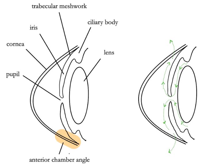
IOP is determined by the balance of production and removal of aqueous humour. This is secreted by the ciliary processes into the posterior chamber and passes through the pupil into the anterior chamber. It leaves the eye from the periphery of this space, the anterior chamber angle, by flowing through the trabecular meshwork, ultimately draining into the episcleral veins (Figure 7).
Obstruction to the outflow of aqueous humour from the anterior chamber causes IOP to rise. This leads to optic nerve axon loss, although the exact mechanism for this is unclear.
Classification of glaucoma
Primary glaucoma is classified by the cause of the outflow obstruction:
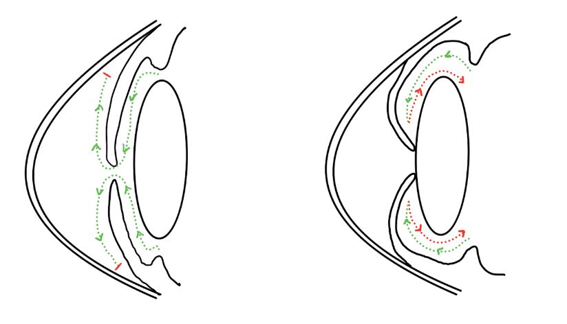
- Open-angle glaucoma: the anterior chamber angle remains open, but outflow is impaired nonetheless, perhaps secondary to structural changes in the trabecular meshwork (Figure 8).
- Closed-angle glaucoma: the peripheral iris is pushed against the trabecular meshwork, blocking outflow. In acute cases, this is often secondary to pupillary block, where the iris comes into contact with the lens, causing a buildup of aqueous humour, which in turn causes the iris to bow outwards, closing the anterior chamber angle (Figure 9).
Secondary glaucoma has various causes including drugs, trauma and inflammation.
Fundoscopy findings in glaucoma
Fundoscopy typically shows cupping of the optic disc (a raised cup to disc ratio).

Clinical findings in open-angle glaucoma
The prevalence of primary open-angle glaucoma increases with age, though is equally common in males and females. Family history is a risk factor, as is afro-caribbean heritage. It is often asymptomatic until relatively advanced disease at which point there may be painless visual loss.
Typical examination findings include a normal appearance of the sclera and cornea (in contrast to acute closed-angle glaucoma). Peripheral visual field loss may be present in advanced disease.
Management of open-angle glaucoma
Management of open-angle glaucoma aims to reduce intraocular pressure. February 2023 NICE guidelines suggest the following:
- First line:
- Laser trabeculoplasty, during which a laser is used to improve drainage of aqueous humour through the trabecular meshwork
- If trabeculoplasty is not available or the patient prefers medical therapy, a generic prostaglandin analogue (PGA) should be prescribed, such as bimatoprost or latanoprost
- Second line:
- An alternative PGA
- Topical beta blockers, such as timolol
- Other topical medications including miotics, sympathomimetics, or carbonic anhydrase inhibitors
Note that topical non-selective beta blockers have been shown to increase asthma morbidity and should therefore be avoided in asthmatic patients.
Acute primary closed-angle glaucoma
Acute primary closed-angle glaucoma is rarer than open-angle glaucoma. The condition is three times more common in females than males, and hypermetropia (being long sighted) is also a risk factor. Acute closed-angle glaucoma is important as a potentially sight-threatening emergency, although it is much more common for patients to have a chronic process which may well be asymptomatic.
Clinical findings in closed-angle glaucoma
Acute closed-angle glaucoma presents with severe discomfort, which may be in the form of a headache rather than eye pain. A typical history includes onset of pain after sitting in a darkened room as dilatation of the pupil can precipitate angle closure. There may also be photophobia, eye watering and loss of vision. The patient may be systemically unwell with nausea, vomiting or abdominal pain (via the oculo-trigemino-vago-abdominal reflex).
The patient may previously have had symptoms of intermittent closed-angle glaucoma, such as self-resolving episodes of eye ache, blurred vision and seeing halos around lights.
Typical examination findings include a red eye, cloudy cornea, a fixed dilated pupil and reduced acuity (Figure 11).
Management of closed-angle glaucoma
This includes immediate ophthalmology referral and admission.
Initial management is medical, with the aim of relieving symptoms and lowering IOP. This includes IV acetazolamide and topical beta-blockers (e.g. timolol).
Definitive management is laser iridotomy to create a hole in the iris through which aqueous humour can flow, equalising pressure in the anterior and posterior compartments, which pushes back the iris and opens up the anterior chamber angle. The contralateral eye is often treated too, prophylactically, as it will likely share anatomical features predisposing to the condition.
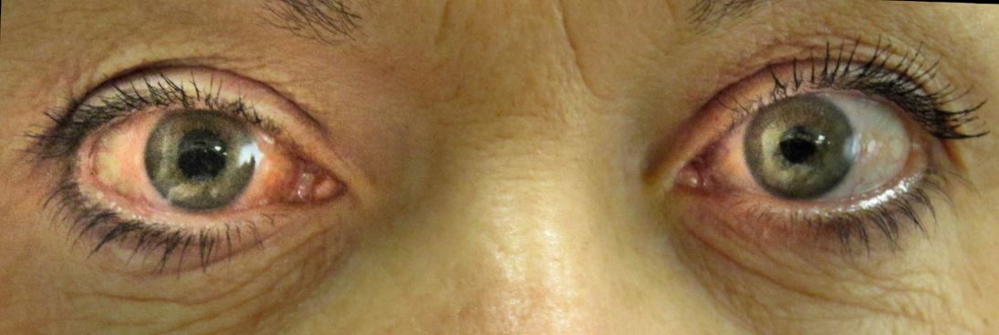
Image Source: Wikimedia Commons (2020). Acute angle closure glaucoma., Licensed under a creative commons licence. Available for free at: https://en.m.wikipedia.org/wiki/File:Acute_angle_closure_glaucoma.JP, via Wikimedia Commons (Accessed: 18 January 2024).
References
- James B, Bron AJ, Parulekar MV. Lecture notes. Ophthalmology. 12th ed. Chichester, West Sussex, UK: Wiley Blackwell; 2017
- Amerasinghe N. Open-angle glaucoma [Internet]. BMJ Best Practice. 2018 [cited 2022 Jan 29]. Available from: https://bestpractice.bmj.com/topics/en-gb/373
- Khondkaryan A, Francis BA. Angle-closure glaucoma [Internet]. BMJ Best Practice. 2019 [cited 2021 Nov 13]. Available from: https://bestpractice.bmj.com/topics/en-gb/372
- Morales DR, Dreischulte T, Lipworth BJ, Donnan PT, Jackson C, Guthrie B. Respiratory effect of beta-blocker eye drops in asthma: population-based study and meta-analysis of clinical trials. Br J Clin Pharmacol. 2016 Sep;82(3):814-22. doi: 10.1111/bcp.13006. Epub 2016 Jun 17. PMID: 27161880; PMCID: PMC5338116
- National Institute for Health and Care Excellence. Scenario: Primary open angle glaucoma and ocular hypertension [Internet]. NICE. 2023 [cited 2023 Jun 24]. Available from: https://www.nice.org.uk/guidance/ng81/chapter/Recommendations#treatment
Uveitis
Overview of uveitis
The term uveitis encompasses inflammation of one or all parts of the uvea (the layer between the retina and sclera). All types of uveitis should be referred to ophthalmology as they carry a risk of blindness.
Anatomical classification is useful both for understanding clinical features and because different aetiologies cause different patterns of inflammation:
- Anterior uveitis (iritis): iris and ciliary body
- Posterior uveitis: posterior vitreous, retina, choroid, retinal vasculature, optic nerve
- Panuveitis: all segments of uveal tract
Uveitis is most commonly idiopathic, but in many cases it is associated with systemic disease. Management is specific to the underlying cause, and systemic signs and symptoms should be sought to make a unifying diagnosis.
Clinical features of uveitis
Ocular signs and symptoms depend on the anatomical distribution of inflammation:
| Feature | Anterior uveitis | Posterior uveitis |
|---|---|---|
| Pain | Dull pain, tender to palpate through eyelid | Painless |
| Visual | Photophobia, slightly decreased acuity | Decreased acuity, floaters, flashing lights |
| Other | Red eye, tearing | May be asymptomatic |
Table 5 Presentation of uveitis
Acute anterior uveitis can be recurrent and is most commonly unilateral. Patients may present with rapid onset of eye pain, redness and photophobia. Visual acuity is usually only mildly impaired but this may be variable, dependent on severity. Conjunctival hyperaemia is usually present, and miosis may be seen as a result of pupillary sphincter spasm. Cells may be present in the anterior chamber and hypopyon describes a cream-coloured exudate made up of inflammatory cells which may settle in the inferior part of the anterior chamber.
The rest of this section will explore key causes of uveitis.
Bacterial causes of uveitis
Tuberculosis
Infection with Mycobacterium tuberculosis predominantly affects the respiratory system but can also have extrapulmonary manifestations. Tuberculosis can cause a range of ocular pathology such as anterior uveitis, vitritis choroiditis and cystoid macular oedema. Posterior uveitis with choroiditis is the most common finding, though anterior uveitis with granulomatous findings can often be seen. Ocular symptoms may be present in the absence of systemic upset and may, therefore, prompt further investigations for suspected tuberculosis. Treatment of the systemic infection is necessary and should be referred to a respiratory specialist.
Syphilis
Anterior uveitis may be found in a small proportion of people infected with the spirochaete Treponema pallidum and may cause unilateral or bilateral disease. It is predominantly associated with secondary syphilis, and therefore, the patient may have a history of a painless genital or anal ulcer or a maculopapular rash. If suspected, the patient should be referred to a sexual health specialist for systemic assessment (HIV status should also be established) in addition to an ophthalmologist for management of uveitis.
Bacterial endophthalmitis
Bacterial endophthalmitis is a sight-threatening bacterial infection of the vitreous or aqueous humour of the eye and is an ophthalmic emergency. It is most commonly caused by exogenous contamination from surgery (most commonly Staphylococcus epidermidis when acute) or trauma to the eye. Spread from endogenous sources is less common and the risk factors are largely the same as fungal sources.
Symptoms may include pain, blurry vision and redness of the eye. Slit lamp examination may reveal a hazy cornea, chemosis, hypopyon and it may be difficult to visualise the fundus due to vitritis. Roth spots are retinal haemorrhages with white centres, and if present may indicate infective endocarditis as a potential source. Vitreous sampling for identification of the pathogen is helpful, and immediate intravitreal antibiotics are required in addition to treatment of systemic infection where present.
Viral causes of uveitis
Herpes simplex virus anterior uveitis
Herpes simplex-associated uveitis can occur in the presence of keratitis or in isolation, but there will often be a history of cold sores, keratitis or genital lesions. Patchy iris atrophy may be seen on slit lamp examination, and the affected pupil may be slightly larger. Keratic precipitates (cellular inflammatory deposits on the corneal endothelium) may be present, and IOP may be raised. It is usually treated with a combination of topical steroids, topical cycloplegic (paralyses the ciliary muscles, preventing accommodation) and an oral antiviral.
Varicella zoster virus anterior uveitis
Anterior uveitis can occur in around half of patients affected by herpes zoster ophthalmicus and usually occurs 1-3 weeks after the rash. In immunocompromised patients, infection with primary varicella zoster infection (chickenpox) may also lead to uveitis. Management is with topical steroids, mydriatics and systemic antivirals such as aciclovir.
Cytomegalovirus
Infection with cytomegalovirus (CMV) is more likely to be clinically significant in immunocompromised patients. Uveitis associated with CMV is usually unilateral. The mainstay of treatment is with antivirals such as ganciclovir and valganciclovir.
Fungal causes of uveitis
Rarely, fungal infections can cause endophthalmitis, which involves inflammation of the vitreous or aqueous humour. These may be exogenous, introduced by spread from fungal keratitis or surgeries, or endogenous, for example as a result of intravenous drug use or systemic infection, or immunocompromise. Aspergillus spp. are most commonly implicated in exogenous cases, followed by Candida albicans. The reverse is true for endogenous cases, with Candida spp more prominently associated.
Culture of intraocular fluid is the gold standard for diagnosis. Slit lamp examination may reveal ‘cotton wool’ or ‘fluffy’ chorioretinal infiltrates. The mainstay of treatment is systemic and, if required, intravitreal anti-fungals. Aspergillus spp. endophthalmitis tends to progress more rapidly than candidal infection and is therefore treated more aggressively.
Parasitic causes of uveitis
Toxoplasmosis
The causative organism is Toxoplasma gondii. Cats are the definitive host so contamination from cat litter into food is the most common route of infection. Infection may be congenital after maternal exposure, or acquired. Toxoplasmosis causes a posterior uveitis. Serology for Toxoplasma may be carried out to aid diagnosis.
Toxocariasis
Toxocariasis is an infestation by a common roundworm found in dogs, so may be transmitted by contaminated soil or food. Usually, it is unilateral. Variable findings include chronic endophthalmitis and a posterior pole or peripheral granuloma seen during fundoscopy. Red reflex may be reduced in the affected eye.
Onchocerciasis (river blindness)
Caused by the helminth Onchocerca volvulus. Systemic features are mostly an intensely itchy maculopapular rash and ‘leopard skin’, which describes hypo-and hyperpigmentation mostly affecting the shins. Anterior uveitis may be an early feature, and it may be possible to visualise live microfilariae in the anterior chamber if the patient is face down prior to examination. Treatment is most commonly with ivermectin, which targets larvae.
Uveitis and rheumatic disorders
Ankylosing spondylitis
Ankylosing spondylitis is an HLA-B27-associated spondyloarthropathy characterised by joint inflammation, calcification and ultimately ossification, with a predilection for the axial skeleton. It commonly affects the sacroiliac joint and also may lead to spinal fusion in later stages. Onset is usually between 20-30 years and is significantly more common in males.
Patients typically present with chronic lower back pain or stiffness (for over 3 months) which is worse in the mornings and does not improve with rest but improves with exercise and a trial of NSAIDs. A family history of ankylosing spondylitis may also be present. Enthesitis and arthritis are common and usually occur asymmetrically and in the lower limbs. Extra-articular manifestations are varied in patients with ankylosing spondylitis; importantly, the lifetime prevalence of anterior uveitis is 30-40%, which means patients may present with symptoms of a unilateral red, painful eye. It is also associated with psoriasis and inflammatory bowel disease.
Cases of suspected ankylosing spondylitis should be referred to rheumatology for further assessment. ESR, CRP and HLA-B27 testing may be appropriate in addition to imaging of the sacroiliac joints and spine. NSAIDs, exercise and physiotherapy are the mainstay of treatment, but biological treatments may be considered in refractory cases. All patients with uveitis should be referred urgently to an ophthalmologist.
Reactive arthritis
This is the triad of urethritis, eye sequelae such as conjunctivitis and uveitis, and arthritis. The prevalence of HLA-B27 positivity in reactive arthritis varies but is estimated to be between 30-50%. Reactive arthritis is more common in males. It may present in the days or weeks following either a bacterial genitourinary or gastrointestinal infection.
Joint pain and swelling is most common in the knee or ankle and is often worse in the mornings. Enthesitis (e.g. Achilles tendonitis and plantar fasciitis) or dactylitis may also be a feature. Skin features include keratoderma blennorrhagica on the palms or soles and painless mouth ulceration. Ocular features include self-limiting conjunctivitis, acute anterior uveitis and occasionally episcleritis. Management is typically with NSAIDs, corticosteroids and disease-modifying anti-rheumatic drugs (DMARDs).
Psoriatic arthritis
Characterised by arthritis which usually develops around 10 years after the onset of preceding psoriasis. Patients can present with asymmetrical oligoarticular or monoarticular joint involvement and may also have dactylitis and sacroiliitis. Nail changes such as pitting or onycholysis may be present. Acute anterior uveitis develops in around 7% of patients with psoriatic arthritis, with conjunctivitis also being associated. Psoriatic arthritis is also associated with HLA-B27. Management of psoriatic arthritis includes NSAIDs and corticosteroids, with DMARDs and biological treatments considered for more severe cases.
Sarcoidosis
Sarcoidosis is one of the most common systemic associations of uveitis. It is a multi-systemic inflammatory disorder of unknown cause, characterised by non-caseating granuloma formation. It is more common in females and has a bimodal age of onset between 20-40 years, and again in older age. Respiratory symptoms such as a dry cough and shortness of breath are common and attributed to variable disease progression from hilar adenopathy to significant pulmonary fibrosis. Malaise and arthralgia may also be present. Skin manifestations are reasonably common and may be specific and granulomatous or non-specific, such as erythema nodosum.
Ocular involvement (usually bilateral) may be seen in 20-75% of patients, most commonly granulomatous anterior uveitis. There are several key findings in ocular sarcoid; ‘mutton fat’ keratic precipitates are large grey-white inflammatory deposits of cells on the corneal endothelium and are a key sign of ocular sarcoid. Trabecular meshwork nodules, vitreous opacities (which may have a ‘snowball’ or ‘string of pearls’ appearance) and optic disc granulomas may also be seen.
Findings in sarcoidosis include elevated serum ACE (angiotensin converting enzyme) levels, deranged liver enzymes (most commonly ALP is raised) and bilateral hilar lymphadenopathy on chest X-ray. Tuberculosis should be ruled out. Treatment of sarcoidosis is by respiratory specialists. Spontaneous remission can occur (with likelihood inversely related to the severity of the disease), and therefore, treatment is not always indicated. If treated, this is usually with corticosteroids.
Behçet’s disease
Behçet’s disease is a multi-system vasculitis characterised by aphthous ulcers of the mouth and genitals, ocular manifestations, and skin lesions such as erythema nodosum. There are a range of CNS, gastrointestinal and other vasculitic manifestations, including pulmonary artery aneurysm. Ocular manifestations are usually bilateral and affect the entire uveal tract (panuveitis), which can be recurrent and may involve a range of sight-threatening complications such as retinal vasculitis and angle-closure glaucoma.
Typically, Behçet’s affects younger patients aged 20-40 years old, with a relatively equal gender distribution. It is more commonly found in people of Turkish, Mediterranean, Israeli or East Asian descent. HLA-B51 is a frequent association.
Investigations may reveal HLA-B51 positivity, a positive pathergy skin test and raised inflammatory markers. Full autoimmune screening should be carried out, and further investigations as appropriate to the clinical findings. Treatment is often with corticosteroids and immunosuppressive therapies.
Other systemic associations of uveitis
Other systemic associations of anterior uveitis include inflammatory bowel disease (uveitis is more common in patients who are also positive for HLA-B27), IgA nephropathy and juvenile idiopathic arthritis.
References
- Adil A, Goyal A, Quint JM. 2023. Behcet Disease. StatPearls [Internet]. Treasure Island (FL): StatPearls Publishing; [Updated 2023 Feb 22]. [cited 2023 Jun 9]. Available from: https://www.ncbi.nlm.nih.gov/books/NBK470257/
- Bowling B. Kanski’s Clinical Ophthalmology, A Systematic Approach. 8th ed. Elsevier Limited. 2016
- Carter JD. 2022. Reactive Arthritis. BMJ Best Practice. Last updated 2022 Feb 4 [cited 2023 Jun 15]. Available at: https://bestpractice.bmj.com/topics/en-gb/597
- Cheeti A, Chakraborty RK, Ramphul K. 2023. StatPearls. Treasure Island (FL). StatPearls Publishing. Updated 2023 Jan 2 [cited 2023 Jun 12]. Available from https://www.ncbi.nlm.nih.gov/books/NBK499831
- Clement J. 2018. Psoriatic Arthritis. BMJ Best Practice. Last updated 2023 May 19 [cited 2023 Jun 15]. Available from: https://bestpractice.bmj.com/topics/en-gb/524
- Denniston AK, Murray PI (editors). Oxford Handbook of Ophthalmology. [Internet]. 4th edition. Oxford: Oxford University Press. 2018 [cited November 13 2021]. Available from: https://oxfordmedicine.com/view/10.1093/med/9780198804550.001.0001/med-9780198804550.
- Durand ML 2017. Bacterial and Fungal Endophthalmitis. Clin Microbiol Rev. Jul;30(3):597-613. doi: 10.1128/CMR.00113-16. PMID: 28356323; PMCID: PMC5475221
- Huang JJ, Elia M. Uveitis [Internet]. BMJ Best Practice. 2021 [cited 2021 Nov 13]. Available from: https://bestpractice.bmj.com/topics/en-gb/407
- Lin P. 2015. Infectious Uveitis. Curr Ophthalmol Rep. 2015. (3):170-183. doi: 10.1007/s40135-015-0076-6. Epub 2015 Jun 13. PMID: 26618074; PMCID: PMC4659396.
- National Institute of Health and Care Excellence (NICE). 2016. Pulmonary Sarcoidosis: Infliximab. Evidence Summary [ES2]. [2016 Dec 20]. [cited 2023 Jun 18]. Available at: https://www.nice.org.uk/advice/es2/chapter/full-evidence-summary
- National Institute of Health and Care Excellence (NICE). 2017. Spondyloarthritis in over 16s: Diagnosis and management. NICE guideline [NG65]. Last updated May 2019 [Cited 2023 Jun 16] Available at: https://www.nice.org.uk/guidance/ng65
- Rosenbaum J. Uveitis: Etiology, clinical manifestations, and diagnosis [Internet]. Thorne J, Romain P, editors. UpToDate. 2021 [cited 2021 Nov 13]. Available from: https://www.uptodate.com/contents/uveitis-etiology-clinical-manifestations-and-diagnosis.
- Salmon, JF. Kanski’s Ophthalmology E-Book: A Systematic Approach. 9th Ed. Elsevier Health Sciences. 2019
- Stolwijk C, van Tubergen A, Castillo-Ortiz JD. 2015. Prevalence of extra-articular manifestations in patients with ankylosing spondylitis; a systematic review and meta-analysis. Annals of the Rheumatic diseases 74 [1], 650-73 DOI: 10.1136/annrheumdis-2013-203582
- Taurog JD, Chhabra A, Colbert RA. 2016.Ankylosing spondylitis and axial spondyloarthritis. New England Journal of Medicine 375 [26] 2563-2574. DOI 10.1056/NEJMra1406182
- Yazici Y. 2020. Behcet’s Syndrome. BMJ Best Practice. Last updated 2020 Aug 6 [cited 2023 Jun 15]. Available from: https://bestpractice.bmj.com/topics/en-gb/376
Complete Guide to Clinical Genetics ST3 Application and Recruitment (2027)
Written by Medibuddy. Last updated: October 2026. Clinical Genetics is

Complete Guide to Clinical Radiology ST3 Application and Recruitment (2027)
Recruitment paused: NHS England is not running Clinical Radiology ST3+ recru

Complete Guide to Paediatric Surgery ST3 Application and Recruitment in United Kingdom (2026)
Written by Medibuddy. Last updated: October 2026. If you’re planning


Take your subscriptions with you
Our mobile app allows you to access your interview and exam question banks wherever you are.

