


 Close
Close
 15th February 2025
Upper Airways Disease and Pulmonary Function Tests
Anaphylaxis and angioedema
Overview of anaphylaxis
Anaphylaxis is a severe type I (IgE-mediated) hypersensitivity reaction. Excessive release of histamine from mast cells and basophils results in the following features:
- Angioedema – non-pitting swelling of deep tissues such as eyelids, lips, tongue and airway
- Skin changes – urticarial rash, flushing (absent in 20% of cases)
- Respiratory – bronchospasm, stridor
- Cardiovascular – distributive shock
The patient may also have reduced consciousness and arrhythmias, including supraventricular tachycardia.
The commonest medications to cause anaphylaxis are: antibiotics (particularly penicillins and cephalosporins), chemotherapy agents, contrast media, NSAIDs and neuromuscular blocking agents.
Management of anaphylaxis
Immediate management
The latest Resuscitation Council guidelines (2021) emphasise the importance of an A-to-E approach with early adrenaline administration and prioritising repeated use of adrenaline over the administration of other drugs. Adrenaline is usually given as 0.5 mg IM repeated up to every 5 minutes. Alternatively, it can be given in 50 mcg boluses IV in experienced hands.
Intravenous crystalloid fluid resuscitation should be aggressive, with fluid requirements sometimes being up to 5 litres.
Nebulised salbutamol and ipratropium can be given if there is wheeze present. Antihistamines may be used to treat cutaneous symptoms following initial resuscitation. The routine use of corticosteroids is no longer advised, though they may be used if asthma plays a role in the presentation or for refractory cases.
Refractory anaphylaxis
This is where there is ongoing cardiorespiratory compromise after two doses of IM adrenaline. The most important ongoing management is to administer further adrenaline, preferably by IV infusion. Other possible treatments include:
- Nebulised adrenaline (1 mg in 5 ml) for airway obstruction
- Tracheal intubation
- IV salbutamol or aminophylline for persistent bronchospasm
- Corticosteroids
Further management and follow up
Five percent of patients experience a biphasic anaphylaxis reaction and, as such,, patients should be observed for 6-12 hours following resolution of symptoms.
Serum mast cell tryptase should be measured in any patient where anaphylaxis is considered. Ideally, three samples should be taken: the first as soon as feasible, the second 1-2 hours after onset and the third 24 hours after complete resolution. A normal tryptase level does not exclude anaphylaxis.
All patients should be educated about the condition and given an adrenaline auto-injector, which comes in doses of 150-500 mcg. They should be referred to a specialist allergy clinic for skin prick testing and follow up.
Non-anaphylactic angioedema
Presentation
Non-anaphylactic angioedema, which is primarily thought to be mediated by bradykinin excess, is compared to anaphylaxis below:
| Anaphylaxis | Non-anaphylactic angioedema (bradykinin-mediated) | |
|---|---|---|
| Aetiology | Allergen-triggered histamine release | Excess bradykinin
Often due to a problem with C1-inhibitor (Various aetiologies – see below) |
| Distribution | Usually symmetrical
Primarily head & neck |
Usually asymmetrical
May include gut and genitals |
| Timescale | Presents over minutes | Presents over minutes to hours
May last days |
| Associated features | Urticaria, pruritus
Distributive shock Wheeze |
Abdominal pain & diarrhoea |
Table 1 Anaphylactic versus non-anaphylactic angioedema
Classification
To differentiate between causes of non-anaphylactic angioedema, complement studies may be helpful. The classification is as follows:
Medication-induced: most commonly due to ACE inhibitors or angiotensin-II receptor blockers; also gliptins, sirolimus, tacrolimus and alteplase.
Hereditary angioedema (HAE): typically presents before age 20, usually with a family history. In type 1 HAE, there is a low level of the C1-inhibitor protein, whereas in type 2 HAE, there is a normal C1-inhibitor level, but the protein has reduced function.
Acquired angioedema: typically presents after age 40. Anti-C1-inhibitor antibodies may be found in plasma, and C1q levels are typically low. It may be caused by lymphoproliferative disorders (MGUS, haematological malignancy) or auto-immune disease.
Idiopathic: no other cause is found, and complement studies are normal.
Management of acute attacks
For severe episodes of non-anaphylactic angioedema, two specific drugs exist: ecallantide and icatibant. Both of these are given by subcutaneous injection and take 2-4 hours to work.
Replacement of C1-inhibitor may also be given, especially in the hereditary forms of the disease. This can be achieved by giving either C1-inhibitor concentrate or fresh frozen plasma (FFP).
References
- Chapman SJ, Robinson GV, Shrimanker R, Turnbull CD, Wrightson JM. Oxford handbook of respiratory medicine. 4th ed. London, England: Oxford University Press; 2021
- Campbell RL, Ellis AK. Differential diagnosis of anaphylaxis in adults and children [Internet]. Kelso JM, editor. UpToDate. 2022 [cited 2022 Dec 9]. Available from: https://www.uptodate.com/contents/differential-diagnosis-of-anaphylaxis-in-adults-and-children
- Campbell RL, Kelso JM. Anaphylaxis: Emergency treatment [Internet]. Walls RM, editor. UpToDate. 2022 [cited 2022 Dec 9]. Available from: https://www.uptodate.com/contents/anaphylaxis-emergency-treatment
- Guyer AC, Banerji A. ACE inhibitor-induced angioedema [Internet]. Saini S, editor. UpToDate. 2022 [cited 2022 Dec 9]. Available from: https://www.uptodate.com/contents/ace-inhibitor-induced-angioedema
- Zuraw B, Farkas H. Hereditary angioedema: Epidemiology, clinical manifestations, exacerbating factors, and prognosis [Internet]. Saini S, editor. UpToDate. 2022 [cited 2022 Dec 9]. Available from: https://www.uptodate.com/contents/hereditary-angioedema-epidemiology-clinical-manifestations-exacerbating-factors-and-prognosis
- Zuraw B, Farkas H. Hereditary angioedema (due to C1 inhibitor deficiency): Pathogenesis and diagnosis [Internet]. Saini S, editor. UpToDate. 2022 [cited 2022 Dec 9]. Available from: https://www.uptodate.com/contents/hereditary-angioedema-due-to-c1-inhibitor-deficiency-pathogenesis-and-diagnosis
- Emergency treatment of anaphylactic reactions: Guidelines for healthcare providers [Internet]. Resuscitation Council UK. [cited 2022 Dec 9]. Available from: https://www.resus.org.uk/library/additional-guidance/guidance-anaphylaxis/emergency-treatment
Obstructive sleep apnoea and obesity hypoventilation
Obstructive sleep apnoea syndrome (OSA)
OSA is the third most common respiratory problem in the UK after asthma and COPD, with 2.5% of women and 5% of men in Western populations having severe enough disease to warrant treatment.
Pathophysiology and risk factors
Loss of pharyngeal dilator tone during REM (rapid eye movement) sleep results in occlusion of the upper airway, leading to apnoea lasting at least 10 seconds, with transient hypoxia and hypercapnia. The patient partially wakes, leading to an increase in airway tone to allow ventilation. The cycle repeats hundreds of times per night, leading to fragmented sleep.
Sympathetic stimulation during apnoeas with resultant hypertension is thought to explain the increased risk of poor cardiovascular outcomes, which is independent of obesity.
Risk factors include obesity, large neck circumference, large tonsils, hypothyroidism, acromegaly, increasing age, smoking, and use of sedatives or alcohol. Although obesity is a risk factor, OSA is also common in those without a raised BMI.
Presentation and diagnosis
Presentation may include loud snoring, daytime sleepiness, morning headaches, mood change, and poor memory and concentration. Their partner may have observed apnoeic episodes.
The STOP-BANG score and Epworth Sleepiness Score are frequently used screening tools.
Polysomnography (sleep study) is the diagnostic investigation of choice; it is comprised of six continuous elements: oximetry, EEG, EMG, ECG, airflow monitoring and electrooculogram.
The apnoea hypopnoea index (AHI) is the number of apnoeas or hypopnoeas per hour. An AHI > 14 in the presence of symptoms indicates moderate disease.
Polysomnography will differentiate OSA from other causes of daytime sleepiness, such as: central sleep apnoea (for example due to heart failure or stroke), nocturnal seizures, gastro-oesophageal reflux disease (GORD), restless legs syndrome, and periodic limb movement disorder.
Management of OSA
Management options include:
- Conservative – weight loss, smoking cessation, avoidance of alcohol/sedatives, mandibular advancement devices
- CPAP (continuous positive airway pressure) – typically nasal CPAP is used with pressures of 5-15 cmH2O. It works by splinting open the upper airway and has been shown to be highly effective in improving quality of life
- Surgical – pharyngeal surgery, bariatric surgery
Obesity hypoventilation syndrome (OHS)
Diagnosis of OHS
Obesity hypoventilation syndrome is a diagnosis of exclusion, defined as where a patient with BMI > 30 has an awake pCO2 > 6 kPa, with no other explanation.
Patients present non-specifically with features similar to OSA and, in severe cases, may additionally show features of right-heart failure. A minority of patients present acutely with severe type II respiratory failure requiring hospital admission.
Management of OHS
Non-invasive ventilation (NIV) is typically started in the presence of acidosis, somnolence or cor pulmonale.
- OSA is co-morbid in 90% of patients, and for these patients typically nighttime CPAP is the initial mode, with escalation to BiPAP (bilevel positive airway pressure) for those who fail to respond adequately
- For the remaining 10% of patients who have few obstructive events during sleep and for patients who present acutely, nighttime BiPAP is the initial treatment
BiPAP may be started with an inspiratory positive airway pressure (IPAP) of 8 cmH2O and an expiratory positive airway pressure (EPAP) of 4 cmH2O. These pressures can then be titrated up. The EPAP will improve airway opening, and the driving pressure (IPAP – EPAP) will improve tidal volumes.
As per management of OSA, conservative or surgical measures to encourage weight loss also form part of OHS management.
Patients should be assessed and treated for complications of OHS, which include:
- Pulmonary hypertension (present in 60%) – all patients should have ECG and echocardiography
- Hypertension, congestive cardiac failure, insulin resistance
References
- Chapman SJ, Robinson GV, Shrimanker R, Turnbull CD, Wrightson JM. Oxford handbook of respiratory medicine. 4th ed. London, England: Oxford University Press; 2021
- Bourke SJ. Respiratory medicine: Lecture notes, 10th edition: Lecture Notes. 10th ed. Hoboken, NJ: Wiley-Blackwell; 2022
- Kline LR. Clinical presentation and diagnosis of obstructive sleep apnea in adults [Internet]. Collop N, editor. UpToDate. 2022 [cited 2022 Dec 9]. Available from: https://www.uptodate.com/contents/clinical-presentation-and-diagnosis-of-obstructive-sleep-apnea-in-adults
- Piper A, Yee B. Clinical manifestations and diagnosis of obesity hypoventilation syndrome [Internet]. Safwan Badr M, editor. UpToDate. 2022 [cited 2022 Dec 9]. Available from: https://www.uptodate.com/contents/clinical-manifestations-and-diagnosis-of-obesity-hypoventilation-syndrome
- Martin T. Treatment and prognosis of the obesity hypoventilation syndrome [Internet]. Safwan Badr M, editor. UpToDate. 2022 [cited 2022 Dec 9]. Available from: https://www.uptodate.com/contents/treatment-and-prognosis-of-the-obesity-hypoventilation-syndrome
- Safwan Badr M. Central sleep apnea: Risk factors, clinical presentation, and diagnosis [Internet]. Chervin R, editor. UpToDate. 2022 [cited 2022 Dec 9]. Available from: https://www.uptodate.com/contents/central-sleep-apnea-risk-factors-clinical-presentation-and-diagnosis
Pulmonary function tests (PFTs)
Spirometry
During a spirometry test, the patient inhales maximally, then exhales into the spirometer as hard as possible until no breath remains. The main values of interest are as follows, with predicted values calculated from the patient’s height and demographics.
Peak expiratory flow rate (PEFR)
Also measured using a peak flow meter, the PEFR is the maximum rate of expiratory flow the patient can achieve, in L/min, typically attained at the beginning of expiration. A reduction in PEFR is suggestive of airway obstruction, such as COPD or asthma.
In asthma, variability in PEFR during the day is typically observed, with the lowest values usually being recorded early in the morning or after exercise. PEFR is usually reduced but stable in COPD. There is also a rare phenotype of asthma where fixed PEFR is observed, as in COPD.
PEFR is frequently used for monitoring acute asthma in the inpatient setting. BTS guidelines recommend the patient is safe for discharge when PEFR is >75% of best or predicted, with a diurnal variation of less than 25%.
FVC, FEV1 and FEV1/FVC ratio
The forced vital capacity (FVC) is the total volume of air expelled from full inspiration to full expiration. Above 80% of predicted is considered normal.
The forced expiratory volume in one second (FEV1) is the volume of air expired in the first second of forced expiration. Typically, the FEV1 is interpreted using the FEV1/FVC ratio, which is more useful as it is independent of other factors. A ratio of >70% is considered normal, with a ratio of <70% suggesting airway obstruction.
Obstructive pattern
An obstructive pattern of PFTs is where the FEV1/FVC ratio is less than 70%, with normal or low FVC.
This usually indicates COPD or asthma, which can be differentiated by a reversibility test, where the test is repeated following administration of a bronchodilator such as salbutamol. If the FEV1 or FVC increases by more than 12%, the obstruction is said to be reversible or bronchodilator responsive, which is suggestive of asthma, although a small degree of reversibility can also be seen in COPD.
Alternatively, diurnal variability in PEFR can be used to differentiate the two, as mentioned above.
Restrictive pattern
A restrictive pattern of PFTs is where the FVC is reduced, with a preserved FEV1/FVC ratio (>70%). This occurs in three main aetiologies:
Reduced lung compliance/volume, such as lung fibrosis or pneumonectomy.
Muscle weakness, such as myasthenia gravis or myopathy.
Reduced chest wall compliance – see following section.
FVC monitoring
Monitoring of FVC is also used for assessing decline in respiratory function in acute neuromuscular disease, such as in Guillain-Barré syndrome and myasthenic crisis. An FVC of less than 15-20 ml/kg body weight suggests endotracheal intubation may be necessary.
Diseases of the chest wall
Pathology of the chest wall may decrease its compliance and, therefore, cause a restrictive defect. It is classified as follows:
- Congenital: for example, pectus excavatum, pectus carinatum and Poland syndrome. The latter involves a constellation of unilateral features, including incomplete development of the hands, respiratory muscles and costal cartilages
- Age related: it is normal for chest wall compliance to decrease with age, and this can be further exacerbated by kyphosis and scoliosis
- Traumatic and iatrogenic: flail chest and thoracoplasty
- Abdominal processes: obesity and ascites cause a restrictive defect via splinting of the diaphragm
- Ankylosing spondylitis: may lead to fixation of the chest wall in advanced disease
Measures of gas exchange – TLᴄᴏ and Kᴄᴏ
To quantify alveolar gas exchange, the patient breathes a mix of an inert gas (helium) and carbon monoxide (CO), breath holding for 10 seconds, then the CO in exhaled gas is measured. Only the CO will be absorbed into the blood, so the drop in CO concentration is indicative of gas exchange, as recorded in two measures:
- The transfer factor of the lung for carbon monoxide (TLᴄᴏ), also called the diffusing capacity (DLᴄᴏ). This measures the total amount of CO transferred to the blood and therefore the total gas exchange capacity of the lungs
- The carbon monoxide transfer coefficient (Kᴄᴏ) – this is the TLᴄᴏ divided by the total lung volume, giving a measure of the average gas exchange per unit volume of the lung. It is low where there is a problem with gas exchange at the level of the alveolar membrane, for example due to thickening
A low Kᴄᴏ helps differentiate between causes of respiratory disease:
- In a restrictive spirometry pattern, a low Kᴄᴏ suggests an intrapulmonary cause such as fibrosis. It will be normal in extrapulmonary causes such as kyphoscoliosis
- In an obstructive spirometry pattern, a low Kᴄᴏ suggests COPD rather than asthma
A high Kᴄᴏ is seen in two scenarios:
- In the few days after alveolar haemorrhage, due to red blood cells taking up CO. For example in SLE, Wegener’s and Goodpasture’s
- In poor lung expansion, such as due to neuromuscular disease or chest wall restrictive disease
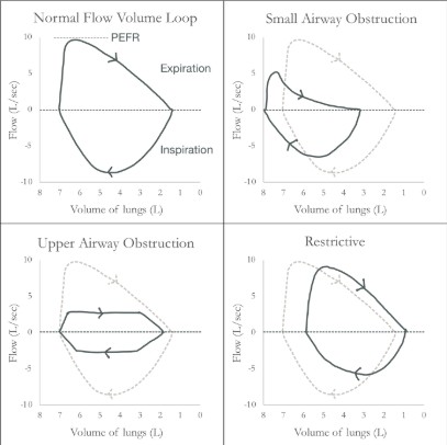
Flow-volume loops
A flow-volume loop is constructed from spirometry involving forced maximal inspiration and expiration. The cycle is clockwise, with an upward deflection indicating expiration and a downward indicating inspiration. The start point of maximal inspiration (total lung capacity, TLC) is the leftmost point of the loop, with the rightmost point representing the minimal lung volume (residual volume, RV).
References
- Chapman SJ, Robinson GV, Shrimanker R, Turnbull CD, Wrightson JM. Oxford handbook of respiratory medicine. 4th ed. London, England: Oxford University Press; 2021
- Bourke SJ. Respiratory medicine: Lecture notes, 10th edition: Lecture Notes. 10th ed. Hoboken, NJ: Wiley-Blackwell; 2022
- Bessant R et al. The pocketbook for PACES. London, England: Oxford University Press; 2012
- British Thoracic Society, Scottish Intercollegiate Guidelines Network. British guideline on the management of asthma. Thorax. 2014;69 Suppl 1:1–192
- Kaminsky DA. Overview of pulmonary function testing in adults [Internet]. McCormack MC, editor. UpToDate. 2022 [cited 2022 Dec 9]. Available from: https://www.uptodate.com/contents/overview-of-pulmonary-function-testing-in-adults
- McCormack MC. Diffusing capacity for carbon monoxide [Internet]. Stoller JK, editor. UpToDate. 2022 [cited 2022 Dec 9]. Available from: https://www.uptodate.com/contents/diffusing-capacity-for-carbon-monoxide
The upper respiratory tract
The mouth and pharynx
Pharyngitis and tonsillitis
These are usually caused by viruses but can also be caused by beta-haemolytic streptococci, H. influenzae, M. pneumoniae or C. pneumoniae. Local spread of infection may lead to quinsy (peritonsillar abscess) or otitis media. Beta-haemolytic streptococci (Streptococcus pyogenes) tend to cause severe infection and may rarely lead to various systemic complications, including glomerulonephritis, rheumatic fever, scarlet fever and toxic shock syndrome. The antibiotic of choice is phenoxymethylpenicillin.
Infectious mononucleosis
Also called glandular fever, this is caused by the Epstein-Barr virus (EBV). In addition to pharyngitis, it may cause fatigue, fever, lymphadenopathy and splenomegaly. Patients have a positive heterophile antibody (Monospot) test, and the blood film shows atypical mononuclear cells. The patient may develop a rash if given certain antibiotics, particularly amoxicillin and ampicillin. Treatment is usually supportive unless there are severe rare complications; these include airway compromise, Guillain-Barré syndrome and splenic rupture.
Ludwig’s angina
This is a rapidly spreading cellulitis that originates in the floor of the mouth, most commonly from a dental infection and spreads to the submandibular space. Causative organisms include viridans streptococci, staphylococci, and oral anaerobes such as Peptostreptococcus, Fusobacterium and Bacteroides. Presenting features include sepsis, airway compromise, drooling, dysphagia and difficulty speaking.
Oropharyngeal candidiasis
Also known as thrush, this occurs most frequently in the immunosuppressed, those who have had recent antibiotics, or those on oral or inhaled steroids. Mild disease is managed with topical antifungals (e.g. nystatin). Oral fluconazole is used for moderate-to-severe disease or with immunocompromise.
Diphtheria
Caused by the Gram-positive bacillus Corynebacterium diphtheriae, respiratory diphtheria has its onset 2 to 5 days after exposure and presents with sore throat, low-grade fever and purulent nasal discharge. Pharyngeal exudates are often noted, and in one third of cases an oral or nasal “pseudomembrane” forms.
The larynx
Disorders of the larynx may present with hoarseness, change in voice, difficulty breathing and chronic aspiration. The investigation may require flexible nasendoscopy (FNE) to visualise the larynx, which can be conducted in the ENT clinic.
Laryngitis
May be acute or chronic (lasting more than four weeks). Acute laryngitis is a common self-limiting illness causing hoarseness, usually caused by respiratory viruses. Chronic laryngitis is caused by irritants such as smoking, alcohol, gastro-oesophageal reflux, and chronic sinusitis with post-nasal drip. Chronic laryngitis can also complicate systemic diseases such as rheumatoid arthritis, pemphigoid, SLE and sarcoid.
Acute epiglottitis
This severe disease is most commonly caused by H. influenzae type B. It can cause life-threatening airway compromise, with a mortality of 7% in adults, but is now rare thanks to widespread vaccination. Presenting features include sepsis, sore throat, drooling, painful dysphagia, respiratory distress and stridor. Support should be sought from ENT and Anaesthetics, and the airway should only be examined where there are facilities to undertake endotracheal intubation. Treatment involves airway management and a third-generation cephalosporin such as ceftriaxone or cefotaxime.
Laryngeal cancer
This is predominantly squamous cell carcinoma related to smoking and alcohol and commonly presents with hoarseness. Flexible nasendoscopy (FNE) typically shows a white plaque-like lesion. Management is usually one or both of radiotherapy and surgery. Following a laryngectomy, it is important to be aware that the patient has no patent upper airway, as the trachea opens directly to the anterior neck.
Polypoid corditis (Reinke’s oedema)
This inflammatory, smoking-related condition of the larynx most commonly affects middle-aged women, presenting with a husky, low-pitched voice.
Vocal cord paralysis
This results from palsy of the vagus or recurrent laryngeal nerve. It presents with hoarseness and carries a risk of aspiration. It may be caused by multiple sclerosis, Lyme disease, Epstein-Barr virus, Herpes Simplex virus, and malignancy on the course of the nerves.
Systemic disease
Rheumatoid arthritis can give rise to vocal cord nodules and may also involve the cricoarytenoid joints, leading to vocal cord immobility. Tuberculosis involving the larynx may also give rise to vocal cord nodules.
References
- Chapman SJ, Robinson GV, Shrimanker R, Turnbull CD, Wrightson JM. Oxford handbook of respiratory medicine. 4th ed. London, England: Oxford University Press; 2021
- Bourke SJ. Respiratory medicine: Lecture notes, 10th edition: Lecture Notes. 10th ed. Hoboken, NJ: Wiley-Blackwell; 2022
- Bruch JM. Hoarseness in adults [Internet]. Kamani DV, editor. UpToDate. 2022 [cited 2022 Dec 9]. Available from: https://www.uptodate.com/contents/hoarseness-in-adults
- Woods CR. Epiglottitis (supraglottitis): Clinical features and diagnosis [Internet]. Edwards MS, Isaacson GC, Neuman MI, editors. UpToDate. 2022 [cited 2022 Dec 9]. Available from: https://www.uptodate.com/contents/epiglottitis-supraglottitis-clinical-features-and-diagnosis
- Pichichero ME. Complications of streptococcal tonsillopharyngitis [Internet]. Sexton DJ, Kaplan SL, editors. UpToDate. 2022 [cited 2022 Dec 9]. Available from: https://www.uptodate.com/contents/complications-of-streptococcal-tonsillopharyngitis
- Wald ER. Peritonsillar cellulitis and abscess [Internet]. Edwards MS et al., editors. UpToDate. 2022 [cited 2022 Dec 9]. Available from: https://www.uptodate.com/contents/peritonsillar-cellulitis-and-abscess
- Chow AW. Ludwig angina [Internet]. Durand ML, editor. UpToDate. 2022 [cited 2022 Dec 9]. Available from: https://www.uptodate.com/contents/submandibular-space-infections-ludwig-angina
- Barraso LF, Samuel Pegram P. Clinical manifestations, diagnosis, and treatment of diphtheria [Internet]. Sexton DJ, editor. UpToDate. 2022 [cited 2022 Dec 17]. Available from: https://www.uptodate.com/contents/clinical-manifestations-diagnosis-and-treatment-of-diphtheria
Complete Guide to Clinical Genetics ST3 Application and Recruitment (2027)
Written by Medibuddy. Last updated: October 2026. Clinical Genetics is

Complete Guide to Clinical Radiology ST3 Application and Recruitment (2027)
Recruitment paused: NHS England is not running Clinical Radiology ST3+ recru

Complete Guide to Paediatric Surgery ST3 Application and Recruitment in United Kingdom (2026)
Written by Medibuddy. Last updated: October 2026. If you’re planning


Take your subscriptions with you
Our mobile app allows you to access your interview and exam question banks wherever you are.

