


 Close
Close
 18th February 2025
Cardiogenic pulmonary oedema
Overview of pulmonary oedema
Pulmonary oedema is the accumulation of excess fluid in the lung’s interstitial and/or alveolar spaces, occurring when fluid is filtered into the lungs faster than it can be removed. This fluid reduces the surface area available for gas exchange, leading to hypoxaemia and acute respiratory failure.
Cardiogenic pulmonary oedema occurs when the left heart is unable to pump effectively, resulting in raised left cardiac filling pressures and back pressure in the lungs manifesting as pulmonary oedema. The most common cause of this is acute decompensated heart failure which will be the focus of this section.
Predisposing and precipitating factors of cardiogenic pulmonary oedema
Predisposing factors are the chronic underlying primary conditions that lead to the development of heart failure. Precipitating events are the triggers that cause decompensation and result in episodes of pulmonary oedema.
This section will not cover all the details and causes of heart failure, but examples of predisposing factors include:
- Left ventricular systolic dysfunction, e.g. from coronary artery disease, valvular heart disease, hypertension or a dilated cardiomyopathy
- Left ventricular diastolic dysfunction, e.g. from left ventricular hypertrophy, hypertrophic or restrictive cardiomyopathies or acute hypertensive crisis
- Left ventricular outflow obstruction, e.g. in aortic stenosis or hypertrophic cardiomyopathy with obstruction
- Valvular disease, e.g. mitral stenosis
Precipitating events can be split into those that increase preload, decrease cardiac contractility or increase afterload:
- Increased preload
- Volume overload: chronic liver disease, chronic kidney disease, nephrotic syndrome, high dietary sodium load, iatrogenic intravenous fluid, blood transfusion
- Acute aortic regurgitation: infective endocarditis, aortic dissection
- Acute mitral regurgitation: chordal rupture, papillary muscle rupture from ischaemia
- Decreased contractility
- Myocardial ischaemia or infarction
- Arrhythmias: atrial fibrillation/flutter, ventricular tachyarrhythmias
- Non-ischaemic: cardiomyopathy dilated cardiomyopathy, Takotsubo cardiomyopathy
- High-output cardiac failure: anaemia, sepsis
- Increased afterload
- Hypertensive crisis
- Left ventricular outflow tract obstruction: progressive aortic stenosis, atrial myxoma or thrombus
These precipitating factors lead to decreased ventricular emptying and cardiac output resulting in increased end-diastolic volume and pressure, increased left atrial pressure and increased pulmonary vascular pressure. This, in turn, causes hydrostatic pressure to exceed oncotic pressure in the pulmonary vasculature, resulting in fluid transudating into the pulmonary interstitium and alveoli. Fluid accumulates once the rate of leakage surpasses the speed of lymphatic drainage. Fluid accumulation leads to impaired gas exchange and hypoxaemia.
These downstream effects precipitate further physiological responses that feed back into a vicious cycle and worsen pulmonary oedema. Hypoxaemia triggers release of catecholamines, increasing systemic vascular resistance and increasing afterload. There is also activation of the renin-angiotensin-aldosterone system, leading to increased salt and water retention, causing increased overall fluid and an increase in pre- and afterload and increases cardiac strain and demand. All these lead back to decreased ventricular emptying, ventricular relaxation and cardiac output, the factors that started the cycle.
Clinical features of cardiogenic pulmonary oedema
Common symptoms include:
- Progressive dyspnoea
- Fatigue
- Orthopnoea
- Paroxysmal nocturnal dyspnoea
- Cough productive of pink frothy sputum
- Abdominal fullness and tenderness
- Ankle swelling
- Sweating and palpitations (from excess catecholamines)
- Other signs from underlying causes, e.g. chest pain from myocardial ischaemia
Common examination findings include:
- Tachypnoea and tachycardia
- Cool, pale and clammy peripherally
- Elevated jugular venous pressure
- Lung auscultation: bibasal crepitations, reduced basal air entry secondary to effusions, cardiac wheeze
- Murmurs if there is underlying valvular heart disease
- Pulsatile hepatomegaly
- Peripheral oedema
Investigations in cardiogenic pulmonary oedema
Acute decompensated heart failure is mainly a clinical diagnosis based on signs and symptoms. Subsequent investigations are used to confirm the suspected diagnosis and investigate underlying causes. Investigations include:
- Electrocardiogram – will often show tachycardia and possible LVH (left ventricular hypertrophy); may show precipitating causes, e.g. acute ischaemic changes or arrhythmia
- Chest x-ray – can rule out other causes of dyspnoea; in pulmonary oedema can show alveolar oedema, cardiomegaly, upper lobe diversion and pleural effusions
- Arterial blood gas – type 1 respiratory failure and assess lactate
- Biochemical tests – full blood count (identify anaemia or infection), renal function and liver function (identify causes of fluid overload), troponin (rule out acute coronary syndrome)
- NT-proBNP – typically raised but may be normal in hyperacute or flash pulmonary oedema or acute MR (mitral regurgitation)
- Echocardiography – can identify ventricular systolic and diastolic function, valvular abnormalities and regional wall motion abnormalities from ischaemia; urgent if a mechanical problem is suspected, e.g. valvular dysfunction, ventricular wall rupture, tamponade
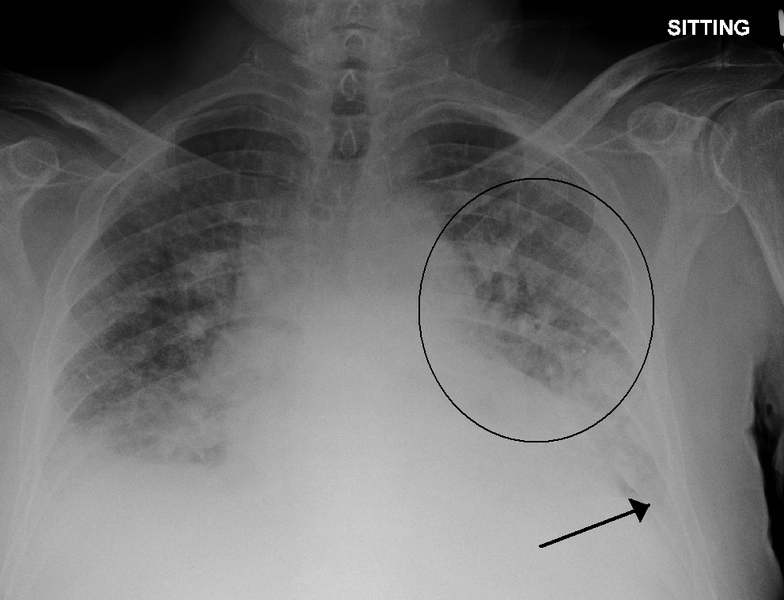
Image Source: Wikimedia Commons (2011). PulmEdema, Licensed under a creative commons licence. Available for free at: https://en.wikipedia.org/wiki/File:PulmEdema.PNG#/media/File:PulmEdema (Accessed: 19 January 2024).
Management of cardiogenic pulmonary oedema
The aims of management are to relieve symptoms and restore haemodynamic stability and tissue perfusion. Cardiogenic pulmonary oedema is a marker of poor long-term outcome, but effective early management can improve short-term survival.
Aspects of acute management include:
- Sitting the patient upright
- Oxygen if hypoxia is present aiming for appropriate target saturations (94-98% or 88-92% in chronic CO2 retainers) – this can be delivered by any method of oxygen delivery
- IV diuretics – this will need monitoring of urine output, patient weight and electrolytes. IV furosemide is often the first choice. A trial bolus of 40 mg would be appropriate initial management in someone who is diuretic naive if the blood pressure will tolerate this. If the patient is normally treated with diuretics then the initial dose should be equal to or greater than their normal dose. Diuretic boluses can be used twice or three times a day. If the patient’s blood pressure is low and they are not tolerating diuretic boluses then a 24-hour infusion can be used, such as 120-240 mg/day. However, infusions should not be used if the patient is not responsive to boluses and there is no evidence that infusions are more effective than boluses at achieving diuresis.
- Vasodilators – The choice of vasodilator depends on the aim. Sodium nitroprusside can be used to reduce arterial tone and reduce cardiac afterload; glyceryl trinitrate can be used to reduce venous tone as an adjunct to diuretics. Routine use of vasodilators is not recommended, only if there is a specific aim as above.
- Continuous positive airway pressure (CPAP) – if there is severe oedema or they are failing to respond to optimal medical therapy then CPAP should be initiated. CPAP can facilitate the delivery of oxygen, splinting open airways and raising intrapulmonary pressure and, therefore, reduce pulmonary oedema. Pressures are normally started at around 5 cmH2O and uptitrated to around 10 cmH2O. If there is associated respiratory acidosis and hypercapnia, the patient may require non-invasive ventilation (NIV) rather than CPAP. There is a risk of hypotension with CPAP as the venous return is reduced.
- Mechanical ventilation – patients who fail to respond to CPAP/NIV or those who have contraindications to CPAP/NIV should be intubated if appropriate and mechanically ventilated
- Additional support – in the presence of shock preventing optimisation of medical therapy and severe systolic dysfunction, the patient may require inotropes (e.g. dobutamine or milrinone) or mechanical support (e.g. intra-aortic balloon pump, left ventricular assist device or extracorporeal membrane oxygenation)
References
- G
- Garan AR. Pathophysiology of cardiogenic pulmonary edema [Internet]. Colucci WS, Dardas TF, editors. UpToDate. 2022 [cited 2023 May 8]. Available from: https://www.uptodate.com/contents/pathophysiology-of-cardiogenic-pulmonary-edema
- Meyer TE. Approach to diagnosis and evaluation of acute decompensated heart failure in adults [Internet]. Colucci WS, Dardas TF, editors. UpToDate. 2023 [cited 2023 May 8]. Available from: https://www.uptodate.com/contents/approach-to-diagnosis-and-evaluation-of-acute-decompensated-heart-failure-in-adults
- Henson VL. Cardiogenic Pulmonary Oedema. Kendall JM, editor. RCEM Learning. 2021 [cited 2023 May 8]. Available from: https://www.rcemlearning.co.uk/reference/cardiogenic-pulmonary-oedema
- Matthay MA, Murray JF. Pulmonary Edema. In: Broaddus C, Mason RJ, Ernst JD, King Jr. TE, Lazarus SC, Murray JF (eds.) et al. Murray & Nadel’s Textbook of Respiratory Medicine. 6th ed. Philadelphia: Saunders; 2016. p. 1096-1117
- Oxford Medical Education. Acute Pulmonary Oedema. Oxford Medical Education. 2015 [cited 2023 May 8]. Available from: https://oxfordmedicaleducation.com/cardiology/acute-pulmonary-oedema/
- Colucci WS. Treatment of acute decompensated heart failure: Specific therapies [Internet]. Gottlieb SS, HoekstraJ, Dardas TF, editors. UpToDate. 2023 [cited 2023 May 8]. Available from: https://www.uptodate.com/contents/treatment-of-acute-decompensated-heart-failure-specific-therapies
- McDonagh TA, Metra M, Adamo M, Gardner RS, Baumbach A, Böhm M et al. 2021 ESC Guidelines for the diagnosis and treatment of acute and chronic heart failure: Developed by the Task Force for the diagnosis and treatment of acute and chronic heart failure of the European Society of Cardiology (ESC) With the special contribution of the Heart Failure Association (HFA) of the ESC. European Heart Journal. 2021;42(36); 3599-3726. Available from: doi: 10.1093/eurheartj/ehab368
Complete Guide to Clinical Genetics ST3 Application and Recruitment (2027)
Written by Medibuddy. Last updated: October 2026. Clinical Genetics is

Complete Guide to Clinical Radiology ST3 Application and Recruitment (2027)
Recruitment paused: NHS England is not running Clinical Radiology ST3+ recru

Complete Guide to Paediatric Surgery ST3 Application and Recruitment in United Kingdom (2026)
Written by Medibuddy. Last updated: October 2026. If you’re planning


Take your subscriptions with you
Our mobile app allows you to access your interview and exam question banks wherever you are.

