


 Close
Close
 18th February 2025
Granulomatosis with polyangiitis
Granulomatosis with polyangiitis (GPA; previously Wegener’s granulomatosis) is an ANCA (antineutrophilic cytoplasmic antibody)-associated vasculitis causing small vessel necrosis affecting the upper and lower respiratory tract, kidneys, skin and nervous system. Typically, both upper airway and pulmonary involvement are present.
Clinical features of GPA
Nasal, sinus and ear disease are found in 90%. They may be present without other manifestations and precede generalised disease by weeks to months. Patients often present with nasal congestion and discharge suggestive of a sinus infection that has failed to resolve with antibiotic therapy.
Specific features include:
- Nasal crusting and obstruction
- Chronic rhinosinusitis – can lead to septal perforation, saddle nose deformity or nasocutaneous fistula, but not nasal polyps
- Purulent or bloody nasal discharge
- Excessive tearing (epiphora)
- Sinus mucoceles
- Conductive and/or sensorineural hearing loss
Pulmonary features include:
- Tracheobronchial disease – subglottic stenosis is most common and tracheostomy may be required
- Lung nodules +/- cavitation (usually asymptomatic) – most common radiological manifestation
- Interstitial lung disease – in around 25% of patients (less common in GPA than microscopic polyangiitis)
- Alveolar haemorrhage (often seen as ground glass changes on CT) – a life-threatening pulmonary manifestation causing rapid-onset dyspnoea, cough, haemoptysis, anaemia and potentially hypoxaemia. Untreated, it can progress quickly to respiratory failure
- Pulmonary artery stenosis is reported rarely and is diagnosed with CTPA
Investigations in GPA
Findings on blood tests include:
- Positive c-ANCA in up to 75% of cases and, less classically, p-ANCA in 25% of cases
- Normochromic, normocytic anaemia
- Raised WCC and platelets (>400 x 109/L)
- Raised ESR and CRP are common, but negative ESR and CRP do not exclude GPA
- Acute creatinine/urea rise (acute kidney injury)
Chest radiograph is typically obtained as an initial investigation. Features include pulmonary nodules that may cavitate, reticular markings, diffuse, hazy opacities, lobar or segmental atelectasis, pleural opacities, and hilar adenopathy.
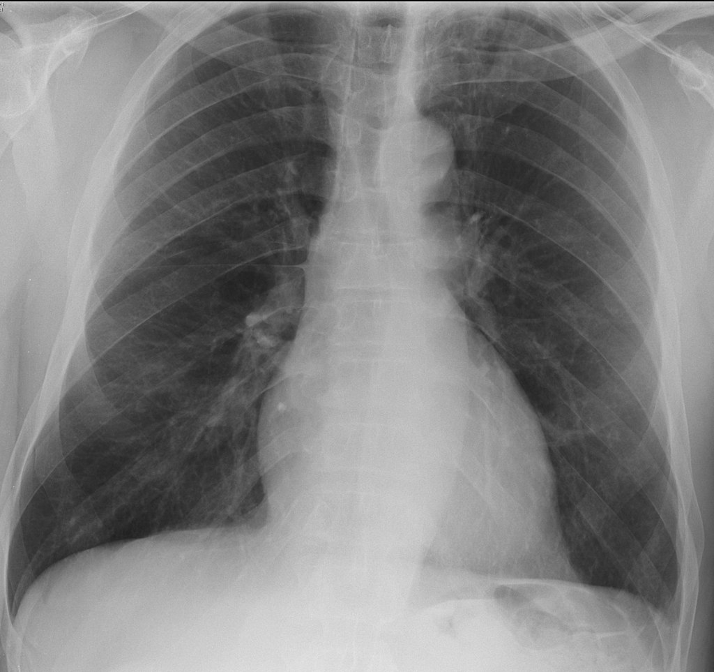
Figure 5: Image Source: Wikimedia Commons (2012). Granulomatosis with polyangiitis, Licensed under a creative commons licence. Available for free at: https://en.wikipedia.org/wiki/Granulomatosis_with_polyangiitis#/media/File:Granulomatosis_with_polyangiitis.jpg (Accessed: 19 January 2024).
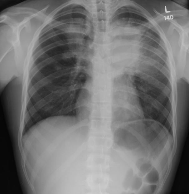
Figure 6: Image Source: Flickr (2011). Pulmonary pathology Licensed under a creative commons licence. Available for free at: https://www.flickr.com/photos/pulmonary_pathology/5958079204/in/photostream/ (Accessed: 19 January 2024).
High-resolution CT should be obtained in patients undergoing evaluation for vasculitis with abnormal chest X-ray. Common findings include:
- Multiple pulmonary nodules
- Patchy or diffuse ground-glass or consolidative opacities (pulmonary haemorrhage or active vasculitis)
- Small, peripheral, wedge-shaped densities (pulmonary microinfarction)
- Enlarged, irregular, stellate-shaped, peripheral pulmonary arteries (‘vasculitis’ sign)
- Tracheal or bronchial stenosis
Other investigations may include:
- CT of the sinuses – to evaluate chronic rhinosinusitis in suspected GPA.
- Bronchoscopy – to assess and take biopsies of endobronchial disease and perform bronchoalveolar lavage (BAL). BAL can identify pulmonary haemorrhage when imaging shows diffuse parenchymal opacities. Subsequent lavage aliquots become increasingly haemorrhagic. Transbronchial biopsy is often too small to make a diagnosis of GPA but can identify alternative diagnoses.
- Lung biopsy – when there are single or multiple pulmonary nodules and extrapulmonary disease is absent or not accessible to biopsy. Transthoracic needle biopsies are usually too small for a definitive diagnosis, so lung tissue must be obtained via video-assisted thoracoscopic surgery (VATS) or thoracotomy.
Management of GPA
Treatment involves an induction regimen of glucocorticoids and rituximab/cyclophosphamide. Plasma exchange may be useful in patients with pulmonary haemorrhage not readily responding to other therapies and optimal supportive care.
In patients with less severe disease without any organ- or life-threatening features (i.e. those with only ENT features, arthritis and/or pulmonary nodules), initial induction therapy with glucocorticoids and weekly methotrexate or, alternatively, mycophenolate mofetil (MMF) monotherapy is suggested. Note that methotrexate is associated with pulmonary fibrosis.
Maintenance therapy typically involves tapering the glucocorticoids and using rituximab or azathioprine as maintenance therapy, usually after a few months.
Complete Guide to Clinical Genetics ST3 Application and Recruitment (2027)
Written by Medibuddy. Last updated: October 2026. Clinical Genetics is

Complete Guide to Clinical Radiology ST3 Application and Recruitment (2027)
Recruitment paused: NHS England is not running Clinical Radiology ST3+ recru

Complete Guide to Paediatric Surgery ST3 Application and Recruitment in United Kingdom (2026)
Written by Medibuddy. Last updated: October 2026. If you’re planning


Take your subscriptions with you
Our mobile app allows you to access your interview and exam question banks wherever you are.

