


 Close
Close
 24th February 2025
Investigation of OA
The mainstay of diagnosis in OA is a comprehensive clinical assessment, where the history and examination findings are correlated, augmented by radiological studies. Of note, the radiological findings of X-rays correlate poorly with the described severity of symptoms and, indeed, the function of the joint in terms of daily living activities. The typical X-ray findings are: loss of joint space, subchondral sclerosis, subchondral cysts and osteophyte formation (see Figure 2).
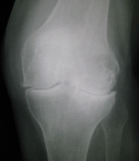
Image source: Wikimedia Commons (2012). Osteoarthritis left knee, Licensed under a Creative Commons licence. Available for free at: https://commons.wikimedia.org/wiki/File:Osteoarthritis_left_knee.jpg (Accessed: 17 January 2024).
Laboratory investigations are rarely required, and if they are, it is to exclude other causes, such as crystal arthropathy (where joint aspiration for microscopy may be indicated) or RA (where inflammatory markers will be raised and auto-antibodies likely detected).
Complete Guide to Clinical Genetics ST3 Application and Recruitment (2027)
Written by Medibuddy. Last updated: October 2026. Clinical Genetics is

Complete Guide to Clinical Radiology ST3 Application and Recruitment (2027)
Recruitment paused: NHS England is not running Clinical Radiology ST3+ recru

Complete Guide to Paediatric Surgery ST3 Application and Recruitment in United Kingdom (2026)
Written by Medibuddy. Last updated: October 2026. If you’re planning


Take your subscriptions with you
Our mobile app allows you to access your interview and exam question banks wherever you are.

