


 Close
Close
 7th May 2025
Ventricular Septal Defects
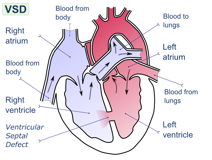
Image Source: Wikimedia Commons (2008). Ventricular Septal Defect, Licensed under a creative commons licence. Available for free at:
https://upload.wikimedia.org/wikipedia/commons/e/e1/Ventricular_septal_defect-en.png?20080222192725 (Accessed 2 May 2025).
Ventricular Septal Defects (VSD) are often congenital abnormalities in the development of the interventricular septum (Figure Three). These anomalies result in an abnormal conduit between the left and right ventricles, which results in an often acyanotic left-to-right shunt. It is important to note that an untreated VSD can result in Eisenmenger syndrome, which results in a cyanotic right-to-left shunt. The key details of VSD are detailed in Table Four.
| Feature | Summary |
|---|---|
| Epidemiology | The most common CHD in infants and children
Isolated VSD occurs in 3.5 per 1000 live births but often close spontaneously. VSD can accompany other congenital defects, not just in isolation Acquired VSD secondary to trauma or myocardial infarction is rare Unusual for VSD to present in adults. |
| Aetiology | Multiple classification systems exist, but the easiest is to think of them as congenital vs acquired.
Congenital
Acquired
|
| Clinical History | Failure to thrive in infancy
Shortness of breath on exertion Risk factors: Maternal alcohol use in pregnancy, Down’s syndrome, family history of CHD Repeated pulmonary infections: in shunt reversal associated with large defects |
| Clinical Examination | Holosystolic Systolic Murmur
Loud pulmonary component of the second heart sound
|
| Diagnostic Investigations | Echocardiogram – to visualise the VSD and confirmation of the left-to-right shunt.
Chest x-ray – may be normal or show cardiomegaly. ECG – dependent on the size of the VSD, not diagnostic but supportive. In medium VSD can show signs of left ventricular hypertrophy. |
| Management | Once a diagnosis has been made utilising an echocardiogram, the pulmonary-systemic blood flow ratio (Qp:Qs) is calculated.
Prophylactic antibiotics should be considered in all VSDs to prevent endocarditis. Congenital Small VSD (Qp:Qs <1) – monitoring and observation, they may spontaneously close. Medium/Large VSD (Qp:Qs ≥1.5)
Acquired Surgical repair |
| Follow Up | Annual follow-up of asymptomatic patients to monitor for development of complications.
Patients who have corrective closure require monitoring for the development of heart block or arrhythmia. Potential complications include aortic regurgitation, heart block, arrhythmia and infective endocarditis. The prognosis is dependent on the size of the VSD.
|
Table 4: This table describes the key features of ventricular septal defects. Sourced and adapted from Aggarwal, Ventricular septal defects: BMJ Best Practice; 2022.
Complete Guide to Clinical Genetics ST3 Application and Recruitment (2027)
Written by Medibuddy. Last updated: October 2026. Clinical Genetics is

Complete Guide to Clinical Radiology ST3 Application and Recruitment (2027)
Recruitment paused: NHS England is not running Clinical Radiology ST3+ recru

Complete Guide to Paediatric Surgery ST3 Application and Recruitment in United Kingdom (2026)
Written by Medibuddy. Last updated: October 2026. If you’re planning


Take your subscriptions with you
Our mobile app allows you to access your interview and exam question banks wherever you are.

