


 Close
Close
 23rd May 2025
The Pituitary Gland
Anatomy and Physiology
The pituitary gland, or hypophysis, is a pea-sized endocrine gland that lies in the sella turcica of the sphenoid bone. It is connected to the hypothalamus by the infundibulum which contains the hypophyseal-portal vessels. The pituitary gland is also known as the “master gland” as it regulates the actions of other endocrine organs throughout the body. Anatomically and embryologically it is divided into the anterior lobe, or adenohypophysis, and posterior lobe, or neurohypophysis.
The adenohypophysis is embryologically derived from an evagination of ectoderm named Rathke’s pouch. It secretes: growth hormone (GH) (Stimulates growth), prolactin (PRL) (stimulates milk production), adrenocorticotropic hormone (ACTH) (stimulates cortisol and androgen production by adrenal cortex), thyroid-stimulating hormone (TSH), follicle-stimulating hormone (FSH) (stimulates production of oestrogen and growth of oocytes), and luteinising hormone (LH) (stimulates production of oestrogen and progesterone by ovaries and testosterone by testes).
The neurohypophysis is continuous with the hypothalamus and embryologically originates from neuroectoderm. It secretes hormones that are produced in the hypothalamus: Antidiuretic hormone (ADH) (increases water reabsorption from kidneys which will increase blood pressure and oxytocin (stimulates uterine contraction in labour and lactation).
The pituitary gland works in close relation to the hypothalamus via the hypothalamic-pituitary-adrenal axis (HPAA). The hypothalamus secretes hormones into hypophyseal-portal veins which reach the pituitary and stimulate secretions of hormones by the adenohypophysis. Dopamine and somatostatin, also secreted by the hypothalamus, inhibit the release of PRL and GH. The pituitary also communicates with the hypothalamus via negative feedback where high levels of hormones secreted by the pituitary inhibit the hypothalamus from secreting the respective hormones that trigger their release.
Disorders of the pituitary gland often arise from levels of these hormones being too high or low.
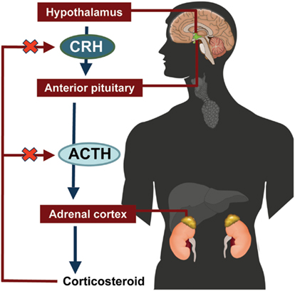
Image Source: Wikimedia Commons (2013). Curid, Licensed under a creative commons licence. Available for free at: https://commons.wikimedia.org/w/index.php?curid=44663207 (Accessed 13 May 2025).
Disorders of Excess Hormone Production
Acromegaly
Acromegaly is an insidious chronic and progressive disorder caused by the overproduction of GH and characterised by features of gigantism. It is usually caused by a somatotroph adenoma in the adenohypophysis. The incidence of acromegaly is between 40-130 cases per million adults. An excess of GH leads to increased secretion of insulin-like-growth factor-1 (IGF-1) leading to multi-systemic symptoms.
Clinical effects
- Facial: Frontal bossing, macroglossia, prognathism, large nose
- Skin: Hyperhidrosis, increased skin thickness
- Respiratory: Sleep apnoea
- Joint: Osteoarthritis, arthropathy, bone enlargement
- Cardiovascular: Hypertension, arrhythmias, valvular heart disease, congestive cardiac failure, ischaemic heart disease
- Metabolic and electrolyte: diabetes mellitus, impaired glucose tolerance, hypercalciuria
- Organomegaly
- Increased height
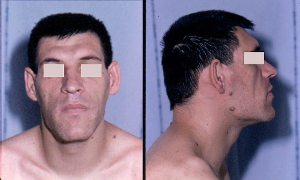
Image Source: Wikimedia Commons (2008). Acromegaly facial Features, Licensed under a creative commons licence. Available for free at: https://commons.wikimedia.org/w/index.php?curid=15072125 (Accessed 13 May 2025).
Investigations
Biochemical
- Excluded in most cases of normal IGF-1 levels
- Random GH >1 microgram/L or >2.5 micrograms/L
- Glucose tolerance test with 75g oral glucose and measuring plasma glucose and GH every 30 mins for 2 hrs. Lack of GH suppression <1 microgram/L (<1 nanogram/mL) suggestive of acromegaly
- Other pituitary hormone plasma levels: cortisol, prolactin, thyroid-stimulating hormone and free thyroxine, follicle-stimulating hormone, luteinising hormone, oestradiol, or testosterone
- Consider genetic testing for AIP
Imaging
- MRI with contrast of pituitary gland
- In <1% of the cases, the above is negative. In this case a CT Thorax, Abdo, Pelvis will likely be needed to locate ectopic hormones producing GH/GHRH
Management
- Transsphenoidal surgery is first-line for enclosed somatotroph microadenomas (< 10mm diameter) and macroadenomas (>10mm diameter)
- If surgery fails, or the tumour is unresectable, somatostatin analogues (SSAs) are used e.g., octreotide with an initial dose of 20mg every month
- If there is mixed GH/PRL release, or IGF-1 levels are <1.5 the upper limit of normal a dopamine agonist, e.g., cabergoline, can be used as an additional treatment
- Pegvisomant, a growth hormone receptor antagonist, is considered in patients who are unsuitable or who have not responded to the above
- If medical and surgical treatments fail, radiotherapy can be attempted
Monitoring
Lifelong monitoring of GH and IGF-1 will be required
Complications
- Cardiovascular (including HTN) (30-40%)
- Sleep apnoea (60-80%)
- Joint (70%)
- Endocrine (50%)
- Pre-cancerous polyps
- Carpal tunnel (64%)
- Hypopituitarism
Prognosis
Surgery is effective in 70-90% of microadenomas and 30-60% of macroadenomas. SSAs control disease in 30-40% of cases.
Cushing Syndrome
Cushing syndrome is a rare disorder characterised by an excess production of cortisol, often secondary to an excess of ACTH production. It has an incidence of 0.7-2.4 per million population per year. It affects females more than males at a ratio of 3:1. Cushing’s disease is Cushing syndrome caused by an ACTH secreting pituitary tumour, affecting more males than females at a ratio of 5:3.
Risk factors
- Hypertension
- Uncontrolled diabetes
- Osteoporosis
- Adrenal masses
Causes
ACTH Dependent causes are due to an overproduction of ACTH which stimulates increased synthesis of cortisol. Causes include:
- Cushing’s disease (70-80%). This has been linked to a mutation in the gene ubiquitin-specific protease 8 on chromosome 15 (USP8) which increases risk of developing pituitary adenoma
- Adrenal adenoma (10%). Linked to mutations in the cAMP/PKA pathway
- Ectopic secretion of ACTH or CRH by non-pituitary tumours (~10%)
- Adrenal carcinoma (1%)
ACTH Independent causes result in overproduction of cortisol by the adrenal glands despite suppressed ACTH levels. Causes include:
- Adrenal adenoma
- Bilateral adrenal hyperplasia
- Adrenal carcinomas (rare). Often presents as a rapidly growing adrenal mass
- Primary pigmented nodular adrenocortical disease (PPNAD)
Exogenous corticosteroids, e.g., prednisolone, can also cause features of Cushings.
Clinical Features
- Facial plethora and facial rounding
- Skin: Purple striae, atrophy, hyperpigmentation (due to increased ACTH rather than cortisol), acanthosis nigricans, cutaneous fungal infections e.g., tinea versicolor
- Metabolic: Diabetes/glucose intolerance, obesity with supraclavicular fullness (due to deposition of subcutaneous fat)
- Cardiovascular: Increased risk of hypertension, venous thromboembolism, heart failure and dilated cardiomyopathy
- Musculoskeletal: Osteoporosis, proximal muscle wasting and weakness,
- Mood changes
- Irregular menstruation
- Signs of androgen excess in females, whose main source of androgens are the adrenals, such as: hirsutism, acne, changes in libido, temporal balding, deep voice, virilization. Note the testes are the main source of androgens for men so they do not show signs of androgen excess.
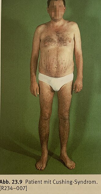
Image Source: Wikimedia Commons (2020). Patient with Cushing-Syndrom, Licensed under a Creative Commons licence. Available for free at: https://commons.wikimedia.org/w/index.php?curid=90182292 (Accessed 13 May 2025).
Diagnosis
- First line is the dexamethasone suppression test. For the low dose test, 1mg of dexamethasone is given at 11pm and plasma cortisol and dexamethasone level are measured at 8am. The latter is measured to ensure adequate levels are achieved, especially in extremely obese patients. For a positive result, morning cortisol levels must be >1.8 micrograms/dL
- If the patient is taking drugs affecting the metabolism of dexamethasone e.g., phenytoin, carbamazepine, rifampicin, and cimetidine, 24-hour urinary free cortisol can be measured.
- Late-night salivary cortisol. Saliva sample taken between 11pm-12am. Can be repeated twice on two separate nights for greater sensitivity. Elevated cortisol levels suggest Cushings.
- Once a diagnosis has been established, morning plasma ACTH testing can be done. Suppressed ACTH (levels <1 picomol/L) suggest ACTH-independent Cushings due to adrenal pathology. No suppression (levels >4 picomol/L) indicates ACTH-dependent Cushings due to pituitary tumour or ectopic source of ACTH production.
- To differentiate between Cushing’s disease and pseudo-Cushing syndrome (physiological hypercortisolism) a CRH stimulation test is done. 0.5mg of oral dexamethasone is administered 6 hourly for 48 hours. Two hours after the last dose, 1 mcg/kg of CRH is given IV. Serum cortisol is measured 15 minutes later. If glucocorticoid suppression is overcome, a diagnosis of Cushing’s disease rather than pseudo-Cushing syndrome is suspected.
Other investigations
- Urine pregnancy test to exclude pregnancy
- Serum glucose
- Elevated plasma dehydroepiandrosterone sulphate (DHEAS) levels could suggest adrenal carcinoma
- Pituitary MRI if ACTH dependent Cushings
- If confirmed pituitary lesion, can attempt inferior petrosal sinus sampling. Blood sample taken from sinus and plasma at the same time. If central/peripheral ACTH ratio is >2:1 the causative pathology lies in the pituitary gland. Otherwise, the source is ectopic
- Adrenal CT if ACTH independent Cushings
- Can consider CT TAP and/or PET scan if considering ectopic source of pathology
Management
Treating ACTH secreting pituitary tumour:
- 1st line: transsphenoidal pituitary adenectomy.
- Note, medical therapy with: somatostatin analogue e.g., Pasiretoride; dopamine agonist e.g., cabergoline; steroidogenesis inhibitor e.g., osilodrostat, or glucocorticoid antagonists e.g., mifepristone or ketoconazole, could be used prior to surgery for severe Cushings
- Post-surgical corticosteroid replacement therapy with hydrocortisone
- Other hormones may also need to be replaced with levothyroxine testosterone/oestradiol, recombinant somatropin, desmopressin
- If hormone replacement fails, repeat transsphenoidal pituitary adenectomy can be considered
- If no success, pituitary radiotherapy can be attempted
- If the disease is still uncontrolled and/or the patient is not suitable for reoperation, a bilateral adrenalectomy can be done. However, this can increase the risk of Nelson syndrome
Treating ectopic ACTH/CRH tumours:
- 1st: Surgical resection/ablation of tumour
- Steroidogenesis inhibitor therapy or a glucocorticoid receptor antagonist can be used prior to surgery in severe Cushing’s, e.g. ketoconazole
- If surgical resection cannot be performed, bilateral adrenalectomy can be considered. After this, permanent post-surgical corticosteroid therapy will be required
- Chemo/radiotherapy, or medical therapy alone, can be considered
Treating adrenal pathology:
- 1st: Unilateral adrenalectomy/tumour resection. If disease is bilateral, a bilateral adrenalectomy is first line.
- Like the above, medical therapy with steroidogenesis inhibitors can be considered before surgery in severe cases
- Chemo/radiotherapy, or medical therapy alone, can be considered
Prognosis and Monitoring
- Patients in the first week post-pituitary adenectomy require monitoring of morning cortisol levels (sample taken 24 hrs post last dose of corticosteroid). Low levels of cortisol predict remission
- Recurrence of ACTH dependent Cushings occurs in 5-26% of patients within 5 years
- Therefore, screening at 6-12 months using one of the diagnostic tests listed above is required. Late night salivary cortisol is the most sensitive test to detect recurrence.
Prolactinoma
Prolactinomas are a type of anterior pituitary adenoma that secrete prolactin (PRL) and lead to hyperprolactinaemia. They are benign tumours of lactotroph cells. Hypersecretion of PRL has inhibitory effects on GNRH and pituitary gonadotrophins.
They commonly occur in women aged 20-50 years. The ratio of incidence between females and males is 10:1.
Aetiology
- Sporadic
- Multiple endocrine neoplasia syndrome type 1 (MEN-1)
- Familial isolated pituitary adenoma (FIPA)
Tumour Classification
This is based on tumour size
- Microadenoma
- <10mm diameter
- Commonest
- Unlikely to get larger
- Macroadenoma
- >10mm diameter
- Often locally invasive to suprasellar/parasellar areas
- Can compress vital structures
- Often benign and do not metastasise to outside the pituitary sella
Clinical Presentation
- Premenopausal women: Hypogonadotropic hypogonadism- infertility, oligo/amenorrhea, galactorrhoea, reduced bone density
- Men: Hypogonadotropic hypogonadism- reduced energy, decreased libido, lowered muscle mass, reduction in body hair, galactorrhoea, decreased bone density
- Postmenopausal women: Tumour often detected as an incidental finding on brain imaging. Or, mass effects from tumour produce symptoms, e.g., unilateral/bi-temporal hemianopia, which prompt investigation
Investigations
- Random serum prolactin level. The concentration often reflects the size of the tumour
- Any PRL level above the normal age range suggests prolactinoma
- PRL > 250 micrograms/L: prolactinoma likely
- PRL > 500 micrograms/L: macroadenoma likely
- Note: medications such as risperidone and metoclopramide can increase PRL levels
- Pituitary MRI
- Formal visual field testing for macroadenomas
- Consider genetic screening in patients <30 years with macroadenomas for
- MEN-1
- Aryl hydrocarbon receptor-interacting protein (AIP) gene mutations present in FIPA
- Consider other tests for hypopituitarism such as IGF-1, ACTH, TSH
- U&Es to assess kidney function as CKD can lead to a decrease in PRL clearance
Management
- Pre-menopausal women with asymptomatic microadenomas: observation
- Pre-menopausal women with symptomatic microadenomas that do not wish to be pregnant:
- 1st: Dopamine agonist e.g., Cabergoline
- 2nd: Combined oral contraceptive pill, especially if only symptom menstrual changes
- 3rd: transsphenoidal surgery
- 4th: sellar radiotherapy
- Pre-menopausal women with symptomatic microadenomas that do wish to be pregnant OR microadenomas in men OR macroadenomas in any patient:
- 1st: dopamine agonist
- 2nd: transsphenoidal surgery
- 3rd: sellar radiotherapy
- Microadenomas in post-menopausal women: observation
Monitoring and follow-up
- Pituitary MRI every year initially
- Regular perimetry
- Regular measurements of PRL levels
- ECHO monitoring for patients on high dose cabergoline due to side effect of valve pathology
Complications
- Bi-temporal hemianopia
- Hypopituitarism
- Cabergoline associated valvular pathology
- Pituitary apoplexy
Craniopharyngioma
Craniopharyngiomas are benign non-glial epithelial tumours arising from remnants of Rathke’s pouch. These tumours often grow above the pituitary gland in the sellar/suprasellar space adjacent to the optic chiasm and often lead to a deficiency of anterior pituitary hormones. They account for 5-10% brain tumours in children and 1-3% of all adult intracranial tumours. The aetiology is unknown.
Classification
Craniopharyngiomas are classified based on pathology:
- Adamantinous type (>80%): Adamantinous epithelium, keratin nodules, poorly demarcated from brain tissue, associated withs CTNNB1 gene mutation
- Squamous papillary type (<20% of tumours): Squamous epithelium with papillaries, well demarcated from brain tissue, rare in children, associate with BRAF oncogene mutation
Clinical Features
- Visual disturbances
- Headache
- Intracranial hypertension: nausea, vomiting, double vision
- Endocrine: Symptoms of hypogonadotrophic hypogonadism (as listed above, galactorrhoea
- Paediatric: macrocephaly and hydrocephalus (prior to fusion of intracranial sutures) and growth failure
Investigations
- Visual field testing
- MRI brain with contrast
- Serum: PRL, GH, IGF-1, LH, FSH, TSH, morning testosterone, morning ACTH and cortisol, U&Es
- Paired urine serum osmolality
- Can consider tissue histology following surgical biopsy/resection
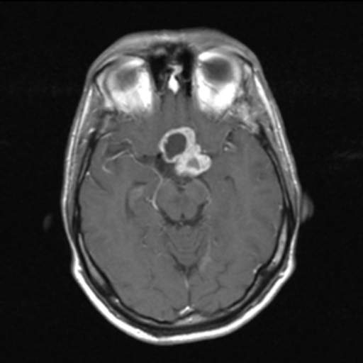
Image Source: Wikimedia Commons (2011). Craniopharyngioma, Licensed under a creative commons licence. Available for free at: https://commons.wikimedia.org/w/index.php?curid=15959020 (Accessed 13 May 2025).
Management
- The exact type of surgical management (biopsy only, cyst aspiration, CSF shunting, or tumour resection) depends on tumour size and location
- Post-operative ophthalmology review will be required
- Hormonal replacement therapy
- Follow-up is done every 6-12 months and involves MRI brain scans as well as endocrinology review
Complications
- Pituitary hormone deficiencies
- Diabetes insipidus
- Visual impairment
- Rarer complications include: seizures, hypothalamic injury, cognitive dysfunction
Disorders of Hormonal Deficiencies
Diabetes Insipidus
Diabetes insipidus (DI) is a disorder that leads to an increased production of dilute urine, polydipsia and can include nocturia. It is rare, with a global prevalence of 1 per 25,000 of the population.
Types of DI:
- Cranial/Central: Impaired production/transportation/release of ADH. Caused by disorders in areas responsible for ADH secretion, such as hypothalamic osmoreceptors, supraoptic/paraventricular nuclei or the superior part of the supraoptic hypophyseal tract. Pathology in the neurohypophysis alone often does not cause DI as ADH produced in the hypothalamus can enter the systemic circulation.
- Nephrogenic: Renal collecting ducts are resistant to ADH
- Gestational: Occurs during pregnancy
Aetiology
Cranial DI
- Transsphenoidal surgery for pituitary adenoma
- Craniopharyngioma
- Lesions to the pituitary stalk
- Traumatic brain injury
- Subarachnoid haemorrhage affecting the anterior communicating artery supplying the anterior hypothalamus
- Congenital hypothalamic-pituitary defects
- Wolfram syndrome: Autosomal recessive progressive neurodegenerative disorder leading to diabetes mellitus, optic atrophy, sensorineural deafness and DI
- Associated with autoimmune disorders e.g., Hashimoto’s thyroiditis
- Medications e.g., phenytoin
- Late complication of meningitis/encephalitis
Nephrogenic DI
- Lithium therapy
- Chronic kidney disease
- Renal amyloidosis
- Renal sarcoidosis
- Associated with chronic hypercalcaemia/ hypokalaemia
- Genetics: mutations AVPR2 receptor (X linked recessive) or AQP2 gene (autosomal recessive)
Initial Investigations
- Paired urine serum osmolality: Low urine (<300 mmol/kg) and high serum osmolality
- Urea and electrolytes, calcium: Assesses renal function and electrolyte abnormalities
- 24 hr urine collection to measure volume: >3L in 24 hrs
- To rule out diabetes mellitus: urine dipstick to test for glucose and serum glucose levels
Diagnostic Tests
- 1st Water deprivation test
- Patient deprived of fluids for 8 hours. Every hour serum osmolality, urine osmolality and urine volume are measured
- Contraindications: Renal insufficiency, uncontrolled diabetes mellitus, uncorrected adrenal or thyroid hormone deficiency
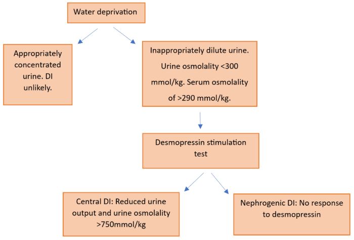
- DI patients are unable to concentrate their urine in response to dehydration so results show inappropriately dilute urine with osmolality <300 mmol/kg and a serum osmolality of >290 mmol/kg
- 2nd Desmopressin stimulation test
- Done after positive water deprivation test
- 2 micrograms desmopressin administered subcutaneously. Every hour for 4 hours serum osmolality, urine osmolality and urine volume are measured
- Patients with central DI will have a reduced urine output and increased urine osmolality (>750 mmol/kg) in response to desmopressin
- Patients with nephrogenic DI show no response to desmopressin
- Hypertonic saline stimulated test can be considered
- Baseline copeptin level >24.1 picomol/L diagnoses nephrogenic DI
- After infusing 3% sodium chloride, a copeptin level >4.9 picomol/L diagnoses central DI
Further investigations to be considered
- MRI brain with contrast
- Genetic testing
- Pituitary hormone levels
Diagram for Diagnosis of Diabetes Insipidus
Management
- Hypernatremia: treat with IVF e.g., 5% dextrose
- Central DI: Desmopressin
- Nephrogenic DI: Adequate fluid intake to match losses, treatment of underlying cause. Can consider low sodium diet (<500mg/day), thiazide diuretics or indomethacin to reduce urine output
Growth Hormone Deficiency in Adults
GH deficiency is a rare disorder caused by inadequate secretion of GH. It can be idiopathic, congenital or acquired. GH deficiency in adults is commonly due to pituitary tumour (78%), extra-pituitary tumour such as craniopharyngioma (13%) and unknown (8%). Therefore, it is important to test adults with known hypothalamic or pituitary disease, such as panhypopituitarism, for GH deficiency.
Clinical manifestations of GH deficiency
- Reduced energy levels
- Reduced lean body mass
- Increase in proportion of fat
- Reduced bone mineral density leading to osteoporosis
- Cardiovascular: increased LDL cholesterol, impaired cardiac function
- Insulin resistance,
- Poor quality of life and increased mortality
Diagnosis
- History of pituitary/hypothalamic disease or panhypopituitarism
- Low serum IGF-1
- If IGF-1 levels are normal, a GHRH-Arginine simulation test can be done to diagnose. This involves taking a blood sample then injecting GHRH and then arginine over 30 minutes. Further blood samples are taken over the next two hours. Facial flushing is a common side effect of GHRH injection. A diagnosis is made if GH remains suppressed. The exact level depends on peak GH levels relative to patient’s BMI
- For further investigations, can consider pituitary imaging if not already completed
Management
Treatment involves injections of recombinant human growth hormone (rHGH). Please note a positive simulation test is required to initiate treatment with rHGH in the National Health Service (NHS), UK. Although patients taking this treatment are unlikely to have marked increases in their energy levels, it can improve muscle strength, bone mineral density, CVS risk factors and has some impact on quality of life
Panhypopituitarism
This is a disorder characterised by the reduced/absent secretion of most or all of the hormones by the pituitary gland. Panhypopituitarism can be due to hypothalamic disease, leading to reduced secretions of releasing hormones, or pituitary disease.
Aetiology
Hypothalamic disease
- Tumour
- Radiation therapy for nasopharyngeal cancer or brain tumours
- Post meningitis
- Traumatic brain injury
- Stroke
Pituitary disease
- Tumours and lesions, e.g., Pituitary adenoma (most common association with panhypopituitarism)
- Pituitary surgery
- Radiotherapy to the pituitary gland
- Hemochromatosis of pituitary gland
- Immunotherapy side effect, e.g., ipilimumab
- Sheehan syndrome
- Pituitary apoplexy: haemorrhage into infarction or existing adenoma
- Pituitary abscess or infection
- Pituitary stalk interruption syndrome- congenital hypopituitarism
- Empty sella
Clinical Features
- ACTH and cortisol deficiency: postural hypotension, tachycardia, anorexia, reduced libido, hypoglycaemia
- TSH deficiency: symptoms of hypothyroidism
- Low levels of gonadotropins lead to hypogonadotropic hypogonadism
- GH deficiency
- PRL deficiency
Investigations
- U&Es
- Serum and urine osmolality
- Morning cortisol, ACTH, testosterone/oestradiol, FSH, LH
- PRL, IGF-1 levels
- Synthetic ACTH stimulation test: serum cortisol levels taken 30- and 60-mins post injection. Suppression of cortisol (<500 nanomol/L) suggests adrenal insufficiency
- Can consider tests for Diabetes insipidus and diabetes mellitus
- Consider neuroimaging
Management
- Treat underlying cause
- Deficient hormones are replaced
- Pituitary apoplexy can lead to acute severe hypopituitarism which is a medical emergency. This is treated with IV hydrocortisone and some cases require emergency surgery.
Other Pituitary Tumours
Non-Functioning Pituitary Adenomas
Pituitary adenomas may be functioning, leading to diseases outlined above, or non-functioning (NFPA). NFPA are classified as microadenomas (<1cm diameter) or macroadenomas (>1cm diameter).
Patients will often present with headache and visual symptoms, such as superior temporal quadrantanopia or temporal hemianopsia, due to mass effect of the tumour. NFPAs can also be detected as an incidental finding on imaging.
To investigate, a panel of serum pituitary hormone levels are taken alongside routine bloods such as FBC and U&Es. Neuroimaging, such as MRI pituitary with contrast, can also be considered.
Management
- Microadenoma and patient asymptomatic: Observation
- Macroadenoma with no mass effect and not compressing optic chiasm: Observation. Can consider hormone replacement therapy if needed.
- Macroadenoma with mass effect and/or compressing optic chiasm: Transsphenoidal surgery to resect tumour. Then hormone replacement therapy. If these strategies fail, radiotherapy, dopamine agonists or somatostatin analogues may be tried
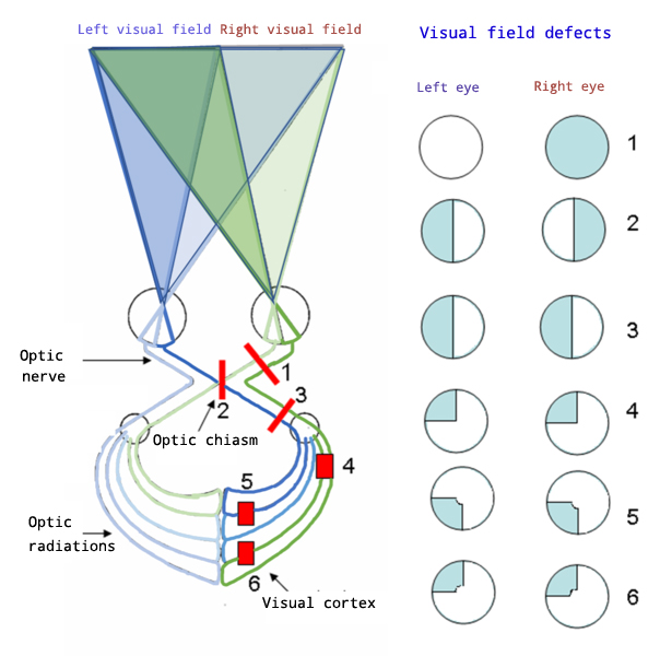
Image Source: Wikimedia Commons (2020). Hemianopsia, Licensed under a creative commons licence. Available for free at: https://commons.wikimedia.org/wiki/File:Hemianopsia_en.jpg (Accessed 13 May 2025).
References
Nussey, S. and Whitehead, S.A. (2002) “Chapter 7 The pituitary gland,” in Endocrinology: An integrated approach. Oxford England: BIOS Scientific Publishers Ltd.
Britannica, The Editors of Encyclopaedia. “syndrome of inappropriate antidiuretic hormone”. Encyclopedia Britannica, 20 Mar. 2023, https://www.britannica.com/science/syndrome-of-inappropriate-antidiuretic-hormone. Accessed 4 April 2023.
Santos Fontanez SE, De Jesus O. Neurohypophysis. [Updated 2023 Feb 12]. In: StatPearls [Internet].
Ross et al. Hypothalamic-pituitary-adrenal axis diagram. Available at: https://commons.wikimedia.org/w/index.php?curid=44663207 (Accessed: September 9, 2023)
Treasure Island (FL): StatPearls Publishing; 2023 Jan-. Available from: https://www.ncbi.nlm.nih.gov/books/NBK560733/
Adelman, D. et al. (2013) “Acromegaly: The disease, its impact on patients, and managing the burden of long-term treatment,” International Journal of General Medicine, p. 31. Available at: https://doi.org/10.2147/ijgm.s38594.
Acromegaly – Symptoms, diagnosis and treatment. BMJ Best Practice. Available at: https://bestpractice.bmj.com/topics/en-gb/522 (Accessed: April 4, 2023).
Chanson and Salenave. Acromegaly facial features. Available at: https://commons.wikimedia.org/w/index.php?curid=15072125 (Accessed: September 9, 2023)
Laurence Katznelson, Edward R. Laws, Jr, Shlomo Melmed, Mark E. Molitch, Mohammad Hassan Murad, Andrea Utz, John A. H. Wass, Acromegaly: An Endocrine Society Clinical Practice Guideline, The Journal of Clinical Endocrinology & Metabolism, Volume 99, Issue 11, 1 November 2014, Pages 3933–3951, https://doi.org/10.1210/jc.2014-2700
Growth Hormone Deficiency in Adults. UpToDate. Available at: https://www.uptodate.com/contents/growth-hormone-deficiency-in-adults (Accessed: April 4, 2023).
Cushings Disease. UpToDate. Available at: https://www.uptodate.com/contents/epidemiology-and-clinical-manifestations-of-cushings-syndrome?search=cushing+syndrome&source=search_result&selectedTitle=3~150&usage_type=default&display_rank=3 (Accessed: April 4, 2023).
Klausur. Pat. Mit Cushing-Syndrom. Avaliable at https://commons.wikimedia.org/w/index.php?curid=90182292 (Accessed: September 9, 2023)
Cushings Disease. BMJ Best Practice. Available at: https://bestpractice.bmj.com/topics/en-gb/522 (Accessed: April 4, 2023).
Prolactinoma. BMJ Best Practice. Available at: https://bestpractice.bmj.com/topics/en-gb/363/treatment-algorithm#! (Accessed: April 4, 2023).
Hyperprolactinemia. UpToDate. Available at : https://www.uptodate.com/contents/management-of-hyperprolactinemia (Accessed: April 4, 2023).
Craniopharyingioma. BMJ Best Practice. Available at: https://bestpractice.bmj.com/topics/en-gb/732 (Accessed: April 4, 2023).
Craniopharyingioma. UpToDate. Available at : https://www.uptodate.com/contents/craniopharyngioma (Accessed: April 4, 2023).
Hellerhoff. Craniopharyngioma. Available at https://commons.wikimedia.org/w/index.php?curid=15959020 (Accessed: September 9, 2023)
Diabetes insipidus. BMJ Best Practice. Available at: https://bestpractice.bmj.com/topics/en-gb/288 (Accessed: April 4, 2023).
Central Diabetes Insipidus. UpToDate. Available at : https://www.uptodate.com/contents/arginine-vasopressin-deficiency-central-diabetes-insipidus-treatment (Accessed: April 4, 2023).
Nephrogenic Diabetes Insipidus. UpToDate. Available at: https://www.uptodate.com/contents/arginine-vasopressin-resistance-nephrogenic-diabetes-insipidus-clinical-manifestations-and-causes (Accessed: April 4, 2023).
Growth Hormone Deficiency. . UpToDate. Available at: https://www.uptodate.com/contents/growth-hormone-deficiency-in-adults (Accessed: April 4, 2023).
Panhypopituitarism. BMJ Best Practice. Available at: https://pmj.bmj.com/content/82/966/259 (Accessed: April 4, 2023).
Panhypopituitarism. UpToDate. Available at: https://www.uptodate.com/contents/treatment-of-hypopituitarism (Accessed: April 4, 2023).
Non-Functioning Pituitary Adenomas. BMJ Best Practice. Available at: https://bestpractice.bmj.com/topics/en-gb/1030 (Accessed: April 4, 2023).
As eo. Hemianopsia. Available at: https://commons.wikimedia.org/wiki/File:Hemianopsia_en.jpg (Accessed: September 9, 2023)
Complete Guide to Clinical Genetics ST3 Application and Recruitment (2027)
Written by Medibuddy. Last updated: October 2026. Clinical Genetics is

Complete Guide to Clinical Radiology ST3 Application and Recruitment (2027)
Recruitment paused: NHS England is not running Clinical Radiology ST3+ recru

Complete Guide to Paediatric Surgery ST3 Application and Recruitment in United Kingdom (2026)
Written by Medibuddy. Last updated: October 2026. If you’re planning


Take your subscriptions with you
Our mobile app allows you to access your interview and exam question banks wherever you are.

