


 Close
Close
 27th May 2025
Primary adrenal insufficiency (Addison’s disease)
Background
In primary adrenal insufficiency (PAI) there is ablation of the adrenal cortex resulting in a deficiency of all steroid hormones. The hypothalamic-pituitary axis (HPA) aims to correct low cortisol levels through increased adrenocorticotropic hormone (ACTH) production, where α-melanocyte-stimulating-hormone is produced as a by-product. Therefore insidious PAI commonly presents with mucocutaneous hyperpigmentation, as well as fatigue, weakness, hypotension, anorexia, nausea and vomiting. It can also present acutely with hypovolaemic shock, acute abdominal pain, fever and vomiting.
Epidemiology
The prevalence of primary adrenal insufficiency is low at 40 cases per million in the UK. In developed countries, autoimmunity is the most common cause of primary adrenal insufficiency (up to 96% of cases), affecting women to a greater degree than men. There is also an association with other autoimmune endocrine disorders including Graves’ disease, type 1 diabetes mellitus and Hashimoto’s thyroiditis.
Aetiology
Anatomic destruction of the adrenal glands can result in acute or chronic PAI:
- Autoimmunity: antibodies formed against 21-hydroxylase.
- Infection: tuberculosis, P. aeruginosa and meningococcal septicaemia, histoplasmosis, HIV, cryptococcosis.
- Surgery: bilateral adrenalectomy e.g. for phaeochromocytoma.
- Haemorrhagic infarction: antiphospholipid syndrome, anticoagulation, Waterhouse-Friderichson syndrome.
- Trauma.
- Infiltrative disease: e.g. amyloidosis, haemochromatosis, sarcoidosis.
- Metastatic: e.g. lung, breast, kidney cancers.
Metabolic failure tends to be more insidious in onset:
- Genetic conditions: e.g. congenital adrenal hyperplasia (described below), adrenoleukodystrophy
- Drug-induced: inhibitors of cortisol production e.g. ketoconazole or etomidate, or inducers of cortisol metabolism e.g. phenytoin, rifampicin.
- ACTH or glucocorticoid resistance.
- Cytotoxic agents.
Diagnosis
Symptoms of PAI include:
- Fatigue
- Weakness
- Myalgia and arthralgia
- Weight loss
- Nausea, vomiting, abdominal pain
Signs of PAI include:
- Mucocutaneous hyperpigmentation – commonly on areas of friction e.g. palms, knuckles.
- Hypotension and postural hypotension.
- Less commonly vitiligo, premature greying, goitre (with Hashimoto’s thyroiditis).
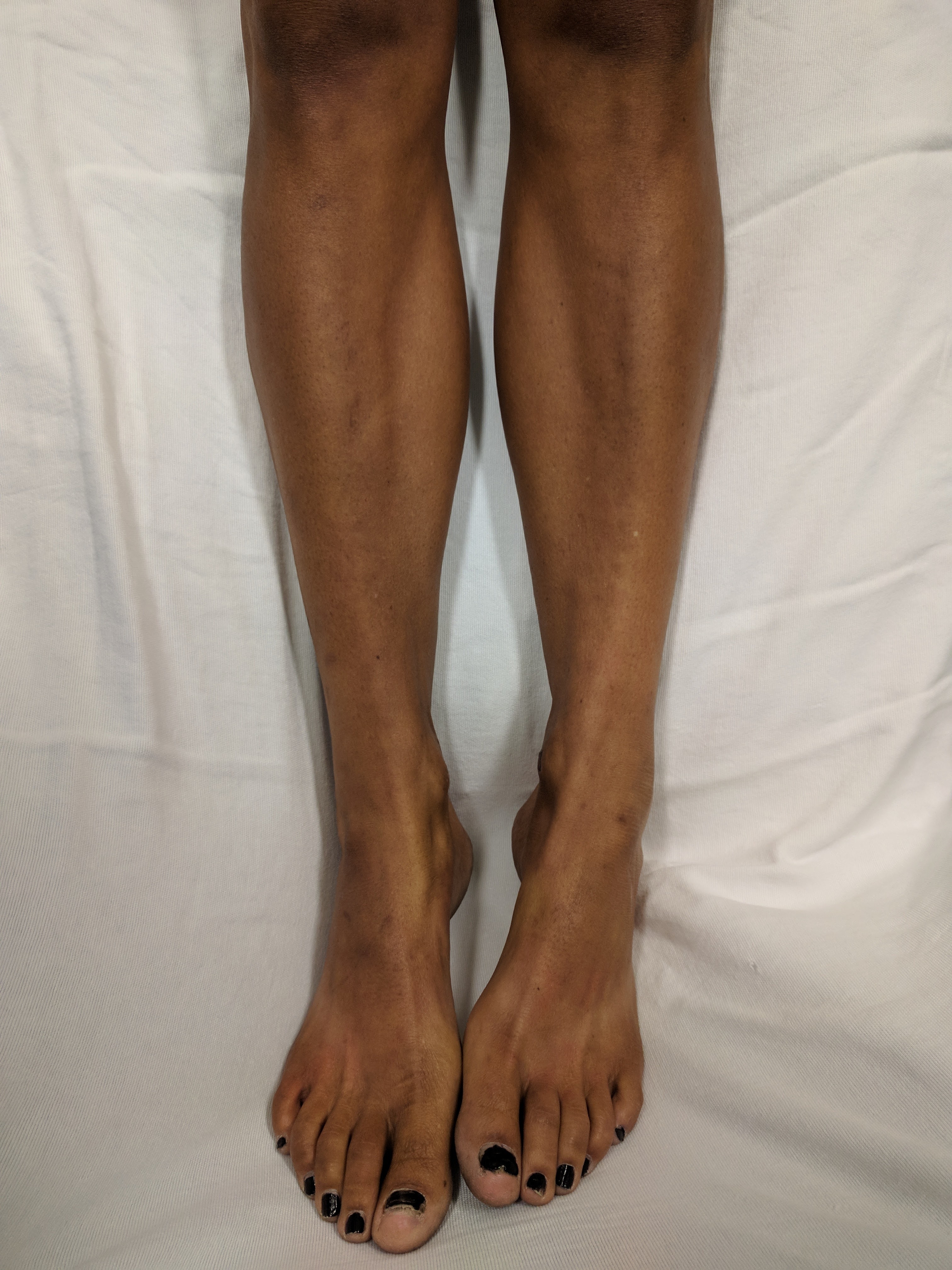
Image Source: Wikimedia Commons (2018) Addison’s Leg’s, Licensed under a creative commons licence. Available for free at: https://upload.wikimedia.org.wikipedia/commons/6/69/Addison%Legs.jpg (Accessed 13 May 2025).
The following laboratory findings are typical in PAI:
| Biochemical test | Findings in PAI |
|---|---|
| Sodium | ↓ in 90% of cases |
| Potassium | ↑ in 50% of cases |
| Calcium | ↑ in 10-20% of cases |
| FBC | Anaemia, mild eosinophilia and lymphocytosis |
| Glucose | ↓ often in children |
| LFTs | ↑ transaminases |
| Morning cortisol | ↓ levels <100 nmol/L warrant urgent admission, 100-500 nmol/L warrants endocrinology referral |
| ACTH | ↑ in primary adrenal insufficiency but ↓ in secondary |
| RAAS | ↑ renin and ↓ aldosterone in PAI |
Table 1: laboratory findings in primary adrenal insufficiency.
Ancillary investigations include an ACTH stimulation (synacthen) test where synthetic ACTH is administered and should result in a rise in cortisol for a physiologic response. However, in primary adrenal insufficiency this does not occur i.e. the cortisol will remain low when re-checked following administration of synacthen, as the adrenal glands do not respond to stimulation.
To investigate the underlying aetiology, the following investigations should be considered:
- Adrenal autoantibody testing.
- Chest x-ray to investigate lung neoplasm or tuberculosis.
- Tuberculosis skin prick or ELISA testing.
- Abdominal x-ray which may show adrenal calcification.
- CT or MRI of adrenal glands.
- MRI of hypothalamus and pituitary if suspicion of a central cause.
Management
In acute adrenal crisis, the mainstay of treatment includes:
- IV hydrocortisone, typically given as 100 mg STAT followed by an infusion of 200 mg/24 hours. N.B. if co-existing hypothyroidism, thyroid hormones must not be replaced prior to glucocorticoids, as this can precipitate adrenal crisis.
- Fluid resuscitation with monitoring of blood pressure, fluid status and electrolytes.
- Correction of hypoglycaemia.
- Treatment of the underlying cause once stable.
- Education regarding symptoms and risk factors for adrenal crisis, issue of a steroid card or medical alert bracelet.
In intercurrent illness, those with PAI will require corticosteroid supplementation. In circumstances of severe stress or high risk of crisis (e.g. acute gastroenteritis, surgery, critical illness), IV hydrocortisone as described above is recommended. However, for minor stressors (e.g. febrile illness, minor procedures requiring fasting), doubling of the patient’s maintenance dose of corticosteroid is recommended.
For the management of PAI in stable patients, oral glucocorticoid (e.g. hydrocortisone or prednisolone) and mineralocorticoid (e.g. fludrocortisone) replacement therapy is given. This is titrated according to clinical symptoms until a maintenance dose is found. Mineralocorticoid dosing will be adjusted based on symptoms such as postural hypotension and salt-craving.
Complications
Long-term PAI can result in acute adrenal crisis with hypovolaemic shock, hypoglycaemia, and electrolyte abnormalities as described above. However it is preventable with good education and dose adjustment during intercurrent illness.
Other complications are related to excessive glucocorticoid replacement, resulting in secondary Cushing’s syndrome, osteoporosis and hypertension. Cushing’s syndrome manifests as weight gain, moon facies and peripheral muscle wasting.
Complete Guide to Clinical Genetics ST3 Application and Recruitment (2027)
Written by Medibuddy. Last updated: October 2026. Clinical Genetics is

Complete Guide to Clinical Radiology ST3 Application and Recruitment (2027)
Recruitment paused: NHS England is not running Clinical Radiology ST3+ recru

Complete Guide to Paediatric Surgery ST3 Application and Recruitment in United Kingdom (2026)
Written by Medibuddy. Last updated: October 2026. If you’re planning


Take your subscriptions with you
Our mobile app allows you to access your interview and exam question banks wherever you are.

