


 Close
Close
 28th May 2025
Type 2 Diabetes Mellitus
Background
Type 2 diabetes mellitus (T2DM) is a progressive metabolic disease characterised by chronic hyperglycaemia caused by insulin resistance and defective insulin secretion. It is an extremely common disease, with an estimated global prevalence of 8.3% in 2019, with the number of cases drastically rising since the 1980s. T2DM can lead to severe macro and microvascular complications if left untreated, which can majorly affect a patient’s length and quality of life, as well as placing a significant burden on the resources of health services. Options for medical management have blossomed since the 1950s with several oral agents developed. Increasingly, however, there has been a shift in emphasis towards prevention of the disease through lifestyle modification.
Epidemiology
From 1980 to 2019 the worldwide prevalence of diabetes mellitus has almost doubled, growing from 4.3% to 8.3% (roughly 7% in the UK). The incidence of newly diagnosed diabetes has plateaued in most countries since the mid-2000s, likely due to increasing patient education and public health strategies. The most significant factors driving the rise in prevalence since this time are therefore the ageing of the global population and better outcomes for patients. These figures do not differentiate between the different types of diabetes, but it is estimated that 90% of cases are T2DM. Within the UK alone roughly one million patients are living with undiagnosed T2DM.
Several risk factors for T2DM have been identified, including:
- Increasing age
- Male sex – ratio of roughly 1.5:1.
- Obesity and inactivity – obesity is the most significant risk factor in the development of T2DM.
- Asian, Black and Afro-Caribbean ethnicity – carries a 2-4x increased risk when compared to Caucasian patients.
- Family history – there is a 2-6x increase in risk for patients with a family history of the disease. If both parents have the condition, then the overall risk for an individual stands at 85%.
- Gestational diabetes – mothers who develop diabetes during pregnancy are 7x more likely to develop T2DM later in life.
- Diet – regularly consuming sugar-rich food and drinks, with low fibre and high glycaemic index, such as sugary snacks and drinks, potatoes, white rice and white bread will significantly increase the risk of developing T2DM.
- Medications – including corticosteroids, thiazide diuretics, beta-blockers, antipsychotics and statins.
- Alcohol – moderate alcohol consumption is protective for T2DM but heavy use increases risk, both due to the effects of the alcohol itself and the increase in consumption of carbohydrates. Drinking heavily also leads to greater variability in blood sugar levels and can lead to hypoglycaemic episodes.
- Smoking – 30-40% increased risk and also independently increases the risk of cardiovascular complications of the disease.
- Metabolic syndrome – describes a pattern of common biochemical and physiological abnormalities that increase a patient’s risk of both T2DM and cardiovascular disease. The syndrome encompasses central obesity, hyperlipidaemia, hypertension, non-alcoholic fatty liver disease, atherosclerosis, insulin resistance and impaired glucose tolerance. It is thought to be a reflection of the modern shift to a more sedentary lifestyle and diet of excess.
- Polycystic ovary disease – known to cause progressive insulin resistance, which can lead to T2DM.
- Psychiatric disease – schizophrenia, bipolar disorder and depression are all associated with an increased risk.
- Sleep disorders – sleep apnoea and restless leg syndrome
Aetiology
The initial pathogenesis of T2DM is characterised by the desensitisation of the liver and skeletal muscle to the action of insulin, aggravated mainly by obesity, increasing age and genetic predisposition. The body’s normal homeostatic response involves increasing the secretion of insulin from beta-cells at the islets of Langerhans in the Pancreas. However, when this relationship decompensates, the beta-cells are unable to secrete sufficient insulin to bring blood glucose under control. Hyperglycaemia has an inhibitory effect on both beta-cell insulin secretion and cell turnover, leading to a reduction in cell function and mass. Therefore, as the disease progresses there is less capacity for insulin production as well as increasing desensitisation of peripheral tissues. Several other processes have been identified that contribute to the development of T2DM, including increased lipolysis, hyperglucagonemia, increased renal glucose reabsorption and resistance to the action of incretin.
Diagnosis
T2DM is often asymptomatic at the time of diagnosis, however, signs and symptoms may include:
- Polyuria
- Polydipsia
- Blurred vision
- Unexplained weight loss
- Recurrent infections (e.g. UTIs, skin infections, balanitis).
- Fatigue
- Acanthosis nigricans (dark skin discolouration present in the skin folds, for example, the axillae or groin) is caused by insulin resistance.
- Hyperosmolar hyperglycaemic syndrome (rare presentation).
Screening is not routinely offered for T2DM but patients and testing is up to the discretion of clinicians. The diagnostic criteria are identical to type 1 diabetes (T1DM) and there is the same choice of three blood tests that may be used. A single positive result is sufficient to diagnose a symptomatic patient with diabetes, but, if a patient is asymptomatic, they require two separate positive results taken on different occasions. The diagnostic criteria is used:
- Fasting glucose ≥ 7 mmol/L
- Random (non-fasting) glucose ≥ 11.1 mmol/L
- HbA1c ≥ 48mmol/mol
HbA1c is a measure of the proportion of glycated haemoglobin of the bloodstream. An HbA1c < 48mmol/mol cannot be used to exclude a diagnosis of diabetes as it gives an aggregate of the preceding three months. Post-splenectomy or in functional hyposplenism (for example in coeliac disease) red blood cells have an increased lifespan and therefore have longer to become glycated, giving an inappropriately high HbA1c. Causes of decreased red blood cell life-span will give a falsely negative result, examples include:
- Haemoglobinopathies (e.g. thalassaemia, sickle cell disease)
- Severe anaemia of any cause
- HIV-positive patients
- Recent blood transfusion
- Gestational diabetes
- Childhood diabetes
Patients who have a fasting glucose of 6.1-7 mmol/L have a diagnosis of impaired fasting glucose (IFG) and should be offered an oral glucose tolerance test. A result of 7.8-11.1 mmol/L gives a diagnosis of impaired glucose tolerance (IGT). Both of these diagnoses fall under the colloquial umbrella term ‘prediabetes’ and place an individual at a much greater risk of developing T2DM.
At the time of diagnosis, patients should be assessed for signs of heart failure, atherosclerotic cardiovascular disease (using QRISK2) and chronic kidney disease as this will affect the potential management of their condition. They should be referred to their local eye screening service for assessment of diabetic retinopathy via a dilated eye examination. Their feet must be checked for signs of diabetic foot disease and if evidence is found then they should be referred to the local food protection service.
Management of Type 2 Diabetes Mellitus
Non-Pharmacological Management
Ensuring the required long-term engagement with the health service and compliance with treatment is integral to successfully managing T2DM. Therefore, there is perhaps a greater emphasis placed on the structured education of patients, family members and carers about the disease than many other medical conditions. NICE recommends that courses should be offered to patients at the time of diagnosis with
Upon initial diagnosis of T2DM, patients should be encouraged to adjust their lifestyle to reduce their sugar levels and avoid medication. Changes must be individualised, taking into account factors such as the risk of hypoglycaemia and comorbidities. Generalisable suggested dietary modifications include:
- Opting for high-fibre, low-glycaemic-index sources of carbohydrates, such as whole grains and vegetables, and avoiding low-fibre, high glycaemic-index sources such as white rice, white bread, potatoes, sugary snacks and drinks.
- Consuming low-fat dairy products where possible.
- Incorporating a portion of oily fish per week into their diet.
- Reducing their amount of saturated and trans-fatty acids.
- Striving to fit 5 portions of fruit or vegetables into their daily diet.
- Avoidance of products that are specifically marketed towards patients with diabetes.
Newly diagnosed patients should be encouraged to engage in regular physical exercise even if they have a normal body weight. Patients who are overweight should target 5-10% weight loss in the initial 6-month period. All patients should be counselled on the risks of smoking and offered help with smoking cessation. Abstinence from alcohol is not required but heavy use is not advisable and patients should be made aware of the erratic effect binge drinking can have on their blood sugar.
Patients living with T2DM with a BMI of over 35kg/m2 should be considered for bariatric surgery, lower than the usual recommended threshold of 40kg/m2 due to there being good evidence that it improves control of the disease. For patients from Asian, Middle Eastern, Black African or African-Caribbean family backgrounds a lower threshold of 32.5kg/m2 is used for referral for assessmen
Monitoring and Targets
HbA1c should be monitored closely in patients newly diagnosed with T2DM, approximately every 3-6 months until the level stabilises and then every 6 months thereafter. The HbA1c target depends on the treatment the patient is taking and whether they have a history of hypoglycaemia:
48 mmol/mol 🡪 Patients who control their blood sugars via lifestyle modifications with or without a single medication that does not cause hypoglycaemia.
53 mmol/mol 🡪 Patients whose treatment regime includes a medication that can cause hypoglycaemic episodes
53 mmol/mol 🡪 Patients already taking one medication but HbA1c has risen to 58 mmol/mol and therefore a second agent is being introduced.
In patients trying to control their sugar levels using lifestyle modifications and a single medication only, an HbA1c of 58 mmol/mol should trigger the introduction of further medication. At each stage where a new medication is started, education should be reinforced, and lifestyle modifications emphasised. HbA1c targets may be loosened on an individual basis in certain situations. These include:
- Where there is a significant risk to their health associated with hypoglycaemic episodes, for example in frailty.
- If the patient has a reduced life expectancy and is therefore unlikely to benefit from the long-term effects of controlling sugar levels.
- The patient has significant comorbidities, so tight control of sugars would not be beneficial.
Self-monitoring of capillary blood glucose is not routinely recommended for T2DM patients unless they are taking insulin or an oral agent that may lead to hypoglycaemic episodes (e.g. sulfonylureas), have a history of hypoglycaemia or are pregnant or trying to conceive. Continuous glucose monitoring, either intermittent scanned (isCGM) or real time (rtCGM) should be considered in T2DM patients injecting insulin multiple times per day and either have a risk of hypoglycaemia or impaired hypoglycaemia awareness. They are also used in patients with impaired cognitive ability who need to but are unable to self-monitor capillary blood glucose. Patients on CGM should still measure their capillary blood glucose to check the accuracy of the CGM and in case of device failure, but this can be done less frequently.
Pharmacological Management
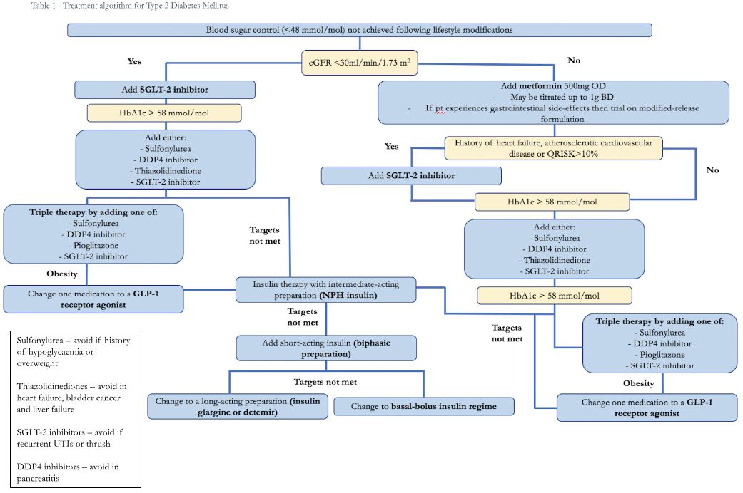
The order of introduction of medications for T2DM hinges on several factors and is not a simple stepwise algorithm. There are multiple therapeutic options that MRCP candidates must know in detail, including their mechanism of action, indication and potential side effects.
Metformin and SGLT-2 inhibitors are used as first-line agents, depending on the clinical scenario. Other medications including DPP-4 inhibitors, sulfonylureas, thiazolidinediones and GLP-1 mimetics may be used if further intervention is required. If glycaemic control is still not achieved, then a third agent can be added, or the patient can be considered for insulin. If three agents are not effective, then the patient should be started on insulin unless there are any contraindications.
Patients who have symptomatic hyperglycaemia (polyuria, polydipsia, headaches, fatigue, thrush or recurrent urinary or skin infections etc) should be considered for rescue treatment with insulin or a sulfonylurea (e.g. gliclazide) until blood glucose is controlled, at which point treatment should be reviewed
Metformin
Metformin is a biguanide medication that has been used as the mainstay of medical treatment for T2DM for a number of years. The standard-release formulation is used first line in patients without CKD, with dosing starting at 500mg OD with the scope to titrate up to 1g BD before considering adding another medication.
Mechanism of action – metformin has multiple glucose-lowering effects:
- Inhibition of gluconeogenesis in the liver through activation of AMP-activated protein kinase (AMPK).
- Opposition to the action of glucagon by inhibiting mitochondrial complex I.
- Increasing systemic sensitivity to insulin through activation of AMPK, increasing the uptake of glucose into the hepatic and skeletal muscle cells.
- Decreasing the amount of carbohydrates absorbed in the small intestine.
Adverse reactions:
- Gastrointestinal upset – this is the most common side-effect of metformin, usually diarrhoea. Modified-release metformin should be trialled before stopping the drug entirely.
- Lactic acidosis – occurs rarely and is normally only seen in patients with significant renal impairment
- Vitamin B12 deficiency – this is due to interaction with intrinsic factor-dependent B12 absorption in the terminal ileum.
Contraindications:
- Renal impairment – the drug is excreted by the kidneys unchanged and the build-up of the drug in the bloodstream increases the risk of lactic acidosis. Metformin should be reviewed and potentially reduced in dose when eGFR falls below 45ml/min and stopped when it falls under 30ml/min. Caution should be taken when the drug is prescribed alongside nephrotoxic drugs when a patient requires x-ray contrast media or in cases of significant dehydration (usually it is advised patients stop metformin 24 hours before the procedure and restart 48 hours after).
- Metabolic acidosis – including lactic acidosis and diabetic ketoacidosis. Caution should also be used in patients with risk factors for developing lactic acidosis such as following periods of tissue hypoxia such as sepsis or following a myocardial infarction.
SGLT-2 Inhibitors (-flozins)
Members of this family of medications, including empagliflozin and dapagliflozin, have been shown to improve outcomes for patients with heart failure and atherosclerotic cardiovascular disease. They are therefore used, alongside metformin, as first-line treatment in these patients, as well as those deemed at high risk of developing cardiovascular disease (QRISK2>10%). Metformin should be started first and if tolerated then an SGLT-2 inhibitor should be added. If metformin is not tolerated, then the SGLT-2 inhibitor can be used as a monotherapy. SGLT-2 inhibitors are also used for patients with CKD, more information can be found in the complications section of this chapter.
Mechanism of action – SGLT-2 inhibitors encourage the renal excretion of glucose via modulation of sodium-glucose co-transporters in the proximal tubule of the nephrons.
Adverse reactions:
- Infection of genitalia and urinary tract – increasing the urinary concentration of glucose makes a more hospitable environment for microorganism growth. Patients who struggle to maintain good personal hygiene are especially at risk. This increases the risk of UTIs, balanitis, prostatitis and Fournier’s gangrene. SGLT-2 inhibitors should be stopped during the treatment of these conditions and reviewed before recommencing.
- Diabetic ketoacidosis – DKA is a very rare but serious reaction that can be difficult to diagnose as these patients are often euglycaemic. The medication should be permanently stopped if this happens.
- Hypoglycaemia – usually occurs when prescribed alongside insulin or a sulfonylurea.
- Hypotension – fluid imbalance leading to hypotension is an uncommon but associated side-effect.
Contraindications – the most important and intuitive is ketoacidosis. They should be used with caution in the elderly and frail due to the increased risk of hypotension and potential falls.
Sulfonylureas
Whilst first-generation sulfonylureas have largely fallen out of favour, second-generation medications are seen commonly, examples include gliclazide and glimepiride. They are often used as a second-line therapy in conjunction with metformin as they have very different mechanisms of action for achieving glycaemic control. However, they are also used as monotherapy in some patients who cannot tolerate metformin.
Mechanism of action – Sulfonylureas antagonise ATP-sensitive potassium channels on the cell membranes of beta-cells at the islets of Langerhans. This inhibits potassium efflux and depolarises the cell membrane causing calcium channels to open and an influx of calcium ions. The increased intracellular calcium stimulates insulin secretion.
Adverse reactions:
- Hypoglycaemia – the mechanism of action of sulfonylureas is unaffected by blood glucose levels so they have the potential to cause dangerous episodes of hypoglycaemia.
- Weight gain – an increase in the circulatory level of insulin means that more glucose will be pushed into cells and turned into fat. Therefore, this class of drugs should be avoided in obese patients.
- GI upset – as with many drugs, nausea, diarrhoea and abdominal pain may be seen whilst taking sulfonylureas.
- Bone marrow suppression – agranulocytosis and pancytopenia are seen rarely.
- Syndrome of inappropriate ADH secretion – SIADH can be caused by first-generation long-acting preparations such as chlorpropamide.
Contraindications – sulfonylureas are principally metabolised by the liver and therefore they shouldn’t be used in severe liver impairment due to increased risk of hypoglycaemia. The same is true for severe renal impairment, they can be used in mild to moderate renal impairment. They should also be avoided in patients with DKA. They are usually avoided in obesity.
DPP-4 inhibitors (-gliptins)
Medications such as sitagliptin and saxagliptin represent a relatively new class of hypoglycaemic agents: DPP-4 inhibitors, the first of which was licensed in 2006. They are generally used as a second-line therapy alongside another medication, usually metformin, but are sometimes used as monotherapy.
Mechanism of action – the hormone glucose-like peptide-1 (GLP-1) is secreted by L-cells in the distal small intestine in response to rising blood sugar levels, stimulating insulin release. The rise in GLP-1 is short-lived as the hormone is quickly broken down by the enzyme DPP-4, thereby producing a spike in insulin after meals. DPP-4 inhibitors delay the degradation of GLP-1, allowing further insulin release and a greater hypoglycaemic effect.
Adverse reactions – DPP-4 inhibitors are usually well tolerated and have few side-effects but some are observed:
- General symptoms – such as headache, dizziness, nausea and constipation are most commonly seen.
- Increased risk of infections – particularly upper respiratory tract and skin infections.
- Pancreatitis – DPP-4 inhibitors are a rare cause of pancreatitis.
- Severe allergic reactions – such as angioedema, anaphylaxis and Steven-Johnson syndrome.
Contraindications – The dose of DPP-4 inhibitor should be reduced in patients with significant renal impairment and they should not be used in patients with a history of pancreatitis.
Thiazolidinediones (-glitazones)
The two currently licensed thiazolidinediones, pioglitazone and rosiglitazone, are another second-line treatment option. They are usually combined with either metformin or a sulfonylurea but can rarely be used as monotherapy.
Mechanism of action – thiazolidinediones regulate gene expression through binding to peroxisome proliferator-activated receptor-gamma (PPAR-gamma), which regulates transcription of genes involved in glucose homeostasis. This leads to increased production of adiponectin and expression of the gene GLUT-4, which increases insulin sensitivity.
Adverse reactions – thiazolidinediones are often avoided in preference for other drug classes because they are associated with several undesirable side effects:
- Heart failure – patients have an increased risk of heart failure and peripheral oedema whilst taking thiazolidinediones for a prolonged period, especially rosiglitazone.
- Weight gain – activation of PPAR-gamma receptors stimulates glucose uptake into adipocytes (where the majority of these receptors are found). Thiazolidinediones also increase appetite.
- Osteoporosis – and the associated risk of fragility fractures.
- Bladder cancer – especially seen in patients taking rosiglitazone.
- Hepatotoxicity – LFTs should be taken at baseline and during treatment as there is a significant risk of acute liver injury.
- Macular oedema – concomitant use of insulin increases this risk.
Contraindications – these generally match the above adverse reactions. Thiazolidinediones should be avoided in patients with a history of heart failure, bladder cancer, obesity, osteoporosis and hepatic impairment.
GLP-1 receptor agonists
There are two categories of GLP-1 receptor agonists, those based on a human GLP-1 structure (for example liraglutide, dulaglutide) and those based on an exendin-4 structure (including exenatide). If patients do not achieve glycaemic control on triple therapy with metformin and two other drugs then one medication can be changed to a GLP-1 receptor agonist if the patient has a history of obesity or cardiovascular disease. Treatment is only continued if they have a satisfactory response after 6 months – a reduction of at least 11 mmol/mol in HbA1c and weight loss of at least 3% of their body weight.
Increasingly, they are being used as a treatment for obesity without type 2 diabetes as they can aid weight loss through appetite suppression and better blood glucose control. These medications are given via subcutaneous injection.
Mechanism of action – GLP-1 receptor agonists mimic the action of the hormone GLP-1 (leading to them also being called incretin mimetics), which spikes after meals and stimulates insulin secretion.
Adverse reactions:
- GI upset – including nausea, dyspepsia, diarrhoea and abdominal pain. The early satiety associated with GLP-1 receptor agonists can develop into nausea if patients continue to eat.
- Immunogenicity – patients can develop antibodies against the medication leading to injection site reactions or angioedema.
- General symptoms – mild tachycardia, headaches and dizziness may occur.
Contraindications – these medications aren’t used in patients of normal weight and should not be given to pregnant patients. They should be avoided where there is hepatic impairment or renal impairment (with a creatine clearance of <30ml/minute)
Insulin
Generally, insulin is considered if patients are not controlled on three oral medications. Patients should be offered a neutral protamine Hagedorn insulin (an intermediate-release preparation) to be administered either once or twice per day, according to an individual’s need. A short-acting insulin should be added to the regime if the HbA1c is over 75 mmol/mol and can be given as a separate injection or in a combined biphasic preparation. A long-acting insulin (detemir or glargine) should be considered in patients who do not meet their HbA1c targets, suffer from significant hypoglycaemia or would benefit from a once-daily injection (for example if they need a carer to administer their insulin). A basal-bolus regime may be used in patients who don’t achieve their targets using a biphasic preparation and are able to administer several injections per day and monitor their own blood glucose.
Metformin should always be continued when patients are started on insulin but other medications should be reviewed (especially those with a capacity to cause hypoglycaemia). All patients should be offered education on insulin administration, blood glucose monitoring, hypoglycaemia awareness and DVLA guidance.
Complications of Type 2 Diabetes
Hypoglycaemia
In T2DM, hypoglycaemia is usually seen as a result of taking either insulin or sulfonylureas, although it can occur without these medications. It is defined as a blood glucose level <3.5mmol/L and can be classified by severity according to the symptoms experienced:
- Mild – hunger, anxiety, palpitations, sweating, perioral paraesthesia.
- Moderate – headache, impaired cognition, reduced level of awareness, drowsiness and features of mild hypoglycaemia.
- Severe – seizures, loss of consciousness and coma.
Some patients will only experience symptoms at a very low blood glucose level, meaning they have impaired hypoglycaemia awareness (IHA). This is especially observed in patients who have had repeated episodes of hypoglycaemia and patients taking certain medications, including beta-adrenergic antagonists (e.g., bisoprolol), beta-adrenergic agonists (e.g. terbutaline), serotonin receptor reuptake inhibitors (e.g. serotonin) and methylxanthines (e.g. caffeine). Patients and, where appropriate, relatives and carers should be educated about the symptoms of hypoglycaemia and advised on emergency management. This is especially important in patients with known IHA as they are more liable to progress to severe hypoglycaemia and will therefore be unable to administer emergency treatment.
The management of hypoglycaemia involves increasing the circulating glucose level as quickly as possible and there are several ways this can be achieved:
- Oral fast-acting carbohydrate – Conscious patients, who can swallow, with blood glucose <4mmol/L with or without symptoms of hypoglycaemia should be given a fast-acting oral carbohydrate such as 40% glucose gel, glucose tablets or glucose liquid. In the community, if these agents are unavailable, then any accessible sugary foods should be used, such as orange juice or candies. The treatment may be repeated up to 3 times at 15-minute intervals if there is not a satisfactory response.
- IM Glucagon – If there is no response to oral agents and the patient doesn’t have intravenous access then they should be given glucagon via intramuscular injection. Glucagon should also be given to unconscious patients and can be administered by appropriately trained family members. Glucagon doesn’t increase the total amount of glucose in the body, rather mobilising stored glucose in the liver, and therefore will be ineffective in patients that have depleted glucose stores. It should therefore be avoided in patients who have undergone a prolonged fast, have adrenal insufficiency or have alcohol-induced hypoglycaemia. It is also less effective in patients who take a sulfonylurea.
- IV Glucose infusion – Patients who have not responded to oral agents and have intravenous access should be started on either a 10% or 20% glucose infusion. 50% glucose should be avoided as it is a hypertonic solution. Patients who are unconscious and haven’t responded to or aren’t suitable for IM glucagon should also be treated with IV glucose. Alcoholic patients should be co-administered IV thiamine to reduce the risk of Wernicke’s encephalopathy (thiamine plays an important role in the intracellular metabolism of glucose).
Once blood glucose >4mmol/L the patient should be given a source of long-acting carbohydrate (such as a slice of toast, 2 biscuits or a glass of milk) if able to eat. Patients who are NBM should be started on a 10% insulin infusion. In T2DM, since most hypoglycemia is seen in response to a sulfonylurea or insulin, the hypoglycaemia can persist for up to 36 hours so blood glucose should be monitored closely.
Diabetic kidney disease (DKD)
Chronic hyperglycemia causes vascular calcification and endothelial damage, contributing to many of the complications seen in T2DM. In DKD, microvascular damage to the nephron arterioles and overstimulation of the local renin-angiotensin system, causing efferent arteriole constriction, leads to intraglomerular hypertension and glomerular hyperfiltration, manifesting as microalbuminuria. The hyperglycaemic environment also triggers intracellular pathways, including through activation of protein kinase C, causing an increase in the production of advanced glycation end products, which lead to oxidative stress and cellular damage. DKD affects all renal cell types, resulting in thickening of the glomerular and tubular basement membranes, mesangial expansion through both proliferation and hypertrophy, tubular hypertrophy and afferent arteriole thickening with endothelial effacement. Clinically, it presents as gradually deteriorating renal function, progressing towards end-stage renal failure.
Unlike in T1DM, where DKD occurs after a long duration of the disease (usually >10 years), DKD may be present on diagnosis with T2DM as it may be undiagnosed for many years. DKD is defined by the presence of albuminuria and deterioration in the estimated glomerular filtration rate (eGFR) without another cause for renal impairment. Not all diabetics will develop DKD, the most notable risk factors are:
- Chronically poor glycaemic control
- Hypertension
- Dyslipidemia
- Family history
- Smoking
- Increasing age
- Race – Afro-Caribbean, Mexican-American and Pima Indian are at an increased risk
DKD usually remains symptom-less until severe, when patients may experience peripheral oedema, anaemia, fatigue, haematuria and progression to end-stage renal failure. Patients are screened on diagnosis using:
- Urinary albumin-creatinine ratio (ACR) – normal ACR is <3mg/mmol. Patients with DKD are categorised into microalbuminuria with an ACR of 3-30mg/g, or macroalbuminuria (overt nephropathy) with an ACR <30mg/mmol. The result should be repeatable on 2 separate occasions.
- eGFR – DKD patients will see a gradual deterioration in eGFR as seen in chronic kidney disease, it can be graded according to the regular CKD values:
Table 2: Staging of chronic kidney disease
| Stage | eGFR (ml/min/1.73m2) | Renal function |
|---|---|---|
| G1 | >90 | Normal |
| G2 | 60-89 | Mild impairment |
| G3 | 30-59 | Moderate impairment |
| G4 | 15-29 | Severe impairment |
| G5 | <15 | Kidney failure |
Adapted from table in NICE CKD treatment summary: https://www.nice.org.uk/guidance/ng203/chapter/Recommendations#classification-of-ckd-in-adults
Definitive diagnosis can be made by renal biopsy but this is not routinely offered to patients and they are normally diagnosed using ACR and clinical assessment to rule out other causes of renal disease. When a biopsy is performed, characteristic changes include mesangial expansion, thickening of the basement membrane and nodular glomerulosclerosis.
Patients diagnosed with DKD (ACR >3mg/mmol) should be offered either an angiotensin-converting enzyme inhibitor (ACEi) (such as ramipril) or an angiotensin receptor blocker (ARB) (such as candesartan), titrated up to the highest dose the patient can tolerate. An SGLT-2 inhibitor should be added when ACR>30mg/mmol and may be considered earlier, they are effective in slowing the progression of DKD. Control of dyslipidaemia, good glycaemic control and smoking cessation are all important in preventing the progression of DKD.
Diabetic eye disease
Patients diagnosed with T2DM should be immediately referred to their local eye screening service. Diabetic retinopathy presents as gradually decreasing visual acuity but in cases where vitreous haemorrhage occurs, patients will experience a sudden partial or complete loss of vision. Several complications of diabetic retinopathy may occur, which are discussed below.
Diabetic retinopathy is caused by progressive microvascular damage in the eye due to chronic hyperglycaemia. Several observable changes are seen in fundoscopy:
- Arteriole microaneurysms – The earliest visible manifestation of diabetic retinopathy, appearing as small red dots on the retina.
- Blot haemorrhages – Weakened capillary walls are more liable to rupture. Haemorrhages may be indistinguishable from microaneurysms by fundoscopy but sometimes have more irregular borders. Fundus fluoresceine angiogram can be used to distinguish between haemorrhages and microaneurysms.
- Hard exudates – It is theorised that increased capillary permeability allows leakage of lipoproteins through the vessel wall to form hard exudates, seen as yellow patches on fundoscopy.
- Cotton wool spots – Small arteriole infarcts result in patches of retinal non-perfusion, seen as white/yellow patches on fundoscopy.
- Intraretinal microvascular abnormalities – communications between arterioles and venules, bypassing capillary beds.
- Venous beading – intermittent dilatations in the retinal venules producing an appearance of beads on a string.
- Neovascularisation – chronic retinal hypoperfusion simulates the production of vascular endothelial growth factor A, which over time will lead to the formation of vessels to supply the retina.
- Vitreous haemorrhage – The new vessels produced are more fragile and liable to larger haemorrhage into the vitreous cavity.
Diabetic retinopathy is categorised by the International Clinical Diabetic Retinopathy Disease Severity Scale:
Table 3: Staging of diabetic retinopathy
| Mild non-proliferative diabetic retinopathy (NPDR) |
|
| Moderate NPDR |
|
| Severe NPDR | Any of:
|
| Proliferative diabetic retinopathy |
|
Adapted from StatPearls: https://www.ncbi.nlm.nih.gov/books/NBK560805/
Patients with diabetic retinopathy may develop several other ophthalmological pathologies:
- Diabetic macular oedema (DMO) – Increased capillary permeability can lead to the accumulation of exudative fluid in the macula, leading to deterioration in central vision. DMO can lead to blindness.
- Posterior vitreous detachment – The detachment of the vitreous membrane from the retina causes the patient reduced visual acuity, floaters and flashers.
- Retinal detachment – Producing a dark shadow to fall over the patient’s vision, which they often describe as curtain-like.
- Amaurosis fugax – Describes a sudden but transient monocular loss of vision due to retinal ischaemia. Diabetic retinopathy increases the risk of retinal artery and vein occlusion, as well as atherosclerosis and dissection.
- Cataracts – Diabetic patients are at a much greater risk of developing cataracts, where the lens becomes thickened and opaque leading to reduced visual acuity and clouding of vision.
Diabetics should be encouraged to attend screening annually and should be referred urgently if they experience a sudden visual loss or neovascularisation or vitreous haemorrhage is seen on fundoscopy. Management of NPDR involves good glycaemic control and modulation of risk factors to prevent progression. Patients with severe NPDR should be considered for anti-VEGF therapy to prevent progression to proliferative retinopathy. Pan-retinal laser photocoagulation can be used once neovascularisation occurs to destroy the vessels before they bleed. Surgery may be offered to relieve intraocular pressure caused by a large vitreous haemorrhage, as well as for cataracts and retinal/vitreous detachment.
Diabetic neuropathy
Damage to the vessels that supply the nerves in the body, through the same mechanism outlined above, leads to diabetic neuropathy affecting both the somatic and autonomic systems. Damage to sensory nerves can cause problems in the feet, making patients more likely to develop ulcers or Charcot arthropathy, discussed in more detail in the diabetic foot disease section. Patients may experience painful neuropathy, typically in the extremities, which should be managed in the same way as neuropathic pain in non-diabetics. They should be offered one of the following: amitriptyline, gabapentin, pregabalin or duloxetine. If one agent is not effective even when titrated to a maximum dose then it should be substituted for an alternative. Patients with severe pain that limits their daily activities (such as sleep) or that causes a deterioration in a comorbidity should be referred to a specialist pain team.
Autonomic neuropathy can cause a variety of symptoms, which can be managed with specific interventions. Other causes of these symptoms must be ruled out before a diagnosis of diabetic autonomic neuropathy can be made:
- Cardiovascular autonomic dysfunction (CAN) – Causing resting tachycardia, reduced heart rate variability, exercise intolerance, orthostatic hypotension, dizziness and syncope. CAN increases an individual’s risk of developing cardiovascular disease, including silent myocardial infarction. ACE inhibitors and beta-blockers are effective treatment options.
- Orthostatic hypotension – Medications should be reviewed as many can exacerbate hypotension, including antihypertensive medications, nitrates, diuretics, alpha-adrenergic blockers (such as tamsulosin), levodopa and tricyclic antidepressants. Other medications that increase the risk of falls should also be reviewed such as opioids, benzodiazepines, ‘Z-drugs’ (zopiclone and zolpidem) and anticholinergics. Patients can be encouraged to increase fluid and salt intake to expand their circulating volume. Medications such as fludrocortisone may be used if the hypotension is symptomatic and resistant to non-pharmacological methods.
- Constipation or diarrhoea – May be treated using laxatives and loperamide respectively and where appropriate.
- Gastroparesis – Causing erratic blood sugars, abdominal bloating and vomiting. There is unfortunately limited evidence for the use of antiemetics for gastroparesis with the most evidence for domperidone. It is recommended that erythromycin and metoclopramide be alternated and domperidone only used in exceptional circumstances given its significant cardiac risk profile and potential interactions with other medications.
- Erectile dysfunction – Men should be offered a phosphodiesterase-5 inhibitor, such as sildenafil, if they have problematic erectile dysfunction.
- Impaired hypoglycaemic awareness – Hypoglycaemic episodes can disrupt autonomic pathways and thereby inhibit the associated neurogenic symptoms.
- Reduced sweating – This leads to dry skin, which can crack and allow entry of microorganisms, causing infections.
- Neurogenic bladder – Patients may have difficulty emptying their bladder, which can cause recurrent urinary tract infections or incontinence.
Diabetic foot disease
Vascular and neurovascular damage associated with T2DM can lead to several problems in the feet and patients must have their feet assessed on diagnosis with T2DM and then annually thereafter. If deemed to be at risk of developing problems, they should be referred to the local foot protection service to be seen 2-8 weeks according to severity. Life-threatening foot problems, requiring immediate referral to the hospital, include ulcers with signs of sepsis or limb ischaemia, evidence of deep-seated soft tissue or bone infection, or gangrene.
Problems that can arise include:
- Peripheral neuropathy – Crude touch, proprioception, fine touch and pain sensation should all be checked as well as motor function. Sensory deficit can lead to unknown injury and development of ulcers, Charcot arthropathy and skin infections.
- Limb ischaemia – The lower limbs should be checked for vascular insufficiency including all pulses. Ankle-brachial pressure index (ABPI) can be used to assess evidence of arterial disease, but results must be viewed in the context of diabetes where calcification of vessels can elevate the measurement. Vascular insufficiency, either arterial or venous, leads to poorer wound healing and exacerbates problems such as ulceration and wound infections.
- Ulceration – A combination of neuropathy and vascular disease can lead to the formation of neuropathic, venous and arterial ulcers. A summary of the difference can be found in the table below.
Table 4: Types of diabetic Ulcers
| Type of Ulcer | Presentation | Management |
|---|---|---|
| Neuropathic |
|
|
| Arterial |
|
|
| Venous |
|
|
Adapted from NICE CKS Diabetic foot problem – prevention and management: https://www.nice.org.uk/guidance/ng19/chapter/Recommendations#diabetic-foot-ulcer, and Dermnet.nz – Diabetic foot ulcers: https://dermnetnz.org/topics/diabetic-foot-ulcer
- Charcot Arthropathy – Loss of sensation allows recurrent small traumas to occur to the foot, which over time cause destruction of the joint. Charcot will present as a unilateral red, hot and swollen joint that can be mistaken for DVT or cellulitis. Imaging will show bony destruction and general inflammation, similar in appearance to osteomyelitis. Due to the loss of sensation, the foot will be much less painful than should be expected or may be completely painless. Management in the acute phase involves immobilising the foot and avoidance of weight-bearing. Surgery may also be considered, although there is no consensus as to whether this should take place in the acute phase or its resolution. The overall aim of treatment is to limit deformity and allow the patient to walk on the affected foot.
- Gangrene – Both wet and dry gangrene may occur. These patients should be referred to vascular surgery for consideration of debridement and amputation whilst being treated with antibiotics.
When a diabetic foot infection is suspected, a sample should be taken from as deep in the wound as possible, ideally a soft tissue or bone sample but failing this a deep swab. X-rays should be taken to assess the extent of the infection and MRI should be used if there is concern about osteomyelitis. Antibiotics should be given empirically until wound samples have been processed:
Table 5: Choosing the right antibiotic for diabetic foot disease
| Situation | Antibiotic of choice |
|---|---|
| Mild diabetic foot infections in adults Antibiotics should be given orally in mild infection |
If unsuitable or penicillin allergic:
Or erythromycin |
| Moderate – severe diabetic foot infection in adults
In severe infection give antibiotics intravenously for at least 48 hours |
With/without gentamicin and/or metronidazole
With or without gentamicin Or (if penicillin allergic);
With or without gentamicin and/or metronidazole Or
+ Metronidazole |
| Pseudomonas aeruginosa suspected |
Or
+ ciprofloxacin And/or gentamicin |
| MRSA suspected |
Or
Or
|
Adapted form NICE CKS Diabetic foot problem – prevention and management: https://www.nice.org.uk/guidance/ng19/chapter/Recommendations#diabetic-foot-ulcer
Macrovascular complications
As discussed previously, hyperglycaemia and insulin resistance lead to an increase in the production of reactive oxygen advanced glycation end products which cause oxidative stress and modulate intracellular signalling. The resulting increase in inflammatory compounds accelerates the formation of atherosclerosis and creates a prothrombin state. Other important risk factors for cardiovascular disease such as obesity and lipid dysregulation are commonly associated with T2DM. These elements lead to a significant increase in risk of:
- Coronary artery disease – including myocardial infarction and heart failure.
- Cerebrovascular disease – including strokes and TIAs.
- Peripheral artery disease – including critical ischaemic limb, acute limb ischaemia and intermittent claudications.
The specific management of these conditions remains the same as for individuals without T2DM and details can be found in their corresponding chapters. From diagnosis, the risk of T2DM patients developing these conditions should be assessed and steps taken to modulate factors such as obesity, glycaemic control, hypertension, smoking and lipid dysregulation.
Skin and mucosal disease
T2DM is associated with several different dermatological pathologies:
- Diabetic dermopathy – Small, slightly indented, red/brown patches that develop on the shins in roughly 1/3rd of diabetic patients. They are benign and can disappear if good glycaemic control is achieved.
- Diabetic bullae – Blistering lesions that can develop typically on the hands and feet and can grow to be quite large. They are benign and self-limiting.
- Acanthosis nigricans – Dark skin discolouration present in the skin folds, for example, the axillae or groin caused by insulin resistance.
- Necrobiosis lipoidica – Waxy, yellow, granulomatous plaques that form on the shins and can develop into ulcers or squamous cell lung cancer.
- Candida infection – Oral, genital and candida intertrigo are all more common in diabetic patients.
- Periodontitis – Microvascular damage at the gums puts diabetics at a higher risk of periodontitis and they should attend regular checks at the dentist.
Hyperosmolar hyperglycaemic syndrome (HHS)
HHS is a dangerous complication of diabetes, seen most commonly in T2DM (accounting for roughly 90-95% of cases). It carries a significant mortality rate, estimated to be around 20%, which is significantly higher than diabetic ketoacidosis (DKA) (most commonly seen in T1DM and covered in that chapter). Despite this, recognition of HHS can prove challenging to clinicians and there is a much greater awareness of DKA. HHS is most commonly seen in patients with T2DM and obesity.
Pathophysiology
In T2DM, the desensitisation of the peripheral tissues to the action of insulin leads to chronic hyperglycaemia, which inhibits pancreatic beta-cell proliferation and secretion of insulin. Therefore, the amount of glucose entering cells and being metabolised is reduced, triggering the compensatory release of glucagon, cortisol, growth hormone and catecholamines to stimulate glycogenolysis and gluconeogenesis, forming a vicious cycle. Greater circulating glucose significantly increases the serum osmolality (Osmolality = 2(Na) + glucose/18 + BUN/2.8) and osmotic gradient, pulling water into the vessels from the extravascular space. Water and electrolytes are lost in the urine leading to dehydration.
The main differentiating feature from DKA is that there is very little production of ketone bodies as there is still some production of insulin, which inhibits ketogenesis. Patients therefore don’t become severely acidotic.
Diagnosis
As in DKA, there is normally a trigger to HHS, most commonly infection but any condition that can place the body under stress. Several medications including corticosteroids, diuretics and atypical antipsychotics may trigger HHS. In contrast to DKA, HHS isn’t commonly seen on diagnosis with T2DM. The symptoms relate to dehydration and any underlying cause:
- Tachycardia with a weak, low-volume pulse. Patients may have palpitations or chest pain if there is a cardiogenic cause.
- Hypotension, reduced skin turgor, reduced capillary refill time, cool and clammy peripheries if severely dehydrated.
- At first polyuria and then oliguria as dehydration worsens.
- Focal neurological signs, reduced consciousness, delirium and coma.
- Signs of infection or sepsis such as fever, hypotension, tachycardia etc.
- Signs of cardiovascular cause such as chest pain or palpitations.
The diagnostic criteria for HHS is a plasma glucose >600mg/dL and plasma osmolarity >320mOsm/L without the presence of ketoacidosis. There is often derangement of electrolytes and their correction is an important part of the management of HHS:
- Pseudohyponatraemia – due to intravascular dilution.
- Hyper/hypokalaemia – decreased insulin can cause the movement of potassium out of cells and into the vasculature. However, as HHS progresses potassium is increasingly lost in the urine.
- Hyperphosphataemia – due to muscle breakdown.
- Hypomagnesaemia – due to intravascular dilution and urinary loss.
- Ketones and bicarbonate – will be normal due to a lack of ketogenesis and normal pH.
Other blood results that may be noted are an increase in creatinine and BUN due to a prerenal azotaemia AKI. White blood cells and acute phase reactants may be raised even in the absence of infection as part of a stress response.
Management
The most important part of treating HHS is aggressive rehydration but there are several factors to consider:
- Rehydration – using an isotonic solution, typically 0.9% saline. An initial bolus should be given followed by an infusion at around 250ml/hour.
- Correction of electrolytes – this should be approached with caution as there is concurrent intravascular dilution and depletion of electrolytes. Potassium replacement should be started when the concentration is 4 – 4.5mmol/L. Other electrolytes should be monitored closely and replaced with specialist input.
- Insulin – this should be avoided initially as it may cause too rapid of a fall in glucose, resulting in cerebral oedema. It may be started later on the advice of an endocrinologist.
- Monitoring – glucose should be closely monitored as if it falls too fast then cerebral oedema will occur. Osmolarity should be monitored as a marker of response to treatment. Electrolytes should be closely monitored and acted upon accordingly.
- Treatment of underlying cause – whatever this may be.
Patients with T2DM must be educated on the symptoms of HHS, common triggering events and when to seek medical attention. If taking insulin, the importance of good compliance should be stressed as to avoid developing HHS.
References
- BMJ best practice – Type 2 diabetes in adults: https://bestpractice.bmj.com/topics/en-gb/24
- BMJ best practice – metabolic syndrome: https://bestpractice.bmj.com/topics/en-gb/212
- NICE CKS – Type 2 diabetes: https://cks.nice.org.uk/topics/diabetes-type-2/
- NHS website – Type 2 diabetes: https://www.nhs.uk/conditions/type-2-diabetes/
- CDC – smoking and diabetes: https://www.cdc.gov/tobacco/campaign/tips/diseases/diabetes.html#:~:text=We%20now%20know%20that%20smoking%20is%20one%20cause%20of%20type%202%20diabetes.&text=In%20fact%2C%20people%20who%20smoke,people%20who%20don’t%20smoke.&text=People%20with%20diabetes%20who%20smoke,and%20with%20managing%20their%20condition.
- Alcohol as a risk factor for type 2 diabetes: A systematic review and meta-analysis: https://pubmed.ncbi.nlm.nih.gov/19875607/
- Pathophysiology of type 2 diabetes mellitus: https://www.ncbi.nlm.nih.gov/pmc/articles/PMC7503727/
- The origins of type 2 diabetes medications https://bjd-abcd.com/index.php/bjd/article/view/1003/1239
- Diabetes UK: https://www.diabetes.org.uk/
- The sleep foundation – https://www.sleepfoundation.org/physical-health/lack-of-sleep-and-diabetes#:~:text=interruptions%20to%20sleep.-,The%20Connection%20Between%20Sleep%20Apnea%20and%20Diabetes,diabetic%20and%20non%2Doverweight%20people.
- Mechanisms of β-Cell Death in Type 2 Diabetes: https://diabetesjournals.org/diabetes/article/54/suppl_2/S108/12857/Mechanisms-of-Cell-Death-in-Type-2-Diabetes
- Metformin–mode of action and clinical implications for diabetes and cancer: https://pubmed.ncbi.nlm.nih.gov/24393785/#:~:text=The%20centre%20of%20metformin’s%20mechanism,opposing%20the%20action%20of%20glucagon.
- NICE BNF – https://bnf.nice.org.uk/
- Association between metformin dose and vitamin B12 deficiency in patients with type 2 diabetes: https://www.ncbi.nlm.nih.gov/pmc/articles/PMC6867725/#:~:text=The%20mechanism%20by%20which%20metformin,absorption%20in%20the%20terminal%20ileum.
- Diabetic ketoacidosis with SGLT2 inhibitors: https://www.bmj.com/content/371/bmj.m4147.full
- Sulfonylureas: https://www.ncbi.nlm.nih.gov/books/NBK513225/
- Dipeptidyl Peptidase-4 (DPP-4) Inhibitors In the Management of Diabetes: https://www.ncbi.nlm.nih.gov/pmc/articles/PMC2957740/
- Thiazolidinediones: https://www.ncbi.nlm.nih.gov/books/NBK551656/#:~:text=There%20are%20two%20thiazolidinediones%2C%20rosiglitazone,%2C%20exercise%2C%20and%20weight%20reduction.
- Glucagon-Like Peptide-1 Receptor Agonists: https://www.ncbi.nlm.nih.gov/books/NBK551568/
- Impaired hypoglycaemia awareness in type 1 diabetes: lessons from the lab: https://www.ncbi.nlm.nih.gov/pmc/articles/PMC6448989/#:~:text=This%20is%20referred%20to%20as,and%20can%20vary%20over%20time.
- The Contribution of Medications to Hypoglycemia Unawareness: https://diabetesjournals.org/spectrum/article/20/2/77/1895/The-Contribution-of-Medications-to-Hypoglycemia
- NICE BNF Treatment summary – Hypoglycaemia: https://bnf.nice.org.uk/treatment-summaries/hypoglycaemia/
- A Glimpse of Various Pathogenetic Mechanisms of Diabetic Nephropathy: https://www.ncbi.nlm.nih.gov/pmc/articles/PMC3700379/
- Diabetic nephropathy – complications and treatment: https://www.ncbi.nlm.nih.gov/pmc/articles/PMC4206379/
- BMJ Best Practice – Diabetic kidney disease: https://bestpractice.bmj.com/topics/en-gb/530
- Frontiers in diabetic retinal disease: https://www.ncbi.nlm.nih.gov/pmc/articles/PMC10350338/
- StatPearls – Diabetic Retinopathy: https://www.ncbi.nlm.nih.gov/books/NBK560805/
- Diabetic autonomic neuropathy: https://pubmed.ncbi.nlm.nih.gov/12716821/#:~:text=Major%20clinical%20manifestations%20of%20DAN,%2C%22%20and%20hypoglycemic%20autonomic%20failure.
- Preventing and treating orthostatic hypotension: As easy as A, B, C: https://www.ncbi.nlm.nih.gov/pmc/articles/PMC2888469/#:~:text=Common%20drugs%20that%20cause%20orthostatic,orthostatic%20hypotension%20in%20predisposed%20patients.
- Macrovascular Complications in Patients with Diabetes and Prediabetes: https://www.ncbi.nlm.nih.gov/pmc/articles/PMC5697393/#:~:text=The%20resulting%20prothrombotic%20state%20and,stroke%2C%20and%20peripheral%20artery%20disease.
- NICE CKS – Diabetic foot problems: prevention and management: https://www.nice.org.uk/guidance/ng19/chapter/Recommendations#care-within-24-hours-of-a-person-with-diabetic-foot-problems-being-admitted-to-hospital-or-the
- Dermnet NZ – Skin problems associated with diabetes mellitus: https://dermnetnz.org/topics/skin-problems-associated-with-diabetes-mellitus
- Statpearls – Hyperosmolar, hyperglycaemic syndrome: https://www.ncbi.nlm.nih.gov/books/NBK482142/
Complete Guide to Clinical Genetics ST3 Application and Recruitment (2027)
Written by Medibuddy. Last updated: October 2026. Clinical Genetics is

Complete Guide to Clinical Radiology ST3 Application and Recruitment (2027)
Recruitment paused: NHS England is not running Clinical Radiology ST3+ recru

Complete Guide to Paediatric Surgery ST3 Application and Recruitment in United Kingdom (2026)
Written by Medibuddy. Last updated: October 2026. If you’re planning


Take your subscriptions with you
Our mobile app allows you to access your interview and exam question banks wherever you are.

