


 Close
Close
 4th June 2025
Blood and lymphatic disorders
Lymphoedema
Background
Lymphoedema is a chronic condition resulting in swelling of one or more parts of the body, when the lymphatic system ceases to function properly. Lymphoedema often affects the arms or legs, however it can occur in any body part – depending on where the lymphatics are disrupted.
Epidemiology
Lymphoedema:
- Up to 250 million people worldwide are affected by lymphoedema
- Lymphoedema is classified as either primary or secondary; secondary lymphoedema is by far the most common form.
- In developing countries, the most common cause of lymphoedema is lymphatic filariasis (a parasitic nematode infection).
- In developed countries, the main cause of lymphoedema is cancer treatment (e.g. lymph node resection or irradiation).
- Primary lymphoedema tends to present in infancy (males) and in adolescence (females) – however it is rare.
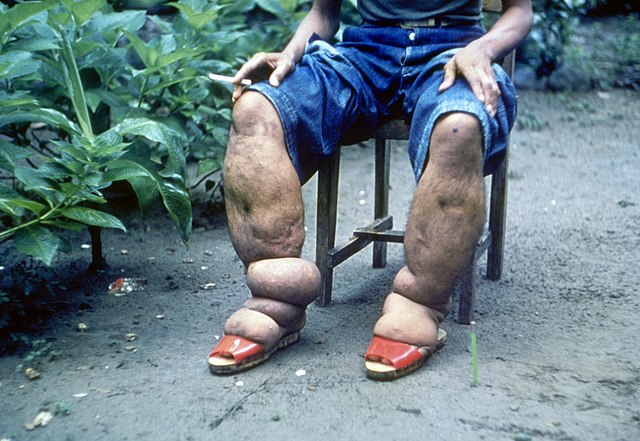
https://upload.wikimedia.org/wikipedia/commons/thumb/5/52/Elephantiasis.jpg/640px-Elephantiasis.jpg
Aetiology
The lymphatic system is a network of vessels and nodes (glands) that form part of both the vascular system and the immune system. As blood circulates around the body, some of the plasma escapes through the capillaries and into the extracellular space. Any fluid that is not reabsorbed into the capillaries is drained via the lymphatic system. Lymphatic vessels are found throughout the body; they carry the extracellular fluid, protein, and waste products (lymph) back to the blood stream and systemic circulation by ultimately draining into the left and right subclavian veins. Along the way, lymph passes through lymph nodes, where microorganisms are presented to immune cells.
Lymphoedema becomes apparent once ≥80% of lymphatic drainage to a body part is impaired (lymphostasis). Accumulation of lymph in the tissues leads to fat hypertrophy, the subcutaneous tissues become thickened and the normal functioning of the immune system is impaired. Over time this leads to further tissue inflammation and fibrosis.
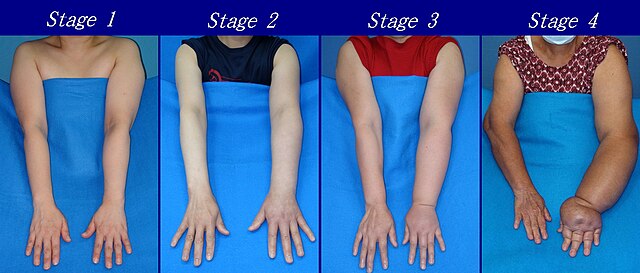
Primary lymphoedema:
- Congenital abnormalities of the lymphatic system lead to reduced function; drainage of lymph is compromised as a result.
- Can be sporadic (most common), familial or as part of a syndrome (e.g. Milroy’s disease, Noonan syndrome and Turner syndrome).
- Most commonly affects the lower limbs.
Secondary lymphoedema:
- Occurs after damage to a previously functioning lymphatic system.
- Causes include: infection, trauma, malignancy and its treatment (radiotherapy and surgery).
- Lymphoedema will become apparent 1-2 years after the initial damage to the lymphatic system.
- E.g. breast cancer: patients frequently undergo axillary lymph node biopsy, dissection or radiotherapy (leading to scarring and fibrosis).
- E.g. lymphatic filariasis: caused by one of three species of parasitic nematodes (roundworms): Wuchereria bancrofti, Brugia malayi or Brugia timori; only Wuchereria bancrofti is found in the Americas.
- Lymphatic drainage is impaired by either direct obstruction (from roundworms) or local tissue inflammation.
Diagnosis
Lymphoedema is a clinical diagnosis based on history and examination. Occasionally laboratory tests will be required if an infective cause (filariasis) is suspected.
History
- Progressive painless swelling (initially distal, then moves proximal)
- Frequently unilateral limb affected
- 90% of cases – lower limb
- Family history – ?primary lymphoedema
- Past travel to filariasis endemic areas
- Malignancy
- Radiotherapy
- Surgical history (especially lymph node biopsy/dissection or surgery in high-risk areas of the axilla/groin)
- Trauma or penetrating injuries to axilla and/or groin
Examination:
- Non-tender swelling and oedema of an extremity
- In early lymphoedema, there may be pitting oedema.
- In late-stage lymphoedema, oedema is usually non-pitting (due to fat hypertrophy and fibrosis of tissue).
- Positive Stemmer’s sign: the examiner is unable to pinch the skin on the dorsum of the patient’s second toe. However a negative Stemmer’s sign doesn’t rule out lymphoedema.
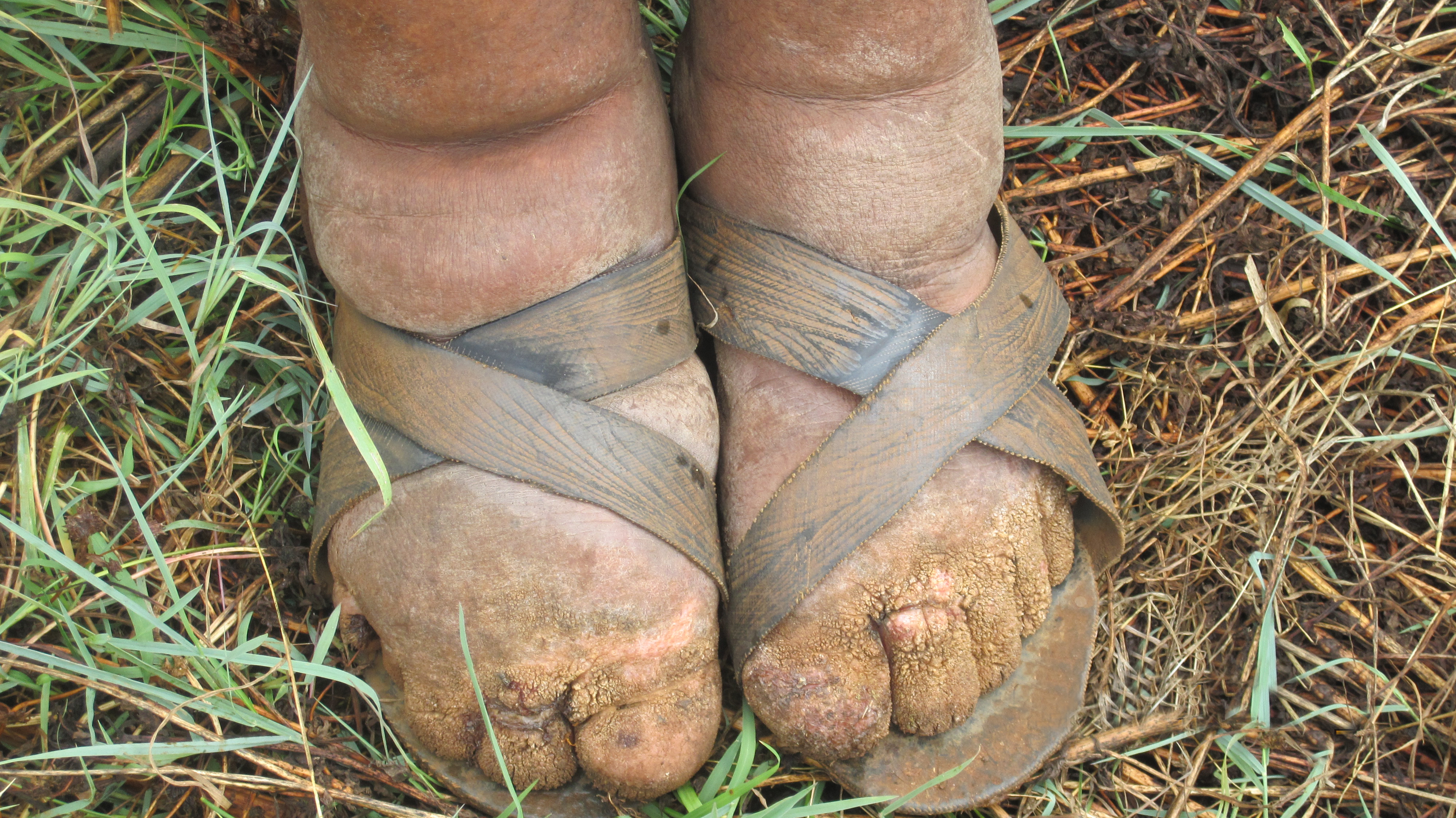
- Skin changes: papillomatosis (projection of dermal papillae in the skin, producing a rough texture) and hyperkeratosis (thickening of the outer skin layer – stratum corneum).
- In advanced lymphoedema, the chronic skin changes can lead to poor wound healing and skin breakdown.
Investigations
- If the diagnosis is unclear, lymphoscintigraphy is an imaging modality that can be used to visualise the lymphatic system and identify abnormalities.
- Lymphoscintigraphy is 96% sensitive and 100% specific for lymphoedema.
- Blood smear (for microfilariae) if lymphatic filariasis is suspected.
- Primary lymphoedema – genetic testing may be performed if syndromic or familial causes are suspected.
Management
Lymphoedema is best managed in a specialist centre with a multi-disciplinary team approach. The mainstay of lymphoedema management is conservative management:
- Elevation of the affected area (although results are limited and effect is usually temporary)
- Exercise – particularly strength training.
- Skin care, such as regular cleaning and moisturising, to maintain healthy skin and reduce the likelihood of infection.
- Protective clothing and shoes to prevent damage to skin.
- Static compression garments or bandaging can help reduce swelling. Garments can be tightened over time, and have been shown to reduce the volume of upper-limb lymphoedema by up to 47%.
- Intermittent pneumatic compression devices (as are used for VTEP in hospitals) may be used alongside static compression garments.
- Combined physical therapy (or CPT) performed by trained therapists; involves skin care, bandaging, manual lymph drainage and exercises.
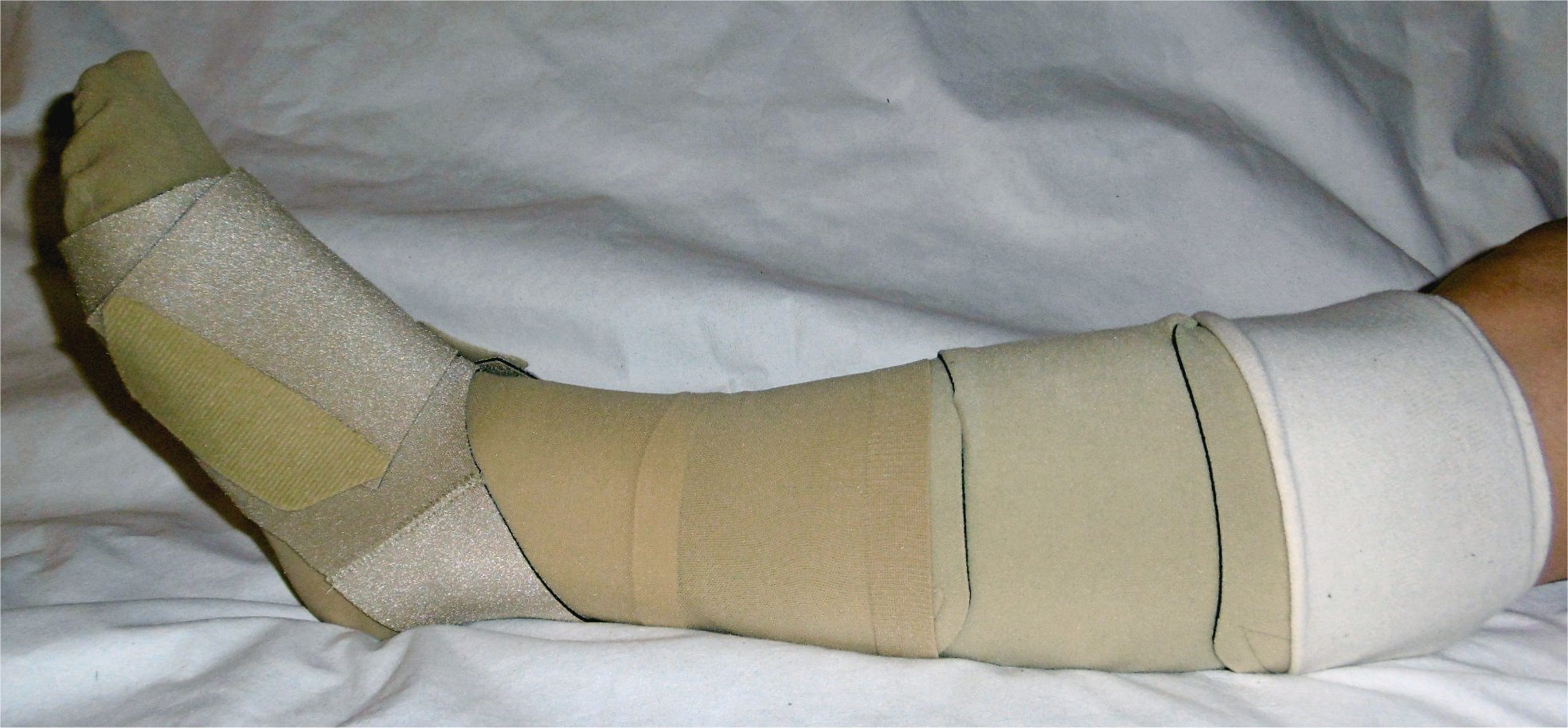
https://commons.wikimedia.org/wiki/File:Klettbandage_mit_Fu%C3%9Fteil.jpg
Medical management
Lymphatic filariasis is treated with combinations of antiparasitic drugs such as diethylcarbamazine and albendazole (depending on the patient’s country of origin), this should be under guidance from an infectious disease specialist.
Patients with underlying infection or cellulitis related to lymphoedema should be treated with antibiotics as for cellulitis, although often require longer courses (and may not be as effective).
Surgical management
In some patients, surgery may be performed if conservative techniques have not helped or they are experiencing severe morbidity associated with their lymphoedema. Surgery can be used to either remove diseased tissue, or repair damaged and/or create new lymph vessels. Surgery is not a curative option and patients will still need to wear compression garments for life.
Complications
- Anxiety and depression is frequently seen in patients with lymphoedema
- Cellulitis
- Impairment in activities of daily living
- Lymphangiosarcoma (a rare malignancy seen in 0.5% lymphoedema patients)
Skin manifestations of anaemia
Aetiology and epidemiology
Anaemia is defined as a low concentration of haemoglobin (Hb); it can be further categorised by mean cell volume (MCV) of the red blood cell (i.e. microcytic, normocytic or macrocytic) and each of these subtypes has their own aetiology. The skin manifestations of anaemia vary depending on the underlying cause. Approximately 33% of the population globally are thought to be anaemic.
Microcytic anaemia
- The most common cause of anaemia globally is iron-deficiency anaemia (IDA) – which is also by far the most common form of microcytic anaemia.
- IDA is more common in low-income countries (reduced dietary iron intake, parasitic worm infections causing blood loss), young children (who require greater amounts of iron for growth) and pre-menopausal women (due to blood loss during menstruation).
- Other uncommon causes of microcytic anaemia include the thalassaemias (genetic diseases of abnormal Hb production, these are more common in people of Mediterranean, Middle-Eastern or Southeast Asian descent), and sideroblastic anaemia. When discussing microcytic anaemia in this topic, this will largely be referring to IDA.
- Aetiology
- Reduced dietary intake of iron (poverty leading to malnutrition, meat-free diet)
- Reduced absorption of iron (inflammatory bowel disease, coeliac disease, helicobacter pylori infection)
- Bleeding (menstruation, gastrointestinal blood loss (malignancy, peptic ulcer disease, haemorrhoids), parasitic intestinal infections)
Normocytic anaemia
- The prevalence of normocytic anaemia increases with age; the most common form of normocytic anaemia is anaemia of chronic disease. Normocytic anaemia has been reported in up to 44% of men older than 85 years (in the USA).
- The causes of normocytic anaemia are numerous, including:
- Anaemia of chronic disease
- Acute blood loss
- Bone marrow failure
- Renal failure
- Pregnancy
Macrocytic anaemia
- The most common cause of macrocytic anaemia is B12 and/or folate deficiency; this will produce a megaloblastic anaemia, where abnormal red blood cell precursors are found in the bone marrow.
- Macrocytic anaemia may also be non-megaloblastic.
- Megaloblastic macrocytic anaemia: causes of B12 and folate deficiency include:
- Vegan diet (the main dietary sources of B12 are animal products such as meat, eggs, fish and dairy)
- Pernicious anaemia (most frequently diagnosed aged 60, associated with other autoimmune conditions, F>M at a ratio of 1.6:1)
- Disease of the terminal ileum (e.g. Crohn’s disease, tapeworm infection, small bowel bacterial overgrowth)
- Causes of a non-megaloblastic macrocytic anaemia:
- Alcohol excess
- Liver disease
- Myelodysplastic syndromes
- Hypothyroidism
Clinical findings in skin manifestations of anaemia
The skin manifestations of anaemia can be subtle and may not be evident until there is a significant drop in haemoglobin.
Skin pallor – including palm creases and mucous membranes (conjunctiva)
- The absence of pallor is not thought to be a reliable clinical sign in anaemia as it may be absent even in the severely anaemic; however the presence of pallor is more useful and can indicate severe anaemia.
- Occurs as blood is directed away from the skin and peripheries, and towards internal organs (to enhance oxygenation when the oxygen-carrying capacity of the blood is reduced).
Angular stomatitis
- It is thought that up to 1/4 of cases of angular stomatitis are secondary to nutritional deficiencies (including iron and B12).
- Overexposure of the delicate skin at the mouth corners (commissures) to saliva leads to skin breakdown, saliva is an irritant and contains many digestive enzymes. It is not completely understood how anaemia leads to angular stomatitis, however it is suggested that nutritional deficiencies reduce the body’s ability to regenerate cells and repair tissues – increasing the risk of conditions such as angular stomatitis.
- Manifests clinically as erythema and cracking and crusting of the mouth corners
Atrophic glossitis – (loss of papillae, leading to a smooth tongue, may be painful)
- Epidemiology: atrophic glossitis is strongly associated with nutritional deficiencies such as IDA and B12 deficiency (causing a macrocytic anaemia). Some studies suggest atrophic glossitis is present in up to 1% of adults and up to 7% of women (due to the increased prevalence of iron deficiency anaemia).
- Aetiology: iron, B12 and folate (along with other nutrients) are required for the production and repair of healthy oral epithelial cells. Therefore anaemia (from such nutritional deficiencies) can cause atrophy of the epithelial surface of the tongue, leading to loss of papillae and atrophic glossitis.
Koilonychia
- Up to 28% of people with IDA have nail changes.
- Kolionychia refers to spoon-shaped nails, and is usually preceded by thinning and flattening of the nails
- Epidemiology: koilonychia can be acquired, inherited or idiopathic, however the most common cause is IDA. Studies have found that up to 5.4% of patients with IDA have koilonychia.
- Aetiology: the pathophysiology of koilonychia is not fully understood, however it is hypothesised that reduced peripheral blood flow (secondary to anaemia) leads to disruption in the normal growth of the connective tissue of the nail bed.
Telogen effluvium
- A form of non-scarring alopecia, associated with increased hair shedding
- Epidemiology: anaemia has long been associated with increased hair loss, however studies have been conflicting. One recent meta-analysis (with over 10,000 total participants) concluded that women with non-scarring alopecia were significantly more likely to be iron deficient.
- Aetiology: as with other manifestations of anaemia, the pathophysiology of hair loss is not fully understood. Hair follicles require significant blood supply and nutrients for hair growth – these may both be compromised in anaemia.
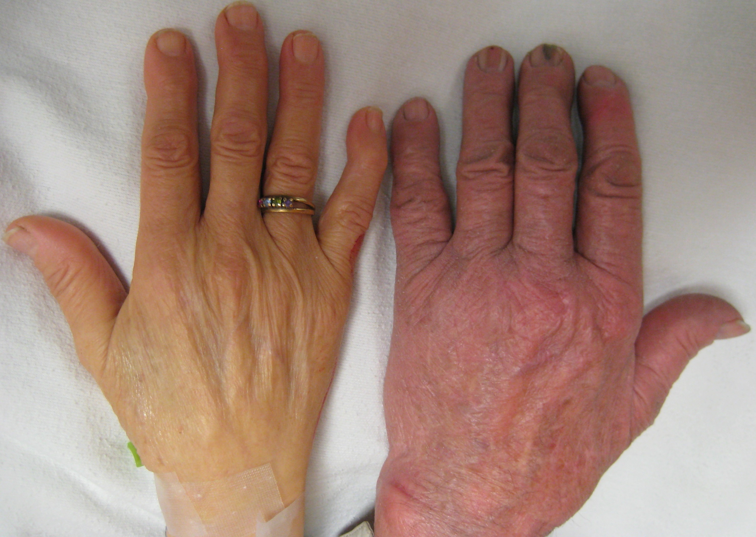
https://en.wikipedia.org/wiki/File:Anemia.JPG
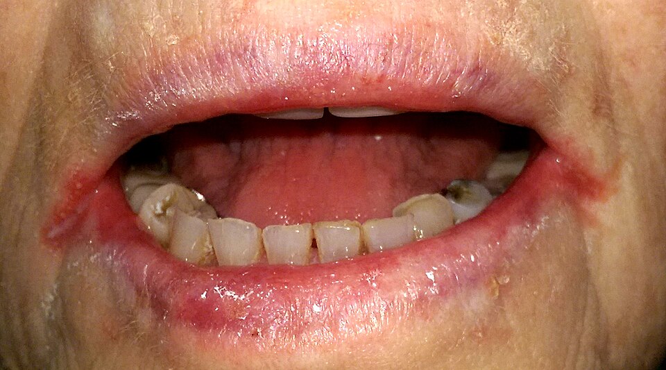
https://en.wikipedia.org/wiki/File:Angular_cheilitis1.jpg
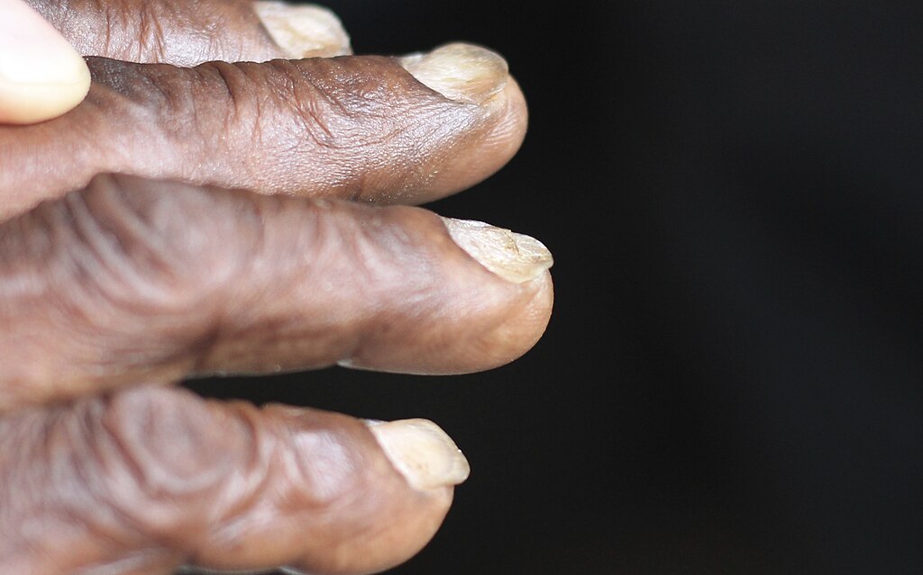
https://en.wikipedia.org/wiki/Koilonychia#/media/File:Koilonychia_iron_deficiency_anemia.jpg
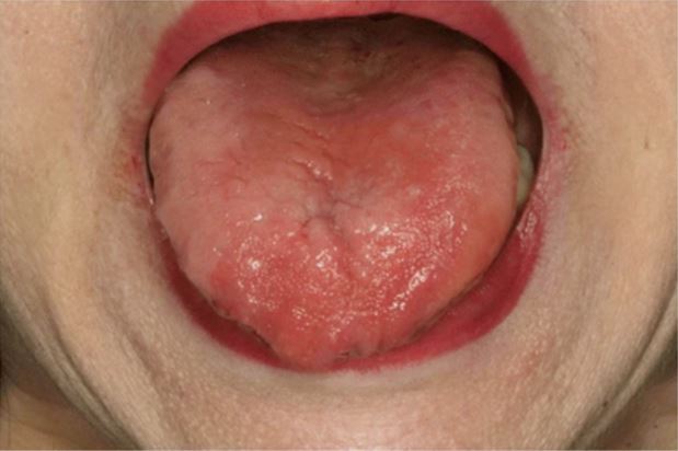
https://upload.wikimedia.org/wikipedia/commons/thumb/3/37/Image_of_the_tongue_in_a_B12-
Medical management
If a patient is found to be anaemic, the underlying cause should be identified and treated (to prevent recurrence). For example in women with IDA secondary to menorrhagia, contraceptive devices and/or tranexamic acid may be used to reduce blood loss.
Patients with IDA will likely require iron replacement to restore iron stores and correct their anaemia.
Treatment options include:
- Increased dietary iron. Haem iron is found in red meat – this is easily absorbed; the absorption of non-haem iron (in vegetables and grains) is increased with vitamin C.
- Oral iron supplementation
- Intravenous iron infusions
- Blood transfusion (in exceptional circumstances)
Complications
Once anaemia is treated, the skin, hair and nail manifestations should slowly recover with time.
Polycythaemia
Polycythaemia refers to an increase in the red cell mass in the blood (erythrocytosis), this can be due to either a primary myeloproliferative disorder (polycythaemia vera – PV) or secondary to chronic hypoxia (e.g. due to underlying lung disease +/- sleep apnoea). Less commonly, polycythaemia may be present due to an erythropoietin-secreting tumour, exogenous steroid use or a congenital disorder. The increase in red cell mass produces clinical signs and symptoms related to hyperviscosity of the blood; we will focus on the skin manifestations of polycythaemia in this sub-chapter.
Epidemiology
Current data indicates that PV is most common in white people aged 60 and above, with the median age of diagnosis at 65 years. PV has been reported more frequently in people of Ashkenazi Jewish descent, and it is very rarely diagnosed in children (fewer than 100 documented cases). However, there is little research data available on PV outside of the USA and Europe.
Skin manifestations of polycythaemia
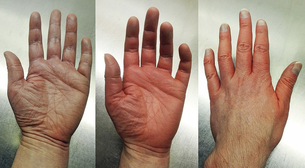
The skin manifestations of polycythaemia are due to the increased red cell volume of the blood (and hence increased circulating blood volume) and hyperviscosity of the blood.
- Plethoric complexion
- As polycythaemia progresses, the venules and capillaries in the skin must distend to accommodate the increase in blood volume
- Leads to a ruddy and plethoric complexion
- The face, palms, earlobes and mucous membranes of the eyes are frequently affected.
- Aquagenic pruritus
- Itchy skin triggered by water contact – is another skin manifestation of polycythaemia.
- Cause unknown but multiple underlying mechanisms have been suggested – such as the release of inflammatory mediators from mast cell degranulation or platelet aggregation.
- Erythromelalgia
- Condition that produces intermittent attacks of redness and a burning pain of the feet and hands.
- It can be a primary condition in itself, or secondary to haematological disorders such as polycythaemia.
- When associated with polycythaemia, it is thought that increased platelet activation causes the formation of thrombi in arterioles within the distal extremities; this results in tissue hypoxia and pain.
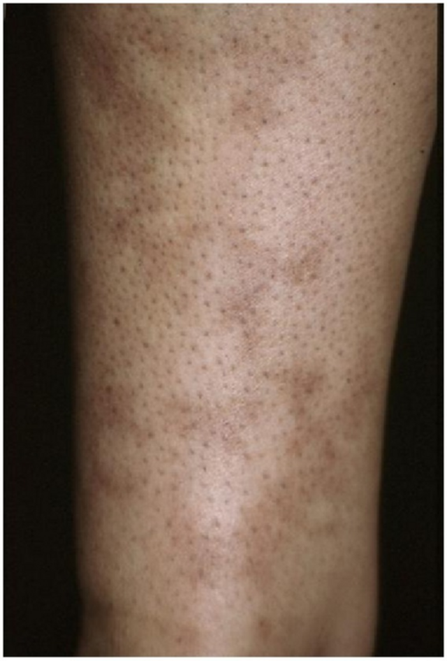
- Livedo reticularis describes
- A net-like (or reticular) skin discolouration, with purplish rings surrounding a paler centre.
- Results from impaired blood flow to the skin; in polycythaemia, this is due to the viscous blood flowing sluggishly through the blood vessels in the skin.
Diagnosis
Dermatological findings in polycythaemia:
Medical management
The management of polycythaemia depends on the underlying cause; there is no cure for PV and so management focuses on symptomatic relief and the prevention of complications. For PV patients under 60 years and at low-risk of thrombosis, phlebotomy and low-dose aspirin are first-line treatments. For PV patients over 60 years and/or at high-risk of thrombosis, cytoreductive therapy (such as with hydroxycarbamide) and low-dose aspirin are first-line. As a result, the reduction in circulating red cell volume should improve some of the skin manifestations of polycythaemia.
For aquagenic pruritus several different treatments have been suggested, such as antihistamines, aspirin, SSRIs and JAK2 inhibitors in refractory cases.
For erythromelalgia secondary to polycythaemia, aspirin is the first-line treatment.
Complications
PV can progress to post-PV myelofibrosis, a form of bone marrow malignancy where the bone marrow becomes scarred. Current evidence suggests that none of the treatment options for PV will reduce the likelihood of progression to myelofibrosis. PV may also transform into an acute leukaemia, the prognoses for these patients are poor.
Cutaneous B-cell lymphoma
Cutaneous B-cell lymphomas (CBCL) are malignancies of B cells that develop in the skin, there should be no extra-cutaneous malignancy at diagnosis. They do not include lymphomas that start elsewhere in the body and then spread to the skin. B-cells are antibody-producing cells that form part of the immune system.
CBCL can be divided into 3 major types:
- Primary cutaneous follicle centre lymphoma (PCFCL)
- Primary cutaneous diffuse large B-cell lymphoma, leg-type (PCDLBCL-LT)
- Primary cutaneous marginal zone B-cell lymphoma (PCMZBCL)
The different forms of CBCL are produced, depending on when (in the development stage of the lymphocyte) the malignant mutation occurs. The very rare forms of CBCL will not be covered in this topic.
Epidemiology
- About 100 people in the UK are diagnosed with CBCL every year.
- PCFCL is the most common form of the 3 types of CBCL.
- PCDLBCL-LT has the poorest prognosis, only 50% of patients are alive after 5 years, it affects half as many men as women.
- PCMZBCL – men are more commonly affected than women; it has been associated with the bacteria Borrelia burgdorferi (known to cause Lyme disease).
Aetiology
The aetiology of CBCL is not entirely known; it has been proposed that CBCL stems from chronic inflammation in the skin (e.g. from bacterial or viral infections) which gradually transforms into a lymphoma. Viruses such as human herpes virus 8 (HHV-8) and Epstein Barr virus (EBV) are known to be strongly associated with some non-cutaneous lymphomas, however the data for causative agents in CBCL is lacking.
CBCL has also been associated with methotrexate, and has been found to improve once methotrexate is stopped – perhaps due to its immunosuppressive effect allowing the unchecked proliferation of B-cells.
Diagnosis
CBCL presents with skin changes including papules, nodules, plaques and tumours. They can range in colour from pink/red to blue/purple. PCFCL and PCMZBCL are often found on the upper extremities, trunk or scalp, whereas PCDLBCL-LT is generally found on the legs.
If CBCL is suspected from the history and examination, a skin biopsy is required for diagnosis; this must be sufficiently deep and include the subcutaneous fat. Histological and cytological examination of the biopsy will be performed, plus immunohistochemistry.
- Primary cutaneous follicle centre lymphoma:
- Histology/cytology – accumulation of germinal centre B-cells (centrocytes and centroblasts) in the dermal and subcutaneous tissues, these form a follicular growth pattern.
- Immunohistochemistry is positive for B-cell antigens (e.g. CD19, CD20 and CD22).
- It is negative for the chromosomal t(14;18) translocation – which is found in generalised follicular lymphoma. This is helpful to distinguish PCFCL from follicular lymphoma that has spread to the skin.
- Primary cutaneous marginal zone B-cell lymphoma:
- Histology/cytology – infiltration of the dermis with marginal zone B-cells (and other immune cells) in a nodular or diffuse pattern.
- Immunohistochemistry is positive for B-cell antigens CD79a, CD20 and Bcl-2.
- Blood tests for Borrelia burgdorferi may be performed, given its association with PCMZBCL.
- Primary cutaneous diffuse large B-cell lymphoma, leg-type:
- Histology/cytology – dermal and subcutaneous fat infiltrates of centroblasts and immunoblasts (B-cell precursors), these may form a sheet pattern.
- Immunohistochemistry is positive for B-cell antigens CD79a, CD20 and IgM.
- Bcl-2 and MUM1 expression is significant for PCDLBCL-LT.
- The presence of palpable lymphadenopathy indicates extra-cutaneous involvement and the need for further imaging – such as a CT or PET-CT scan.
Management
Primary cutaneous follicle centre lymphoma:
- A single lesion or limited area of disease will be treated with radiotherapy or surgery.
- Numerous lesions may be treated with a range of therapies including topical agents, cryotherapy, radiation or systemic treatments.
- Disseminated or very large lesions may be treated with chemotherapy (e.g. R-CHOP).
- PCFCL may sometimes be observed without treatment, as lesions may remain stable in size or may even subside.
Primary cutaneous marginal zone B-cell lymphoma:
- As with PCFCL, a single lesion or limited area of disease may be treated with radiotherapy or surgery (note – relapse rates for radiotherapy are higher for PCMZBCL).
- Numerous lesions may be treated with a range of therapies including chemotherapy or rituximab.
- Disseminated or very large lesions may be treated with chemotherapy (e.g. R-CHOP).
- PCMZBCL in patients with Borrelia burgdorferi can be treated with antibiotics.
Primary cutaneous diffuse large B-cell lymphoma, leg-type:
- Limited or diffuse disease is treated with radiotherapy +/- R-CHOP.
Complications
Primary cutaneous follicle centre lymphoma:
- Close to 95% of patients are alive at 5 years post-diagnosis.
- Disease recurs in >45% patients.
- In 5 – 10% patients, PCFCL will metastasise to extracutaneous sites.
Primary cutaneous marginal zone B-cell lymphoma:
- Up to 99% of patients are alive at 5 years post-diagnosis.
- Metastasis is rare
- PCMZBCL may transform into systemic diffuse large B-cell lymphoma, but this is very rare
Primary cutaneous diffuse large B-cell lymphoma, leg-type:
- Nearly ≥60% of patients relapse after treatment
- In 30% the disease will spread to extracutaneous sites.
Coagulation disorders
Coagulation disorders can produce a range of clinical signs, depending on whether there is excessive bleeding (hypocoagulation), or excessive clotting (hypercoagulation). This chapter will cover the skin manifestations of coagulation disorders.
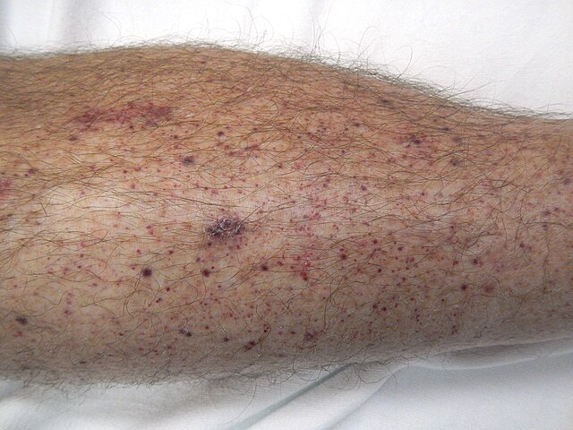
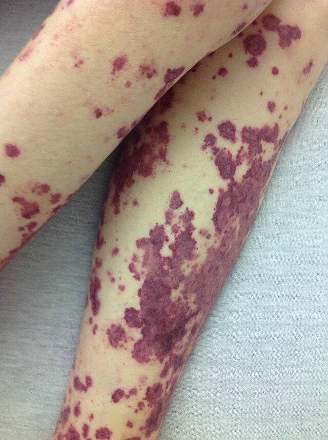
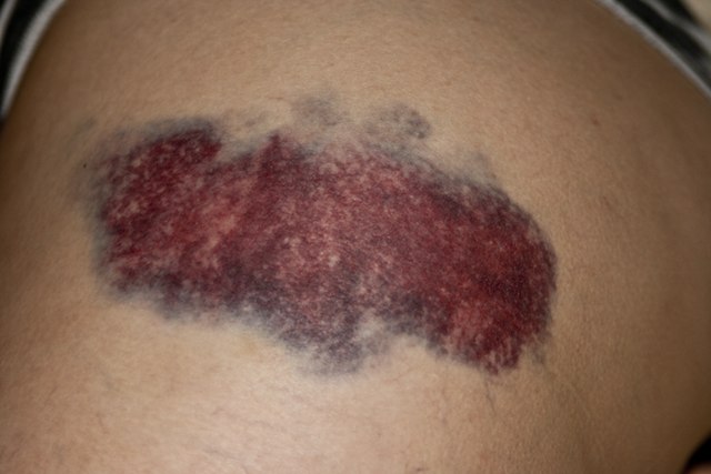
Petechiae, purpura and ecchymoses are descriptive terms for areas of non-blanching, purple-black discolouration – secondary to haemorrhage into the skin. Petechiae are generally <4mm in diameter, purpura are 4-10mm and ecchymoses (colloquially = bruise) are >10mm.
Livedo reticularis (as described previously in ‘polycythaemia’) has multiple causes, including disorders of hypercoagulation; when associated with an underlying disorder it is usually termed ‘secondary livedo reticularis’ or ‘livedo racemosa’. Non-healing ulcers and other atypical wounds (in the absence of arterial or venous disease) can also be a sign of an underlying hypercoagulable state.
Epidemiology
- Occasional petechiae (1-2) have been found in over 25% of well children, and are not considered concerning (unless associated with other clinical signs and symptoms).
- Petechiae and purpura are more commonly seen in young children and older adults.
- Ecchymoses are more common in:
- Older adults, due to thin skin and delicate blood vessel walls.
- Young children due to bumps and falls (note – bruises on the buttocks, chest and abdomen are considered abnormal and may indicate abuse).
- People who take part in contact sports.
- Livedo reticularis:
- A temporary form (termed ‘cutis marmorata’) is found in up to 50% of healthy young children and frequently seen in young women; it is a physiological response to the cold and not harmful.
Aetiology
Petechiae, purpura and ecchymoses result from damage to blood vessels within the skin (causing leakage of blood into surrounding tissues) or thrombocytopaenia (causing spontaneous bleeding without preceding injury).
Causes include:
- Disorders of reduced platelet production, e.g. leukaemia and bone marrow failure.
- Disorders of increased platelet consumption/destruction, e.g. thrombotic thrombocytopaenic purpura, haemolytic uraemic syndrome and idiopathic thrombocytopaenic purpura, SLE.
- Disseminated intravascular coagulation (DIC) can occur secondary to malignancy or severe infection; activation of the coagulation pathways cause widespread clotting (using up coagulation factors and platelets) which then leads to excessive bleeding.
- Drugs affecting platelet function, e.g. NSAIDs, aspirin and clopidogrel.
- Drugs which act on clotting factors, e.g. heparin, warfarin, DOACs.
- Severe clotting disorders such as haemophilia A or B tend not to present with petechiae or purpura (which are more indicative of thrombocytopaenia), but do present with ecchymoses, haematomas and haemarthrosis. This is due to a deficiency of factor VIII (haemophilia A) or IX (haemophilia B), leading to delayed clot formation; any clot that is formed is fragile and frequently dislodged.
- Other disorders causing clotting factor deficiency, e.g. vitamin K deficiency, advanced liver disease, Von Willebrand disease.
Pathological/secondary livedo reticularis can be due to a range of underlying systemic diseases, coagulation disorders which can cause it include:
- Antiphospholipid syndrome – antiphospholipid antibodies produce a prothrombotic state, resulting in blood clots. When small clots form within the skin’s blood vessels, blood flow is disturbed and this produces livedo reticularis.
- Protein C, deficiency, protein S deficiency and antithrombin III deficiency are thought to cause livedo reticularis by a similar prothrombotic mechanism.
- Skin ulcers may also occur due to a similar mechanism of blood vessel obstruction.
Diagnosis
Petechiae, purpura and ecchymoses are diagnosed via a thorough history and clinical examination. Once diagnosed, the underlying cause may require further investigation. This will initially involve blood tests such as FBC, clotting studies and vitamin K levels – or a septic screen if infection is suspected.
https://upload.wikimedia.org/wikipedia/commons/thumb/6/63/Pferdekuss.jpg/640px-Pferdekuss.jpg
Livedo reticularis is diagnosed via history and examination; if there are features suspicious of an underlying coagulation disorder, initial investigations will involve blood tests including:
- Full blood count
- Clotting studies
- Antibody panel (e.g. lupus anticoagulant, anticardiolipin antibodies)
- Occasionally, punch biopsies of the rash may be taken for histopathology and diagnosis
Management
Management of the skin manifestations of coagulation disorders relies on identifying and treating the underlying cause.
There is no specific treatment for petechiae, purpura or ecchymoses, however simple analgesia (such as paracetamol) will help to alleviate pain and discomfort. NSAIDs are not advised as they can worsen existing bleeding.
Secondary livedo reticularis may be treated with anti-platelets or anticoagulants.
Complications
Petechiae, purpura and ecchymoses will self-resolve with time. Livedo reticularis and other skin signs are not harmful in themselves, but untreated coagulation disorders are associated with increased morbidity and mortality.
Hereditary Haemorrhagic Telangiectasia
Hereditary haemorrhagic telangiectasia (HHT) – sometimes known by its other name ‘Osler-Weber-Rendu syndrome’ – is a rare disorder of autosomal dominant inheritance. It is characterised by abnormal vasculature, including telangiectasia of the mucous membranes and skin, and arteriovenous malformations of the lung, liver and central nervous system. This produces characteristic skin signs and an increased tendency to bleed. This chapter will focus on the cutaneous signs of HHT.
Epidemiology
Worldwide, HHT is thought to affect between 1 in 5000 to 1 in 10,000 people. It is more commonly diagnosed in women, however this is likely due to men being less engaged with healthcare services. Given HHT is a multi-system disorder that can present in numerous different ways (e.g. epistaxis, gastrointestinal (GI) bleeds and intracerebral bleeds) medical professionals may fail to link the presentations to a unifying diagnosis; given all of this, HHT is probably under diagnosed.
Aetiology
The majority of HHT cases are caused by a mutation in one of two genes involved in blood vessel development: activin receptor-like kinase 1 gene (chromosome 12) or the endoglin gene (chromosome 9). This mutated gene (inherited in an autosomal dominant pattern) produces abnormal blood vessels without the usual capillaries between arteries and veins (arteriovenous malformations – AVM). When small these are described as telangiectasia (telangiectases – plural), when large arteries and veins are involved they are called AVMs. AVMs are weak and have an increased risk of bleeding.
In HHT, patients will often present with epistaxis due to AVMs within the nose. They can often be spontaneous or after light trauma, however the severity of bleeding may vary hugely between different patients.
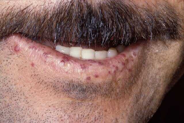
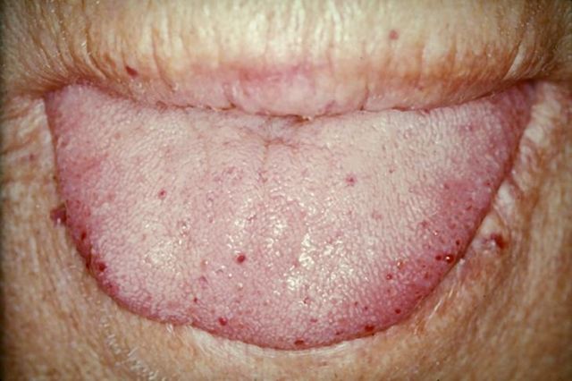
Telangiectases on the mucous membranes, lips, and face produce the most characteristic skin signs of HHT.
Diagnosis
A diagnosis of HHT is made using the ‘Curacao’ diagnostic criteria:
- Epistaxis – must be recurrent and spontaneous
- Telangiectases – multiple, on the mucous membranes, lips, face and hands
- AVMs – internal organ involvement
- First-degree relative with a diagnosis of HHT
For a definite diagnosis of HHT, a patient must meet at least three of the above criteria.
Clinical signs of HHT:
https://upload.wikimedia.org/wikipedia/commons/thumb/2/2dCase_115.jpg/640px-Case_115.jpg
https://upload.wikimedia.org/wikipedia/commons/thumb/7/77/TongueTelang.JPG/640px-TongueTelang.JPG
Medical management
It is not possible to prevent or cure HHT, however the symptoms can be managed. Mild HHT (up to 1/3 of those diagnosed) may not require any treatment.
- Topical intranasal treatment with timolol or sirolimus can reduce the frequency and severity of nosebleeds.
- Tranexamic acid can reduce the severity of bleeds.
- Bevacizumab and thalidomide inhibit angiogenesis and are being investigated for their use in the treatment of HHT.
Surgical management
- Laser therapy for the treatment of skin telangiectases.
- Laser therapy can also be used to manage mild-moderate epistaxes. Severe epistaxis may require skin transplants to the septum (septal dermoplasty).
- Internal organ AVMs often require surgical management.
Complications
- Anaemia
- Epistaxis
- Pulmonary AVMs and pulmonary haemorrhage
- Central nervous system AVMs and stroke or spinal cord bleed
- GI AVMs and GI bleed
- Hepatic AVMs and hepatic bleed – can also lead to hepatic failure or cardiac failure
References
https://ajronline.org/doi/pdf/10.2214/
ajr.178.2.1780405#:~:text=Lymphoscintigraphy%20is%20now%20the%20primary,reliable%20and%20reproducible%20%5B2%5D.
https://bestpractice.bmj.com/topics/en-gb/610
https://www.britannica.com/science/lymphatic-system
https://dermnetnz.org/topics/lymphoedema
https://www.ncbi.nlm.nih.gov/pmc/articles/PMC6635205/
https://www.ncbi.nlm.nih.gov/books/NBK562206/
#:~:text=Introduction-,Hyperkeratosis%20refers%20to%20the%20increased%20thickness%20of%20the%20stratum%20corneum,their%20nucleus%20and%20cytoplasmic%20organelles
https://www.nhs.uk/conditions/lymphoedema/
https://www.who.int/news-room/fact-sheets/detail/lymphatic-filariasis
https://www.aafp.org/pubs/afp/issues/2000/1115/
p2255.html#:~:text=Its%20prevalence%20increases%20with%20age,patients%20hospitalized%20by%20family%20physicians.
https://academic.oup.com/qjmed/article/111/4/271/4665617
https://bestpractice.bmj.com/topics/en-gb/94
https://dermnetnz.org/topics/angular-cheilitis
https://dermnetnz.org/topics/iron-deficiency
https://karger.com/sad/article/8/2/83/826740/Iron-Deficiency-and-Nonscarring-Alopecia-in-Women
https://www.ncbi.nlm.nih.gov/books/NBK536929/
https://www.ncbi.nlm.nih.gov/pmc/articles/PMC5838265/
#:~:text=It%20results%20from%20malnutrition%20or,underlying%20nail%E2%80%90connective%20tissue%203.
https://otorhinolaryngologypl.com/api/files/view/5025.pdf
https://patient.info/doctor/macrocytosis-and-macrocytic-anaemia#nav-2
https://pubmed.ncbi.nlm.nih.gov/10555095/
https://www.sciencedirect.com/science/article/pii/S0929664619303481
https://www.sciencedirect.com/topics/medicine-and-dentistry/
pallor#:~:text=Pallor%20is%20the%20most%20commonly,blood%20flow%20to%20vital%20organs.
https://www.wjoanemia.com/doi/WJOA/pdf/10.5005/jp-journals-10065-0011
Wilkinson, I., Raine, T., Wiles, K., Goodhart, A., Hall, C., O’Neill, H., 2017. Oxford handbook of clinical medicine, Oxford: Oxford University Press.
https://bestpractice.bmj.com/topics/en-gb/178?q=Polycythaemia%20vera&c=suggested
https://dermnetnz.org/topics/livedo-reticularis
https://www.ncbi.nlm.nih.gov/pmc/articles/PMC3803410/pdf/
QBullNorthwestUnivMedSch-29-1-8_3.pdf
https://www.ncbi.nlm.nih.gov/books/NBK557660/
https://www.ncbi.nlm.nih.gov/books/NBK557787/
#:~:text=Abnormal%20aggregation%20and%20consumption%20of,important%20role%20in%20this%
20pathophysiology.
https://pubmed.ncbi.nlm.nih.gov/20597963/
https://pubmed.ncbi.nlm.nih.gov/94209/
https://www.sciencedirect.com/science/article/abs/pii/
S0031302516354915#:~:text=The%20mechanism%20of%20water%2Dinduced,be%20responsible%20f
or%20the%20pruritus.
https://dermnetnz.org/topics/cutaneous-b-cell-lymphoma
https://dermnetnz.org/topics/primary-cutaneous-follicle-centrelymphoma#:~:text=Primary%20cutaneous%20follicle%20centre%20lymphoma%20(PCFCL)
%20is%20a%20low%2D,at%20the%20time%20of%20diagnosis.
https://dermnetnz.org/topics/cutaneous-marginal-zone-lymphoma
https://lymphoma-action.org.uk/types-lymphoma-skin-lymphoma/skin-cutaneous-b-cell-lymphoma
https://www.ncbi.nlm.nih.gov/pmc/articles/PMC4631235/
https://adc.bmj.com/content/86/4/291
https://bestpractice.bmj.com/topics/en-gb/468/aetiology
https://dermnetnz.org/topics/bleeding-and-bruising
https://dermnetnz.org/topics/livedo-reticularis
https://dermnetnz.org/topics/purpura
https://dermnetnz.org/topics/skin-manifestations-of-haematological-diseases
https://www.ncbi.nlm.nih.gov/books/NBK1404/
https://www.ncbi.nlm.nih.gov/pmc/articles/PMC6797011/
Dabiri, G., et al. 2016, ‘Coagulation disorders and their cutaneous presentations: Diagnostic work-up and
treatment’, Journal of the American Academy of Dermatology, vol 74(5):795-804.
Weir, N.U., et al. 1995, ‘Livedo reticularis associated with hereditary protein C deficiency and recurrent
thromboembolism’, British Journal of Dermatology, vol 132, 283-285.
https://curehht.org/understanding-hht/diagnosis-treatment/diagnostic-criteria-hht/
https://dermnetnz.org/topics/hereditary-haemorrhagic-telangiectasia
https://www.ncbi.nlm.nih.gov/pmc/articles/PMC6443364/
https://www.sciencedirect.com/science/article/pii/S1098360021045937
Complete Guide to Clinical Genetics ST3 Application and Recruitment (2027)
Written by Medibuddy. Last updated: October 2026. Clinical Genetics is

Complete Guide to Clinical Radiology ST3 Application and Recruitment (2027)
Recruitment paused: NHS England is not running Clinical Radiology ST3+ recru

Complete Guide to Paediatric Surgery ST3 Application and Recruitment in United Kingdom (2026)
Written by Medibuddy. Last updated: October 2026. If you’re planning


Take your subscriptions with you
Our mobile app allows you to access your interview and exam question banks wherever you are.

