


 Close
Close
 4th June 2025
Coagulation disorders
Coagulation disorders can produce a range of clinical signs, depending on whether there is excessive bleeding (hypocoagulation), or excessive clotting (hypercoagulation). This chapter will cover the skin manifestations of coagulation disorders.
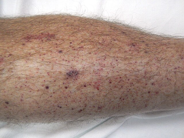
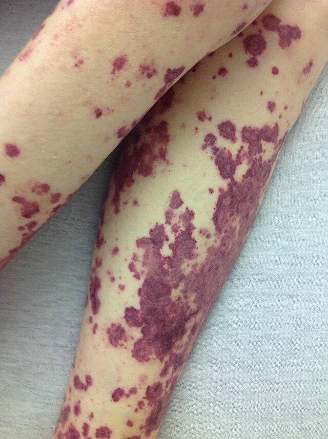
Petechiae, purpura and ecchymoses are descriptive terms for areas of non-blanching, purple-black discolouration – secondary to haemorrhage into the skin. Petechiae are generally <4mm in diameter, purpura are 4-10mm and ecchymoses (colloquially = bruise) are >10mm.
Livedo reticularis (as described previously in ‘polycythaemia’) has multiple causes, including disorders of hypercoagulation; when associated with an underlying disorder it is usually termed ‘secondary livedo reticularis’ or ‘livedo racemosa’. Non-healing ulcers and other atypical wounds (in the absence of arterial or venous disease) can also be a sign of an underlying hypercoagulable state.
Epidemiology
- Occasional petechiae (1-2) have been found in over 25% of well children, and are not considered concerning (unless associated with other clinical signs and symptoms).
- Petechiae and purpura are more commonly seen in young children and older adults.
- Ecchymoses are more common in:
- Older adults, due to thin skin and delicate blood vessel walls.
- Young children due to bumps and falls (note – bruises on the buttocks, chest and abdomen are considered abnormal and may indicate abuse).
- People who take part in contact sports.
- Livedo reticularis:
- A temporary form (termed ‘cutis marmorata’) is found in up to 50% of healthy young children and frequently seen in young women; it is a physiological response to the cold and not harmful.
Aetiology
Petechiae, purpura and ecchymoses result from damage to blood vessels within the skin (causing leakage of blood into surrounding tissues) or thrombocytopaenia (causing spontaneous bleeding without preceding injury).
Causes include:
- Disorders of reduced platelet production, e.g. leukaemia and bone marrow failure.
- Disorders of increased platelet consumption/destruction, e.g. thrombotic thrombocytopaenic purpura, haemolytic uraemic syndrome and idiopathic thrombocytopaenic purpura, SLE.
- Disseminated intravascular coagulation (DIC) can occur secondary to malignancy or severe infection; activation of the coagulation pathways cause widespread clotting (using up coagulation factors and platelets) which then leads to excessive bleeding.
- Drugs affecting platelet function, e.g. NSAIDs, aspirin and clopidogrel.
- Drugs which act on clotting factors, e.g. heparin, warfarin, DOACs.
- Severe clotting disorders such as haemophilia A or B tend not to present with petechiae or purpura (which are more indicative of thrombocytopaenia), but do present with ecchymoses, haematomas and haemarthrosis. This is due to a deficiency of factor VIII (haemophilia A) or IX (haemophilia B), leading to delayed clot formation; any clot that is formed is fragile and frequently dislodged.
- Other disorders causing clotting factor deficiency, e.g. vitamin K deficiency, advanced liver disease, Von Willebrand disease.
Pathological/secondary livedo reticularis can be due to a range of underlying systemic diseases, coagulation disorders which can cause it include:
- Antiphospholipid syndrome – antiphospholipid antibodies produce a prothrombotic state, resulting in blood clots. When small clots form within the skin’s blood vessels, blood flow is disturbed and this produces livedo reticularis.
- Protein C, deficiency, protein S deficiency and antithrombin III deficiency are thought to cause livedo reticularis by a similar prothrombotic mechanism.
- Skin ulcers may also occur due to a similar mechanism of blood vessel obstruction.
Diagnosis
Petechiae, purpura and ecchymoses are diagnosed via a thorough history and clinical examination. Once diagnosed, the underlying cause may require further investigation. This will initially involve blood tests such as FBC, clotting studies and vitamin K levels – or a septic screen if infection is suspected.

https://upload.wikimedia.org/wikipedia/commons/thumb/6/63/Pferdekuss.jpg/640px-Pferdekuss.jpg
Livedo reticularis is diagnosed via history and examination; if there are features suspicious of an underlying coagulation disorder, initial investigations will involve blood tests including:
- Full blood count
- Clotting studies
- Antibody panel (e.g. lupus anticoagulant, anticardiolipin antibodies)
- Occasionally, punch biopsies of the rash may be taken for histopathology and diagnosis
Management
Management of the skin manifestations of coagulation disorders relies on identifying and treating the underlying cause.
There is no specific treatment for petechiae, purpura or ecchymoses, however simple analgesia (such as paracetamol) will help to alleviate pain and discomfort. NSAIDs are not advised as they can worsen existing bleeding.
Secondary livedo reticularis may be treated with anti-platelets or anticoagulants.
Complications
Petechiae, purpura and ecchymoses will self-resolve with time. Livedo reticularis and other skin signs are not harmful in themselves, but untreated coagulation disorders are associated with increased morbidity and mortality.
Complete Guide to Clinical Genetics ST3 Application and Recruitment (2027)
Written by Medibuddy. Last updated: October 2026. Clinical Genetics is

Complete Guide to Clinical Radiology ST3 Application and Recruitment (2027)
Recruitment paused: NHS England is not running Clinical Radiology ST3+ recru

Complete Guide to Paediatric Surgery ST3 Application and Recruitment in United Kingdom (2026)
Written by Medibuddy. Last updated: October 2026. If you’re planning


Take your subscriptions with you
Our mobile app allows you to access your interview and exam question banks wherever you are.

